Review
. 2020 Mar 17:3:100046.
doi: 10.1016/j.jtauto.2020.100046. eCollection 2020.

Immunometabolism in the pathogenesis of systemic lupus erythematosus

Affiliations
Review

Immunometabolism in the pathogenesis of systemic lupus erythematosus

Chen-Xing Zhang et al. J Transl Autoimmun. .

Abstract

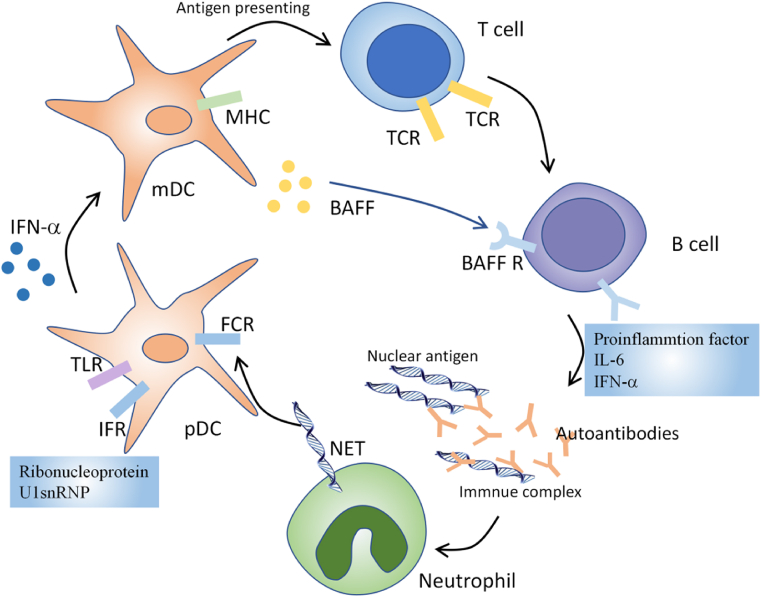
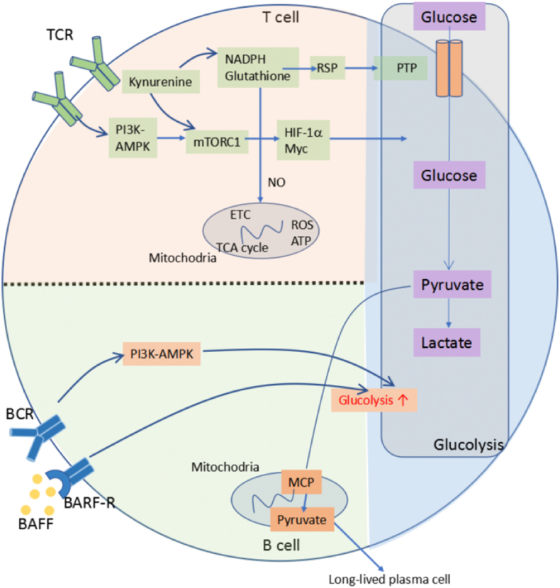
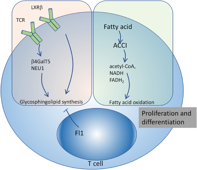
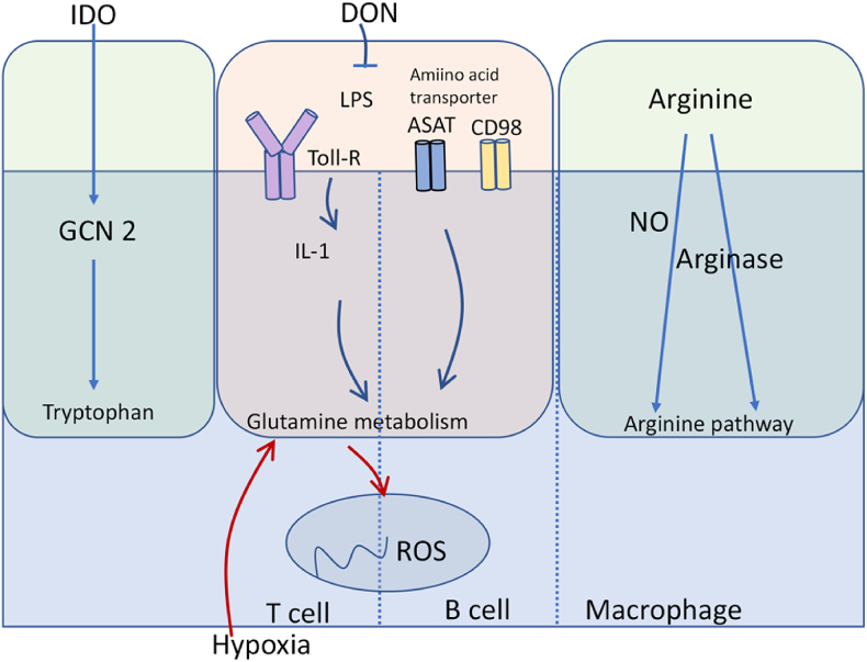
Systemic lupus erythematosus (SLE) is a typical autoimmune disease characterized by chronic inflammation and pathogenic auto-antibodies. Apart from B cells, dysregulation of other immune cells also plays an essential role in the pathogenesis and development of the disease including CD4+T cells, dendritic cells, macrophages and neutrophils. Since metabolic programs control immune cell fate and function, they are critical checkpoints in an effective immune response and are involved in the etiology of autoimmune disease. In addition, mitochondria and oxidative stress are both involved in cellular metabolism and is also essential in immune response. In this review, apart from the disturbed immune system, we will discuss mitochondrial dysfunction, oxidative stress, abnormal metabolism (including glucose, lipid and amino acid metabolism) of immune cells as well as epigenetic control of metabolism reprogramming to elucidate the underlying pathogenic mechanisms of systemic lupus erythematosus.

Keywords: Immune response; Metabolic programs; Pathogenesis; Systemic lupus erythematosus (SLE).


Conflict of interest statement

The authors declare that they have no known competing financial interests or personal relationships that could have appeared to influence the work reported in this paper.

Figures

Fig. 1
Fig. 1
Disturbed immune system in SLE. Nuclear particles activate Toll like receptors (TLR) on antigen-presenting cells, mainly dendritic cells and promote their maturation. Persistent activation of dendritic cells induces T cell activation and proliferation. Activated T cells then lead to mature autoreactive B cells. Furthermore, ribonucleoprotein and U1snRNP can induce type I IFN secretion by pDCs in SLE, which promotes the differentiation of activated B cells into plasmablasts and antibody-secreting plasma cells. Autoantibodies can bind to nuclear antigens, form immune complex and activate innate immune cells, which is a positive feedback loop and amplifies the pathogenic processes in SLE.
Fig. 2
Fig. 2
Glucose metabolic pathways in immune cells. The glucose metabolic pathway includes both glycolysis and oxidative phosphorylation. T cell receptor (TCR) stimulation activates mechanistic target of rapamycin complex 1 (mTORC1) through PI3K-AMPK pathway. Low levels of NADPH and glutathione leads to increased levels of mitochondrial reactive oxygen species (ROS) and decreased levels of ATP. It also contributes to mTORC1 activation, directly or through elevated levels of kynurenine. mTORC1 activation facilitates glucose metabolism through hypoxia-inducible factor 1α (HIF-1α) and Myc. In B cells, B cell activating factor (BAFF) and B cell receptor (BCR) signals increase glucose metabolism and glycolysis. This promotes pyruvate influx into the mitochondria, which is essential for the survival of long-lived plasma cells.
Fig. 3
Fig. 3
Lipid metabolic pathways in immune cells. Fatty acid oxidation pathway converts fatty acids into multiple intermediates (including acetyl-CoA, NADH and FADH2) for energy generation. Fatty acid synthesis is essential for activation-induced proliferation and differentiation of effector T cells.
Fig. 4
Fig. 4
Amino acid metabolic pathways in immune cells. Amino acids and their metabolism play a vital role in immune function. Glutamine has been demonstrated to be necessary for IL-1 induction upon LPS stimulation. Indoleamine-2,3-dioxygenase (IDO), is responsible for tryptophan catabolism through General control nonderepressible 2 (GCN2). The arginine pathway is mainly modulated by Arg-degrading enzymes such as NO synthase.

References

    1. Tsokos G.C. Systemic lupus erythematosus. N. Engl. J. Med. 2011;365:2110–2121. doi: 10.1056/NEJMra1100359. - DOI - PubMed
    1. Sang A., Zheng Y.Y., Morel L. Contributions of B cells to lupus pathogenesis. Mol. Immunol. 2014;62:329–338. doi: 10.1016/j.molimm.2013.11.013. - DOI - PMC - PubMed
    1. Herrada A.A., Escobedo N., Iruretagoyena M., Valenzuela R.A., Burgos P.I., Cuitino L., Llanos C. Innate immune cells contribution to systemic lupus erythematosus. Front. Immunol. 2019;10:772. doi: 10.3389/fimmu.2019.00772. - DOI - PMC - PubMed
    1. O’Neill L.A., Kishton R.J., Rathmell J. A guide to immunometabolism for immunologists. Nat. Rev. Immunol. 2016;16:553–565. doi: 10.1038/nri.2016.70. - DOI - PMC - PubMed
    1. Pearce E.L., Poffenberger M.C., Chang C.H., Jones R.G. Fueling immunity: insights into metabolism and lymphocyte function. Science. 2013;342:1242454. doi: 10.1126/science.1242454. - DOI - PMC - PubMed