[Preprint]. 2020 Jul 27:2020.07.27.222943.
doi: 10.1101/2020.07.27.222943.

Epitope-resolved profiling of the SARS-CoV-2 antibody response identifies cross-reactivity with an endemic human CoV

Affiliations

Epitope-resolved profiling of the SARS-CoV-2 antibody response identifies cross-reactivity with an endemic human CoV

Jason T Ladner et al. bioRxiv. .

Update in

Abstract

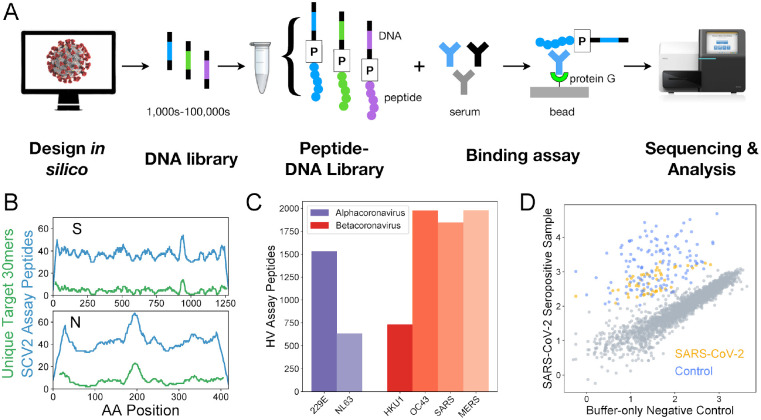
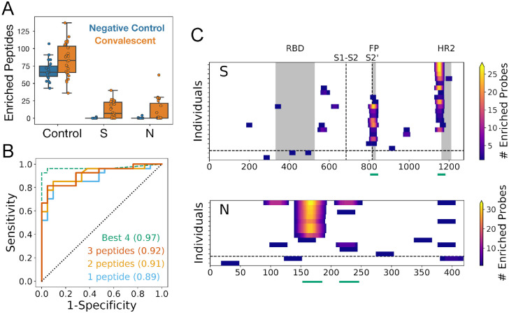
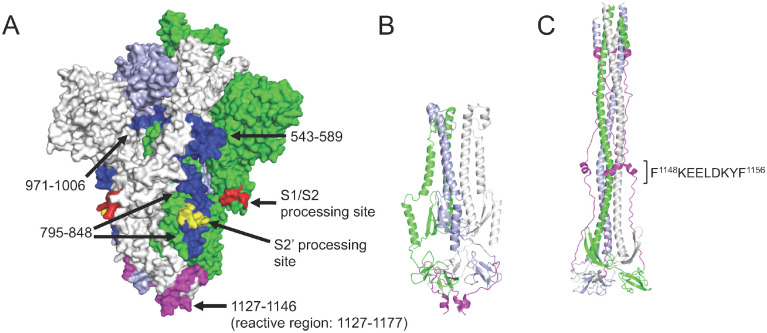
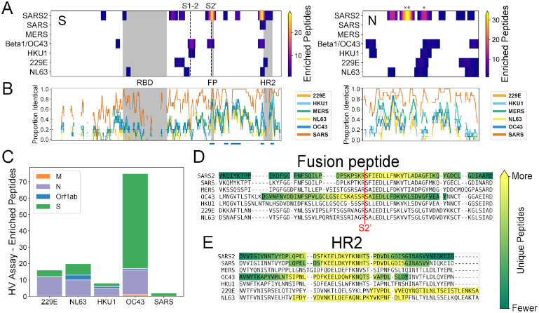
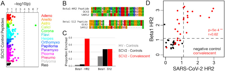
A high-resolution understanding of the antibody response to SARS-CoV-2 is important for the design of effective diagnostics, vaccines and therapeutics. However, SARS-CoV-2 antibody epitopes remain largely uncharacterized, and it is unknown whether and how the response may cross-react with related viruses. Here, we use a multiplexed peptide assay ('PepSeq') to generate an epitope-resolved view of reactivity across all human coronaviruses. PepSeq accurately detects SARS-CoV-2 exposure and resolves epitopes across the Spike and Nucleocapsid proteins. Two of these represent recurrent reactivities to conserved, functionally-important sites in the Spike S2 subunit, regions that we show are also targeted for the endemic coronaviruses in pre-pandemic controls. At one of these sites, we demonstrate that the SARS-CoV-2 response strongly and recurrently cross-reacts with the endemic virus hCoV-OC43. Our analyses reveal new diagnostic and therapeutic targets, including a site at which SARS-CoV-2 may recruit common pre-existing antibodies and with the potential for broadly-neutralizing responses.


Figures

Figure 1.
Figure 1.. Epitope-resolved CoV serology using a highly-multiplexed peptide-based assay (‘PepSeq’).
A) Platform for customizable highly-multiplexed peptide-based serology, comprising the following steps: (i) in silico design, (ii-iii) generation of a library of DNA-barcoded peptides from oligonucleotide templates using bulk in vitro reactions (transcription, ligation of a Puromycin (P)-containing adapter, translation, reverse transcription), (iv) serum binding assay and protein G capture, and (v) sequencing and analysis of the distribution of binders by their DNA barcodes. B) Peptide coverage depth across the SARS-CoV-2 spike (S) and nucleocapsid (N) proteins within the ‘SCV2’ peptide library. Peptide coverage depth (blue) correlates well with amino acid sequence diversity within the target SARS-CoV-2 sequences (green), calculated as the number of unique 30mers. C) Number of peptides within the HV library that were designed from each of the six human coronaviruses known prior to 2019. D) Example scatter plot illustrating SCV2 PepSeq assay results for a single serum sample. This plot shows normalized sequence read counts (log10 scale) for each peptide in the SCV2 library. Assay results using an antibody-free negative control are shown on the x-axis, while the results from a SARS-CoV-2 convalescent serum sample are shown on the y-axis. Grey circles represent unenriched peptides, with a strong correlation between the two assays, based on the starting abundance of the different peptides. Colored circles represent SARS-CoV-2 (orange) and non-SARS-CoV-2 control (blue) peptides that have been enriched through interaction with serum antibodies.
Figure 2.
Figure 2.. PepSeq identifies recurrent reactivities to SARS-CoV-2 peptides and classifies exposure status with high accuracy.
A) Boxplots showing the number of enriched SCV2 library peptides from assays with negative control (blue, n=21) and SARS-CoV-2 convalescent (orange, n=27) samples, divided into three different categories: non-SARS-CoV-2 control peptides (Control), and SARS-CoV-2 Spike (S) and Nucleocapsid (N) peptides. All three of these comparisons are statistically significant (t-test, p<0.05). Individual data points are shown as circles, the limits of the boxes correspond to the 1st and 3rd quartiles, the black line inside each box corresponds to the median and the whiskers extend to points that lie within 1.5 interquartile ranges of the 1st and 3rd quartiles. B) ROC curves for prediction of SARS-CoV-2 exposure based on peptide-level Z-scores calculated for all SCV2 library peptides (solid lines) and for a subset of four peptides identified through a decision tree analysis (dashed line). Positivity of the assay was determined by the enrichment of peptides designed from SARS-CoV-2, and the full library analysis was run with three different thresholds for the number of enriched peptides required for a sample to be considered positive. For the analysis using only the “Best 4” peptides, only a single enriched peptide was required for a positive result. For all analyses, the AUC (shown in parentheses) was ≥0.89. C) Heat maps showing the locations of enriched SARS-CoV-2 peptides within the S and N proteins. Each row represents a single serum/plasma sample and each plot includes only samples with at least one enriched peptide. Each position is colored according to the number of enriched peptides that overlap that position. The horizontal dashed line separates COVID-19 convalescent samples (top) from negative control samples (bottom). The vertical dashed lines in the S protein plot represent the S1-S2 and S2’ cleavage sites, respectively. Grey boxes indicate selected functional regions: receptor binding domain (RBD), fusion peptide (FP) and heptad repeat 2 (HR2). Green lines below each plot indicate the positions of the “Best 4” peptides from panel (B).
Figure 3.
Figure 3.. Recurrent Spike protein epitopes correspond to accessible and functionally-important sites within the protein structure.
A) Space-filling SARS-CoV-2 Spike protein structure (PDB id: 6VYB) (Walls et al., 2020) showing the native trimer (monomers shown in green, gray and light blue) with the four recurrent epitope regions targeted by COVID-19 convalescent IgG (Figure 2C) highlighted in blue or magenta. Each epitope is identified by its amino acid range within the S protein sequence (GenBank: YP_009724390.1). The epitope at positions 1127–1177 (magenta) includes a region that is unresolved in the structure (1147–1177). Protease processing sites are highlighted in red and yellow, including the S2’ site that occurs within the 795–848 epitope. B) Ribbon model of the SARS-CoV-2 Spike S2 subunit after protease processing, using the same color scheme as in (A). C) Ribbon model of the 6-helical bundle (post-fusion) conformation of the S2 subunit, built based on the Cryo-EM structure of mouse hepatitis virus (PDB id: 6B3O) (Walls et al., 2017). The 1127–1177 region is again highlighted in magenta (with the minimal epitope region shown), and a comparison with (B) shows the dramatic conformational rearrangement that occurs at this site.
Figure 4.
Figure 4.. Recurrent SARS-CoV-2 epitopes correspond to conserved regions of Spike S2 that are also targeted in the response to other CoVs.
A) Heat maps illustrating the relative locations of enriched SCV2 (from COVID-19 convalescent samples) and HV (from pre-pandemic controls) library peptides within the S (left) and N (right) proteins and across all human-infecting coronaviruses. Results have been aggregated across all tested samples and the color at each location indicates the number of unique enriched peptides. The vertical dashed lines in the S protein plot represent the S1-S2 and S2’ cleavage sites, respectively. Above the N plot, ‘**’ and ‘*’ indicate the 1st and 2nd most commonly immunogenic regions of this protein in COVID-19 convalescent samples, respectively. B) Comparison of amino acid sequence identity between SARS-CoV-2 and the other six human CoVs across the same S and N alignments used in panel (A). A sliding window of 15 amino acids was used and gaps represent windows with ≥30% indels. Blue bars under the S plot indicate regions ≥15 amino acids long that exhibit ≥70% identity between SARS-CoV-2 and hCoV-OC43 and/or hCoV-HKU1. Grey boxes in panels (A) and (B) indicate selected functional domains: receptor binding domain (RBD), fusion peptide (FP) and heptad repeat 2 (HR2). C) Protein-level distribution of enriched HV library peptides across five HCoVs and 33 pre-pandemic control samples. A single peptide could be counted multiple times if enrichment was independently observed in multiple samples. C-D) Multiple sequence alignments of the immunodominant and most widely-recognized protein regions of SARS-CoV-2, including representative sequences from each of the seven human coronaviruses. Regions containing enriched peptides are highlighted by colored backgrounds, with bright yellow indicating residues contained within the most unique enriched peptides and dark green indicating those contained within the least unique enriched peptides (colorbar scales are distinct among species). SARS-CoV-2 reactivity was determined using the SCV2 peptide library, while reactivity for the other coronaviruses was determined using the HV peptide library.
Figure 5.
Figure 5.. Spike HR2 antibodies elicited by SARS-CoV-2 strongly cross-react with the homologous region of Betacoronavirus 1.
A) Fisher’s exact test p-values measuring the correlation between donor SARS-CoV-2 status and enrichment for each of 373 control peptides. These peptides were designed from 55 virus species that belong to 14 different families (colors, labels correspond to family names with the omission of “-viridae”), and they recognize epitopes that we previously identified as commonly reactive in the general population. Dashed vertical line shows the Bonferroni-corrected threshold for significance. B) Sequence alignments between SARS-CoV-2 (SARS2) and the Betacoronavirus 1 (beta1-CoV) strain, hCoV-OC43 (OC43), at two Spike protein regions covered by SCV2 library control peptides designed from beta1-CoV (Beta1) sequences. Residues are colored according to amino acid properties: small non-polar (orange), hydrophobic (green), polar (pink), negatively charged (red) and positively charged (blue). C) Proportion of samples reactive to the two Betacoronavirus 1 peptides shown in panel (B). Two separate sets of negative controls are shown, those assayed with the HV peptide library (grey, n=33) and those assayed with the SCV2 peptide library (black, n=21). Results from COVID-19 convalescent samples are shown in red (n=27). D) Quantitative comparison of reactivities to homologous HR2 peptides from SARS-CoV-2 (x-axis) and beta1-CoV (y-axis) across the SCV2-characterized donor cohort. Axes represent log10(2 + Z-scores) and dashed lines indicate threshold for significance (Z-score ≥ 11).

References

    1. Amanat F., Stadlbauer D., Strohmeier S., Nguyen T.H.O., Chromikova V., McMahon M., Jiang K., Arunkumar G.A., Jurczyszak D., Polanco J., et al. (2020). A serological assay to detect SARS-CoV-2 seroconversion in humans. Nat. Med. - PMC - PubMed
    1. Callow K.A., Parry H.F., Sergeant M., and Tyrrell D.A. (1990). The time course of the immune response to experimental coronavirus infection of man. Epidemiol. Infect. 105, 435–446. - PMC - PubMed
    1. Casadevall A., and Pirofski L.-A. (2020). The convalescent sera option for containing COVID-19. J. Clin. Invest. 130, 1545–1548. - PMC - PubMed
    1. Chang C.-K., Hsu Y.-L., Chang Y.-H., Chao F.-A., Wu M.-C., Huang Y.-S., Hu C.-K., and Huang T.-H. (2009). Multiple nucleic acid binding sites and intrinsic disorder of severe acute respiratory syndrome coronavirus nucleocapsid protein: implications for ribonucleocapsid protein packaging. J. Virol. 83, 2255–2264. - PMC - PubMed
    1. Chi X., Yan R., Zhang J., Zhang G., Zhang Y., Hao M., Zhang Z., Fan P., Dong Y., Yang Y., et al. (2020). A neutralizing human antibody binds to the N-terminal domain of the Spike protein of SARS-CoV-2. Science. - PMC - PubMed

Publication types