[Preprint]. 2020 Jul 24:2020.06.25.172403.
doi: 10.1101/2020.06.25.172403.

Virus-Receptor Interactions of Glycosylated SARS-CoV-2 Spike and Human ACE2 Receptor

Affiliations

Virus-Receptor Interactions of Glycosylated SARS-CoV-2 Spike and Human ACE2 Receptor

Peng Zhao et al. bioRxiv. .

Update in

Abstract

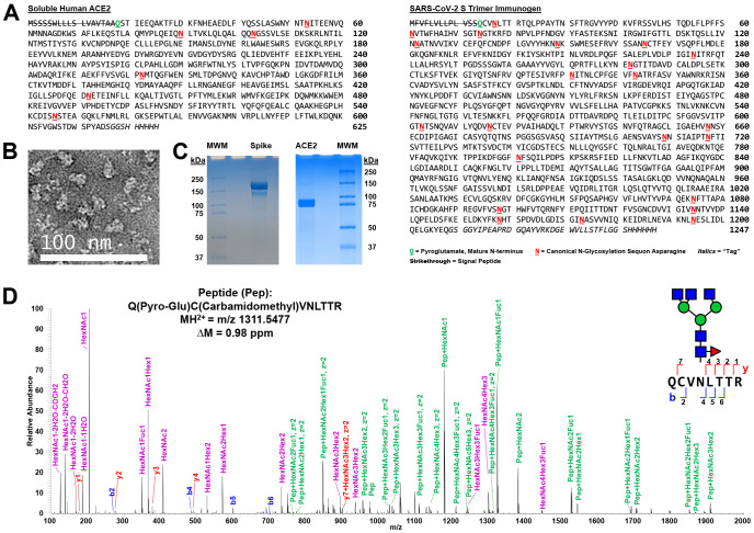
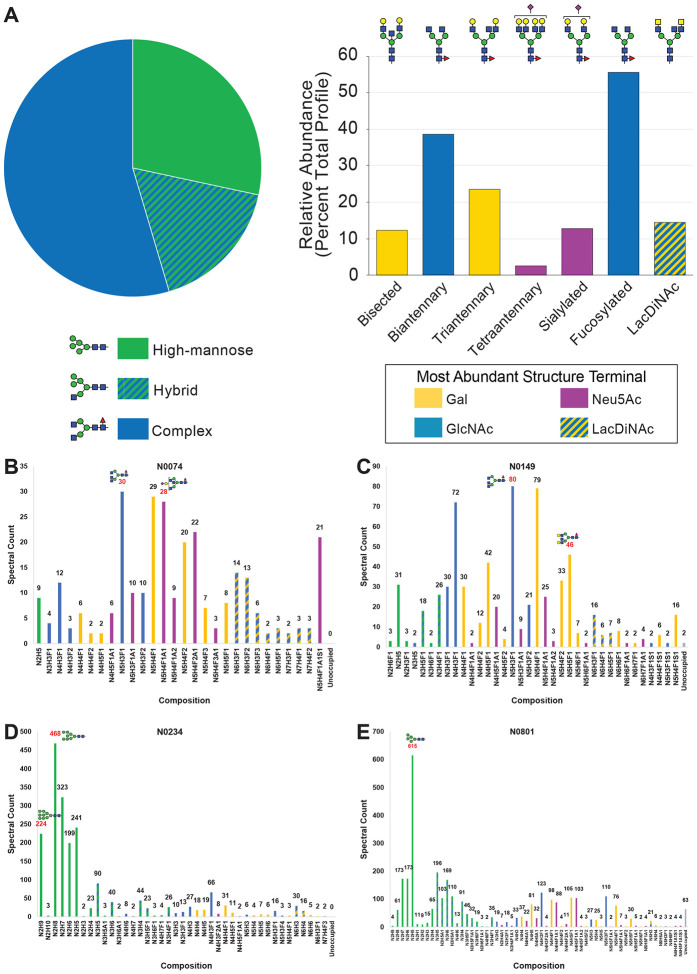
The current COVID-19 pandemic is caused by the SARS-CoV-2 betacoronavirus, which utilizes its highly glycosylated trimeric Spike protein to bind to the cell surface receptor ACE2 glycoprotein and facilitate host cell entry. We utilized glycomics-informed glycoproteomics to characterize site-specific microheterogeneity of glycosylation for a recombinant trimer Spike mimetic immunogen and for a soluble version of human ACE2. We combined this information with bioinformatic analyses of natural variants and with existing 3D-structures of both glycoproteins to generate molecular dynamics simulations of each glycoprotein alone and interacting with one another. Our results highlight roles for glycans in sterically masking polypeptide epitopes and directly modulating Spike-ACE2 interactions. Furthermore, our results illustrate the impact of viral evolution and divergence on Spike glycosylation, as well as the influence of natural variants on ACE2 receptor glycosylation that, taken together, can facilitate immunogen design to achieve antibody neutralization and inform therapeutic strategies to inhibit viral infection.

Keywords: 3D-modeling; ACE2; COVID-19; SARS-CoV-2; Spike protein; coronavirus; glycoprotein; glycosylation; mass spectrometry; molecular dynamics.


Conflict of interest statement

DECLARATION OF INTERESTS The authors declare no competing interests.

Figures

Figure 1.
Figure 1.. Expression and Characterization of SARS-CoV-2 Spike Glycoprotein Trimer Immunogen and Soluble Human ACE2.
A) Sequences of SARS-CoV-2 S immunogen and soluble human ACE2. The N-terminal pyroglutamines for both mature protein monomers are bolded, underlined, and shown in green. The canonical N-linked glycosylation sequons are bolded, underlined, and shown in red. Negative stain electron microscopy of the purified trimer (B) and Coomassie G-250 stained reducing SDS-PAGE gels (C) confirmed purity of the SARS-CoV-2 S protein trimer and of the soluble human ACE2. MWM = molecular weight markers. D) A representative Step-HCD fragmentation spectrum from mass spectrometry analysis of a tryptic digest of S annotated manually based on search results from pGlyco 2.2. This spectrum defines the N-terminus of the mature protein monomer as (pyro-)glutamine 0014. A representative N-glycan consistent with this annotation and our glycomics data (Fig. 2) is overlaid using the Symbol Nomenclature For Glycans (SNFG) code. This complex glycan occurs at N0017. Note, that as expected, the cysteine is carbamidomethylated and the mass accuracy of the assigned peptide is 0.98 ppm. On the sequence of the N-terminal peptide and in the spectrum, the assigned b (blue) and y (red) ions are shown. In the spectrum, purple highlights glycan oxonium ions and green marks intact peptide fragment ions with various partial glycan sequences still attached. Note that the green-labeled ions allow for limited topology to be extracted including defining that the fucose is on the core and not the antennae of the glycopeptide.
Figure 2.
Figure 2.. Glycomics Informed Glycoproteomics Reveals Substantial Site-Specific Microheterogeneity of N-linked Glycosylation on SARS-CoV-2 S.
A) Glycans released from SARS-CoV-2 S protein trimer immunogen were permethylated and analyzed by MSn. Structures were assigned, grouped by type and structural features, and prevalence was determined based on ion current. The pie chart shows basic division by broad N-glycan type. The bar graph provides additional detail about the glycans detected. The most abundant structure with a unique categorization by glycomics for each N-glycan type in the pie chart, or above each feature category in the bar graph, is indicated. B – E) Glycopeptides were prepared from SARS-CoV-2 S protein trimer immunogen using multiple combinations of proteases, analyzed by LC-MSn, and the resulting data was searched using several different software packages. Four representative sites of N-linked glycosylation with specific features of interest were chosen and are presented here. N0074 (B) and N0149 (C) are shown that occur in variable insert regions of S compared to SARS-CoV and other related coronaviruses, and there are emerging variants of SARS-CoV-2 that disrupt these two sites of glycosylation in S. N0234 (D) contains the most high-mannose N-linked glycans. N0801 (D) is an example of glycosylation in the S2 region of the immunogen and displays a high degree of hybrid glycosylation compared to other sites. The abundance of each composition is graphed in terms of assigned spectral counts. Representative glycans (as determined by glycomics analysis) for several abundant compositions are shown in SNFG format. The abbreviations used here and throughout the manuscript are N for HexNAc, H for Hexose, F for Fucose, A for Neu5Ac, and S for Sulfation. Note that the graphs for the other 18 sites and other graphs grouping the microheterogeneity observed by other properties are presented in Supplemental Information.
Figure 3.
Figure 3.. SARS-CoV-2 S Immunogen N-glycan Sites are Predominantly Modified by Complex N-glycans.
N-glycan topologies were assigned to all 22 sites of the S protomer and the spectral counts for each of the 3 types of N-glycans (high-mannose, hybrid, and complex) as well as the unoccupied peptide spectral match counts at each site were summed and visualized as pie charts. Note that only N1173 and N1194 show an appreciable amount of the unoccupied amino acid.
Figure 4.
Figure 4.. 3D Structural Modeling of Glycosylated SARS-CoV-2 Spike Trimer Immunogen Reveals Predictions for Antigen Accessibility and Other Key Features.
Results from glycomics and glycoproteomics experiments were combined with results from bioinformatics analyses and used to model several versions of glycosylated SARS-CoV-2 S trimer immunogen. A) Sequence of the SARS-CoV-2 S immunogen displaying computed antigen accessibility and other information. Antigen accessibility is indicated by red shading across the amino acid sequence. B) Emerging variants confirmed by independent sequencing experiments were analyzed based on the 3D structure of SARS-CoV-2 S to generate a proximity chart to the determined N-linked glycosylation sites. C) SARS-CoV-2 S trimer immunogen model from MD simulation displaying abundance glycoforms and antigen accessibility shaded in red for most accessible, white for partial, and black for inaccessible (see Supplemental movie A). D) SARS-CoV-2 S trimer immunogen model from MD simulation displaying oxford class glycoforms and sequence variants. * indicates not visible while the box represents 3 amino acid variants that are clustered together in 3D space. E) SARS-CoV-2 S trimer immunogen model from MD simulation displaying processed glycoforms plus shading of Thr-323 that has O-glycoslyation at low stoichiometry in yellow.
Figure 5:
Figure 5:. Glycomics Informed Glycoproteomics of Soluble Human ACE2 Reveals High Occupancy, Complex N-linked Glycosylation.
A) Glycans released from soluble, purified ACE2 were permethylated and analyzed by MSn. Structures were assigned, grouped by type and structural features, and prevalence was determined based on ion current. The pie chart shows basic division by broad N-glycan type. The bar graph provides additional detail about the glycans detected. The most abundant structure with a unique categorization by glycomics for each N-glycan type in the pie chart, or above each feature category in the bar graph, is indicated. B – G) Glycopeptides were prepared from soluble human ACE2 using multiple combinations of proteases, analyzed by LC-MSn, and the resulting data was searched using several different software packages. All six sites of N-linked glycosylation are presented here. Displayed in the bar graphs are the individual compositions observed graphed in terms of assigned spectral counts. Representative glycans (as determined by glycomics analysis) for several abundant compositions are shown in SNFG format. The abbreviations used here and throughout the manuscript are N for HexNAc, H for Hexose, F for Fucose, and A for Neu5Ac. The pie chart (analogous to Figure 3 for SARS-CoV-2 S) for each site is displayed in the upper corner of each panel. B) N053. C) N090. D) N103. E) N322. F) N432. G) N546, a site that does not exist in 3 in 10,000 people.
Figure 6:
Figure 6:. 3D Structural Modeling of Glycosylated Soluble Human ACE2.
Results from glycomics and glycoproteomics experiments were combined with results from bioinformatics analyses and used to model several versions of glycosylated soluble human ACE2. A) Soluble human ACE2 model from MD simulations displaying abundance glycoforms, interaction surface with S, and sequence variants. N546 variant is boxed that would remove N-linked glycosylation at that site (see Supplemental movie B). B) Soluble human ACE2 model from MD simulations displaying processed glycoforms and interaction surface with S.
Figure 7:
Figure 7:. Interactions of Glycosylated Soluble Human ACE2 and Glycosylated SARS-CoV-2 S Trimer Immunogen Revealed By 3D-Structural Modeling and Molecular Dynamics Simulations.
A) Molecular dynamics simulation of glycosylated soluble human ACE2 and glycosylated SARS-CoV-2 S trimer immunogen interaction (see Supplemental simulations 1–3). ACE2 (top) is colored red with glycans in pink while S is colored white with glycans in dark grey. Highlighted are ACE2 glycans that interacts with S that are zoomed in on to the right. B) Zoom in of ACE2-S interface highlighting ACE2 glycan interactions using 3D-SNFG icons (60) with S protein (pink) as well as ACE2-S glycan-glycan interactions. C) Zoom in of dynamics trajectory of glycans at the interface of soluble human ACE2 and S (see Supplemental movies C and D).

References

    1. Zhou P., Yang X. L., Wang X. G., Hu B., Zhang L., Zhang W., Si H. R., Zhu Y., Li B., Huang C. L., Chen H. D., Chen J., Luo Y., Guo H., Jiang R. D., Liu M. Q., Chen Y., Shen X. R., Wang X., Zheng X. S., Zhao K., Chen Q. J., Deng F., Liu L. L., Yan B., Zhan F. X., Wang Y. Y., Xiao G. F., and Shi Z. L. (2020) A pneumonia outbreak associated with a new coronavirus of probable bat origin. Nature 579, 270–273 - PMC - PubMed
    1. Lu R., Zhao X., Li J., Niu P., Yang B., Wu H., Wang W., Song H., Huang B., Zhu N., Bi Y., Ma X., Zhan F., Wang L., Hu T., Zhou H., Hu Z., Zhou W., Zhao L., Chen J., Meng Y., Wang J., Lin Y., Yuan J., Xie Z., Ma J., Liu W. J., Wang D., Xu W., Holmes E. C., Gao G. F., Wu G., Chen W., Shi W., and Tan W. (2020) Genomic characterisation and epidemiology of 2019 novel coronavirus: implications for virus origins and receptor binding. Lancet 395, 565–574 - PMC - PubMed
    1. Zhong N. S., Zheng B. J., Li Y. M., Poon, Xie Z. H., Chan K. H., Li P. H., Tan S. Y., Chang Q., Xie J. P., Liu X. Q., Xu J., Li D. X., Yuen K. Y., Peiris, and Guan Y. (2003) Epidemiology and cause of severe acute respiratory syndrome (SARS) in Guangdong, People’s Republic of China, in February, 2003. Lancet 362, 1353–1358 - PMC - PubMed
    1. Xia X. (2020) Extreme genomic CpG deficiency in SARS-CoV-2 and evasion of host antiviral defense. Mol Biol Evol - PMC - PubMed
    1. Zhang T., Wu Q., and Zhang Z. (2020) Probable Pangolin Origin of SARS-CoV-2 Associated with the COVID-19 Outbreak. Curr Biol 30, 1346–1351 e1342 - PMC - PubMed

Publication types