. 2020 Aug 1;76(Pt 8):778-789.
doi: 10.1107/S2059798320009274. Epub 2020 Jul 27.

Structural and biochemical characterization of a glutathione transferase from the citrus canker pathogen Xanthomonas

Affiliations

Structural and biochemical characterization of a glutathione transferase from the citrus canker pathogen Xanthomonas

Eduardo Hilario et al. Acta Crystallogr D Struct Biol. .

Abstract

The genus Xanthomonas comprises several cosmopolitan plant-pathogenic bacteria that affect more than 400 plant species, most of which are of economic interest. Citrus canker is a bacterial disease that affects citrus species, reducing fruit yield and quality, and is caused by the bacterium Xanthomonas citri subsp. citri (Xac). The Xac3819 gene, which has previously been reported to be important for citrus canker infection, encodes an uncharacterized glutathione S-transferase (GST) of 207 amino-acid residues in length (XacGST). Bacterial GSTs are implicated in a variety of metabolic processes such as protection against chemical and oxidative stresses. XacGST shares high sequence identity (45%) with the GstB dehalogenase from Escherichia coli O6:H1 strain CFT073 (EcGstB). Here, XacGST is reported to be able to conjugate glutathione (GSH) with bromoacetate with a Km of 6.67 ± 0.77 mM, a kcat of 42.69 ± 0.32 s-1 and a kcat/Km of 6.40 ± 0.72 mM-1 s-1 under a saturated GSH concentration (3.6 mM). These values are comparable to those previously reported for EcGstB. In addition, crystal structures of XacGST were determined in the apo form (PDB entry 6nxv) and in a GSH-bound complex (PDB entry 6nv6). XacGST has a canonical GST-like fold with a conserved serine residue (Ser12) at the GSH-binding site near the N-terminus, indicating XacGST to be a serine-type GST that probably belongs to the theta-class GSTs. GSH binding stabilizes a loop of about 20 residues containing a helix that is disordered in the apo XacGST structure.

Keywords: GSTs; Xanthomonas; Xanthomonas citri subsp. citri; bromoacetate; citrus canker; dehalogenases; glutathione; glutathione S-transferases.


Figures

Figure 1
Figure 1
Multiple sequence alignment of XacGST with theta-class GST members and their evolutionary relationship. (a) Sequence alignment was performed using ESPript 3.0 (Robert & Gouet, 2014 ▸) utilizing the ClustalW algorithm (Thompson et al., 1994 ▸). Sequence aliases with the corresponding UniProtKB accession numbers for classified theta-class GSTs are as follows: XacGST, Xanthomonas citri substr. citri strain 306, Q8PG02; JaDCM, Janthinobacterium sp. KBS0711 dichloromethane dehalogenase, A0A0M2WQ11; StGST, uncharacterized and unclassified YliJ from Salmonella typhimurium, Q8Z859; EcGSTB, unclassified GstB from Escherichia coli O6:H1 strain CFT073, P0ACA8; DmGSTT1, DmGSTT2, DmGSTT3 and DmGSTT4, Drosophila melanogaster GST T1, T2, T3 and T4, Q7K0B6, A1Z7X7, E1JJS1 and Q8MRM0, respectively; PxGST, Papilio xuthus GST, I4DNY4; BmGST, Bombyx mori GST, B0LB14; AgGSTT1 and AgGSTT2, Aedes aegypti GST T1 and T2, Q8MUQ1 and Q8MUQ2, respectively; hGSTT1 and hGSTT2, Homo sapiens GST theta-1 and theta-2, P30711 and P0CG29, respectively. Strictly conserved residues among GSTs are highlighted with a red background and highly homologous residues are shown in blue boxes. Strictly conserved class-dependent residues from the GSH catalytic site of the serine-type GSTs (theta class) are highlighted with a yellow background. The secondary-structural elements of XacGST (PDB entry 6nv6) and hGSTT2 (PDB entry 4mpg) defined by the crystal structure are indicated as black coils and arrows at the top and bottom of the sequence alignment, respectively. The unstructured loop region (residues 35–53) observed in the GSH-free monomers is represented as a green bar. The residues marked with asterisks in the XacGST sequence are related to GSH binding. (b) Phylogenetic analysis of the amino-acid sequences of theta-class GSTs. The scale bar is equal to 0.5 substitutions per site and the length of each branch is proportional to the average number of substitutions per site as indicated by the scale. The evolutionary history was inferred using the maximum-likelihood method based on the JTT matrix-based model (Jones et al., 1992 ▸) and the evolutionary analyses were conducted in MEGA X (Kumar et al., 2018 ▸).
Figure 1
Figure 1
Multiple sequence alignment of XacGST with theta-class GST members and their evolutionary relationship. (a) Sequence alignment was performed using ESPript 3.0 (Robert & Gouet, 2014 ▸) utilizing the ClustalW algorithm (Thompson et al., 1994 ▸). Sequence aliases with the corresponding UniProtKB accession numbers for classified theta-class GSTs are as follows: XacGST, Xanthomonas citri substr. citri strain 306, Q8PG02; JaDCM, Janthinobacterium sp. KBS0711 dichloromethane dehalogenase, A0A0M2WQ11; StGST, uncharacterized and unclassified YliJ from Salmonella typhimurium, Q8Z859; EcGSTB, unclassified GstB from Escherichia coli O6:H1 strain CFT073, P0ACA8; DmGSTT1, DmGSTT2, DmGSTT3 and DmGSTT4, Drosophila melanogaster GST T1, T2, T3 and T4, Q7K0B6, A1Z7X7, E1JJS1 and Q8MRM0, respectively; PxGST, Papilio xuthus GST, I4DNY4; BmGST, Bombyx mori GST, B0LB14; AgGSTT1 and AgGSTT2, Aedes aegypti GST T1 and T2, Q8MUQ1 and Q8MUQ2, respectively; hGSTT1 and hGSTT2, Homo sapiens GST theta-1 and theta-2, P30711 and P0CG29, respectively. Strictly conserved residues among GSTs are highlighted with a red background and highly homologous residues are shown in blue boxes. Strictly conserved class-dependent residues from the GSH catalytic site of the serine-type GSTs (theta class) are highlighted with a yellow background. The secondary-structural elements of XacGST (PDB entry 6nv6) and hGSTT2 (PDB entry 4mpg) defined by the crystal structure are indicated as black coils and arrows at the top and bottom of the sequence alignment, respectively. The unstructured loop region (residues 35–53) observed in the GSH-free monomers is represented as a green bar. The residues marked with asterisks in the XacGST sequence are related to GSH binding. (b) Phylogenetic analysis of the amino-acid sequences of theta-class GSTs. The scale bar is equal to 0.5 substitutions per site and the length of each branch is proportional to the average number of substitutions per site as indicated by the scale. The evolutionary history was inferred using the maximum-likelihood method based on the JTT matrix-based model (Jones et al., 1992 ▸) and the evolutionary analyses were conducted in MEGA X (Kumar et al., 2018 ▸).
Figure 2
Figure 2
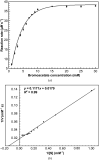
XacGST dehalogenase activity using bromoacetate as a substrate. (a) A plot of the rate of bromoacetate–GSH conjugation versus bromoacetate concentration. The experiments were carried out in triplicate. Error bars refer to the standard deviations. (b) Lineweaver–Burk plot for XacGST, where [S] represents the substrate concentration of bromoacetate and V represents the initial reaction velocity.
Figure 3
Figure 3
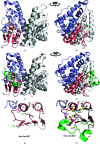
Crystal structures of XacGST and its complex with GSH. (a, b) XacGST dimer (PDB entry 6nxv, chains A and B) showing an N-terminal thioredoxin-like domain (β1–α1–β2–α2–β3–β4–α3 topology, salmon) and a C-terminal all-α-helical domain (α4–α8 topology, light blue). The partner monomer is colored light gray. (c, d) XacGST dimer (PDB entry 6nv6, chains A and B) containing GSH bound in one enzyme active site (PDB entry 6nv6, chain B). (e, f) Enlarged views of the flexible loop region of apo XacGST (e) and the XacGST–GSH complex (f). The loop between strands 2 and 3 contains helix α2 in the GSH-bound monomer and is represented by green or gray dashed lines in GSH-free monomers, where it is disordered. The bound GSH molecule is shown as a ball-and-stick representation and is colored according to atom type: C in yellow, O in red, N in blue and S in orange. Key residues interacting with GSH are highlighted with sticks and labeled. This figure was produced with PyMOL (DeLano, 2002 ▸) and manually modified.
Figure 4
Figure 4
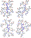
Schematic interactions of GSH with key residues in the G-site of XacGST and other theta-class GSTs. Representations of residues that make hydrogen bonds (green dashed lines with corresponding bond distances) to GSH bound in the G-site are shown. Residues involved in GSH binding are shown as stick models and labeled with residue identities. C, N, O and S atoms are shown as small black, blue, red and yellow solid spheres, respectively. GSH and protein atoms are shown as sticks colored magenta and black, respectively. Residues labeled in blue correspond to residues of the partner monomer in the dimer. (a) XacGST with GSH bound (PDB entry 6nv6, chain B). (b) StGST from S. typhimurium with GSH bound (YliJ; PDB entry 4kh7). (c, d) The theta-class human hGSTT1 mutant W234R (PDB entry 2c3q) and hGSTT2 (PDB entry 4mpg) in complex with S-hexylglutathione and GSH, respectively. Theses figures were prepared with LigPlot 2.1 (Wallace et al., 1995 ▸) and manually modified (amino-acid residue and hydrogen-bond distance labels).

Similar articles

References

    1. Ahmad, S., Thulasingam, M., Palombo, I., Daley, D. O., Johnson, K. A., Morgenstern, R., Haeggström, J. Z. & Rinaldo-Matthis, A. (2015). Biochim. Biophys. Acta, 1854, 1365–1371. - PubMed
    1. Allocati, N., Casalone, E., Masulli, M., Ceccarelli, I., Carletti, E., Parker, M. W. & Di Ilio, C. (1999). FEBS Lett. 445, 347–350. - PubMed
    1. Allocati, N., Federici, L., Masulli, M. & Di Ilio, C. (2009). FEBS J. 276, 58–75. - PubMed
    1. Allocati, N., Federici, L., Masulli, M. & Di Ilio, C. (2012). Biochimie, 94, 588–596. - PubMed
    1. Armstrong, R. N. (1991). Chem. Res. Toxicol. 4, 131–140. - PubMed

MeSH terms

Supplementary concepts