. 2020 Nov 4;15(21):2040-2051.
doi: 10.1002/cmdc.202000457. Epub 2020 Sep 15.

Aromatic Linkers Unleash the Antiproliferative Potential of 3-Chloropiperidines Against Pancreatic Cancer Cells

Affiliations

Aromatic Linkers Unleash the Antiproliferative Potential of 3-Chloropiperidines Against Pancreatic Cancer Cells

Tim Helbing et al. ChemMedChem. .

Abstract

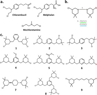
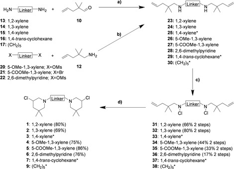
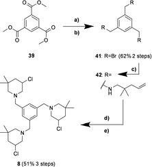
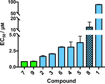
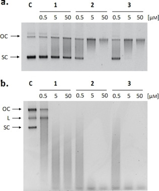
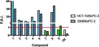
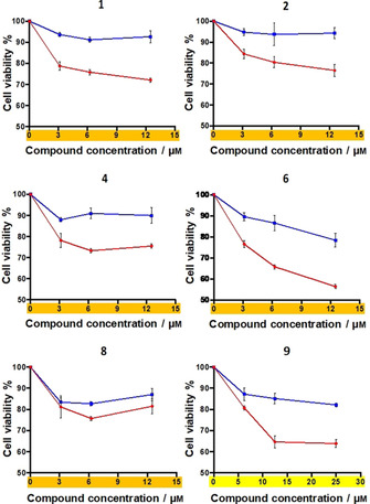
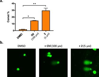
In this study, we describe the synthesis and biological evaluation of a set of bis-3-chloropiperidines (B-CePs) containing rigid aromatic linker structures. A modification of the synthetic strategy also enabled the synthesis of a pilot tris-3-chloropiperidine (Tri-CeP) bearing three reactive meta-chloropiperidine moieties on the aromatic scaffold. A structure-reactivity relationship analysis of B-CePs suggests that the arrangement of the reactive units affects the DNA alkylating activity, while also revealing correlations between the electron density of the aromatic system and the reactivity with biologically relevant nucleophiles, both on isolated DNA and in cancer cells. Interestingly, all aromatic 3-chloropiperidines exhibited a marked cytotoxicity and tropism for 2D and 3D cultures of pancreatic cancer cells. Therefore, the new aromatic 3-chloropiperidines appear to be promising contenders for further development of mustard-based anticancer agents aimed at pancreatic cancers.

Keywords: DNA damage; alkylating agents; aromatic chloropiperidines; pancreatic cancer spheroids; structure-activity relationships.


Conflict of interest statement

The authors declare no conflict of interest.

Figures

Figure 1
Figure 1
a) Chemical structure of chlorambucil, melphalan and mechloretamine. b) General structure of B−CePs. Different connecting linkers (L) are schematized with different colors. c) Chemical structures of the analyzed 3‐chloropiperidines.
Scheme 1
Scheme 1
Synthesis of bis‐3‐chloropiperidines 16. a) NaBH(OAc)3, AcOH, dry CH2Cl2, 0 °C to RT, 16–18 h; b) NaH, dry THF, 0 °C to RT, 20–22 h; c) NCS, dry CH2Cl2, 0 °C to RT, 2.5–3 h; d) TBAI (cat.), dry CHCl3, 60 °C (oil bath temperature), 2–2.5 h (inseparable diastereomeric mixture). *The synthesis of compounds 3, 7 and 9 as well as their corresponding precursors has been described elsewhere. [4b]
Scheme 2
Scheme 2
Synthesis of tris‐3‐chloropiperidine 8. a) LAH, dry THF, 0 °C to RT to reflux, 18 h; b) PBr3, dry Et2O, 0 °C to RT, 24 h; c) 2,2‐dimethylpent‐4‐en‐1‐amine 12, dry CH2Cl2, 0 °C to RT, 68 h; d) NCS, dry CH2Cl2, 0 °C to RT, 2.5 h; e) TBAI (cat.), dry CHCl3, reflux, 4 h.
Figure 2
Figure 2
DNA cleavage EC50 values of analyzed B−CePs. The supercoiled pBR322 plasmid was incubated with increasing concentrations of test compounds at 37 °C for 3 h in BPE buffer. EC50 values were calculated by comparing the intensities of the supercoiled species band to the negative control as a function of compound concentration. Average EC50 values and standard deviations result from two independent experiments. Green bars: aliphatic B−CePs; blue bars: aromatic B−CePs; blue checkered bar: Tri‐CeP.
Figure 3
Figure 3
DNA cleavage assay for compounds 1, 2 and 3. DNA cleavage assay upon incubation with pBR322 after a) 3 and b) 18 h at 37 °C with increasing compound concentrations (0.5, 5, 50 μm). SC: supercoiled plasmid; L: linearized plasmid; OC: open circular plasmid; C: supercoiled pBR322 plasmid control.
Figure 4
Figure 4
Preferential activity indexes of 3‐chloropiperidines. P.A.I are expressed as the ratio between the compound MTT IC50 against HCT‐15 and BxPC‐3 cells (solid bars), as well as 2008 and BxPC‐3 cells (dashed bars). Chl: Chlorambucil. Green bars: aliphatic B−CePs; blue bars: aromatic B−CePs; gray bars: Chl.
Figure 5
Figure 5
BxPC‐3 cell viability upon exposure to increasing concentrations of selected compounds (3 to 12 μm of rigid derivatives 1, 2, 4, 6, 8 and 10 to 25 μm of 9) for 5 h at 37 °C () or 4 °C (), followed by a gentle rinse of wells with PBS and addition of fresh RPMI medium. The MTT assay was performed at 72 h.
Figure 6
Figure 6
An alkaline SCGE assay was performed on BxPC‐3 cells upon incubation with DMSO 0.5 %, chlorambucil (Chl) and compound 2 for 6 h at the indicated concentrations. a) The relative percentage of comets (number of cells forming a comet/total number of cells) detected in two randomly captured fields from two independent experiments per condition. b) Representative images (40x) of treated samples (including negative and positive controls) with comets are also reported. Paired t‐test: * p<0.05, ** p<0.001.

References

    1. None
    1. Saijo N., Cancer Res. 2012, 44, 1–10; - PMC - PubMed
    1. Huang M., Shen A. J., Ding J., Geng M. Y., Trends Pharmacol. Sci. 2014, 35, 41–50. - PubMed
    1. Chabner B. A., T. G. Roberts Jr. , Nat. Rev. Cancer 2005, 5, 65–72. - PubMed
    1. Singh R. K., Kumar S., Prasad D. N., Bhardwaj T. R., Eur. J. Med. Chem. 2018, 151, 401–433. - PubMed

Publication types

MeSH terms

LinkOut - more resources