Review
. 2020 Aug 3;12(15):15856-15874.
doi: 10.18632/aging.103794. Epub 2020 Aug 3.

Pompe disease: pathogenesis, molecular genetics and diagnosis

Affiliations
Review

Pompe disease: pathogenesis, molecular genetics and diagnosis

Simona Taverna et al. Aging (Albany NY). .

Abstract

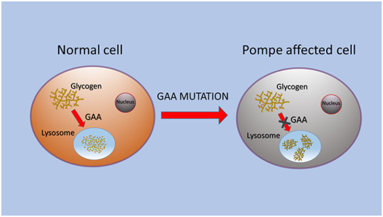
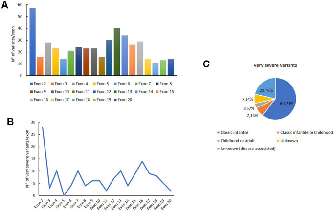
Pompe disease (PD) is a rare autosomal recessive disorder caused by mutations in the GAA gene, localized on chromosome 17 and encoding for acid alpha-1,4-glucosidase (GAA). Currently, more than 560 mutations spread throughout GAA gene have been reported. GAA catalyzes the hydrolysis of α-1,4 and α-1,6-glucosidic bonds of glycogen and its deficiency leads to lysosomal storage of glycogen in several tissues, particularly in muscle. PD is a chronic and progressive pathology usually characterized by limb-girdle muscle weakness and respiratory failure. PD is classified as infantile and childhood/adult forms. PD patients exhibit a multisystemic manifestation that depends on age of onset.Early diagnosis is essential to prevent or reduce the irreversible organ damage associated with PD progression. Here, we make an overview of PD focusing on pathogenesis, clinical phenotypes, molecular genetics, diagnosis, therapies, autophagy and the role of miRNAs as potential biomarkers for PD.

Keywords: GAA; Pompe disease; acid alpha-1,4-glucosidase; glycogen; lysosomal storage disorder.


Conflict of interest statement

CONFLICTS OF INTEREST: The authors declare no conflicts of interest.

Figures

Figure 1
Figure 1
Schematic representation of GAA alteration that caused glycogen storage in lysosomes of PD cells.
Figure 2
Figure 2
Genetic variants distribution into GAA exons. Distribution of variants for each exon (A); distribution of very severe variants for each exon (B); association of the very severe variants with PD phenotypes.
Figure 3
Figure 3
Genetic variants distribution into GAA introns. Distribution of variants for each intron (A); distribution of very severe variants for each intron (B).
Figure 4
Figure 4
Second mutation located in a second allele of GAA gene associated to c.-32-13T>G variant.

References

    1. Platt FM, Boland B, van der Spoel AC. The cell biology of disease: lysosomal storage disorders: the cellular impact of lysosomal dysfunction. J Cell Biol. 2012; 199:723–34. 10.1083/jcb.201208152 - DOI - PMC - PubMed
    1. Ballabio A, Gieselmann V. Lysosomal disorders: from storage to cellular damage. Biochim Biophys Acta. 2009; 1793:684–96. 10.1016/j.bbamcr.2008.12.001 - DOI - PubMed
    1. Martina JA, Raben N, Puertollano R. SnapShot: lysosomal storage diseases. Cell. 2020; 180:602–02.e1. 10.1016/j.cell.2020.01.017 - DOI - PMC - PubMed
    1. de Araujo ME, Liebscher G, Hess MW, Huber LA. Lysosomal size matters. Traffic. 2020; 21:60–75. 10.1111/tra.12714 - DOI - PMC - PubMed
    1. Parenti G, Andria G, Ballabio A. Lysosomal storage diseases: from pathophysiology to therapy. Annu Rev Med. 2015; 66:471–86. 10.1146/annurev-med-122313-085916 - DOI - PubMed

MeSH terms