. 2020 Oct 2:287:198105.
doi: 10.1016/j.virusres.2020.198105. Epub 2020 Jul 31.

Host antiviral protein IFITM2 restricts pseudorabies virus replication

Affiliations

Host antiviral protein IFITM2 restricts pseudorabies virus replication

Jingying Xie et al. Virus Res. .

Abstract

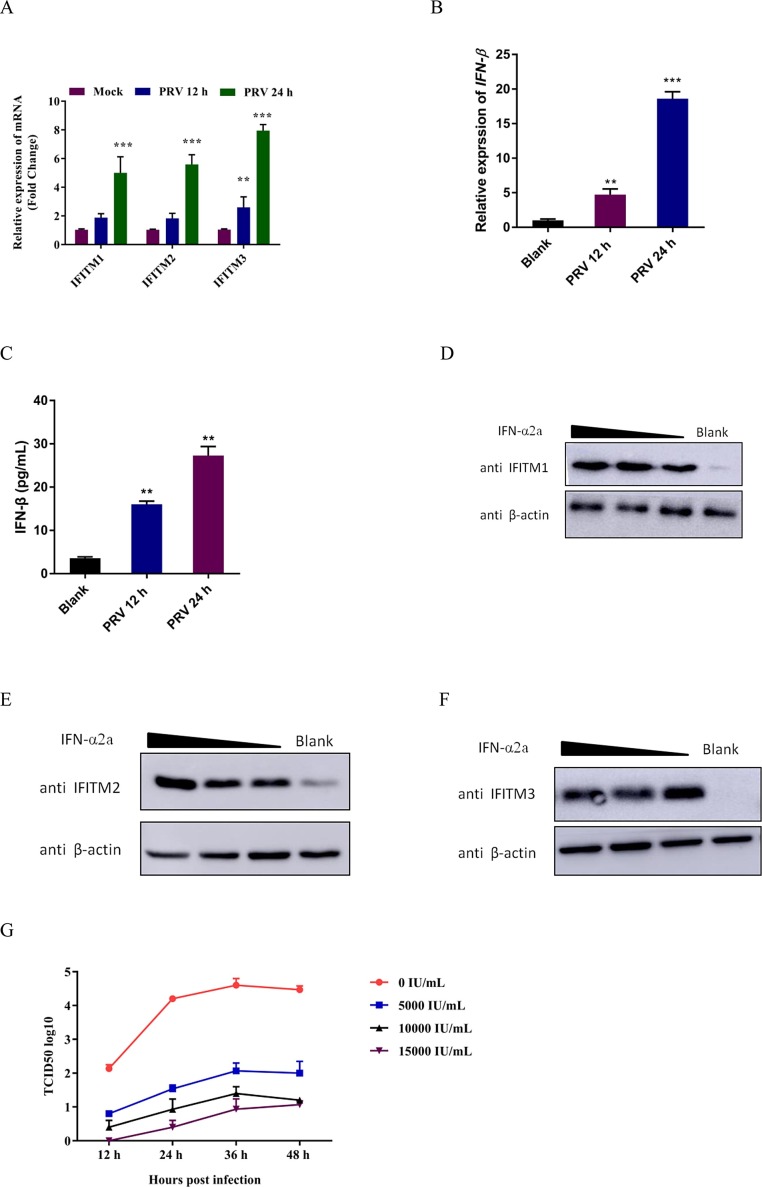
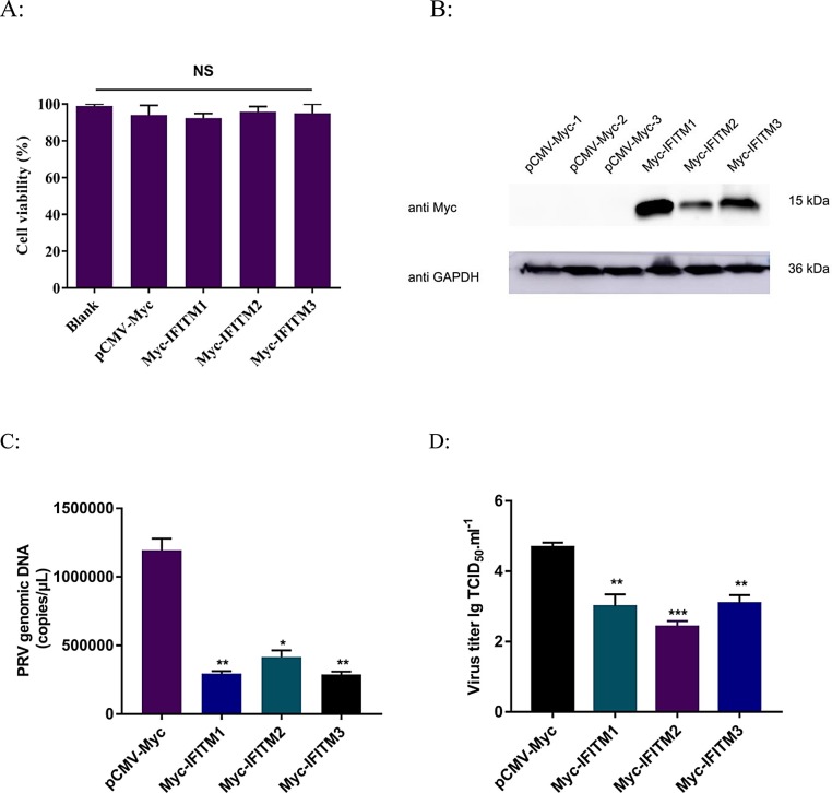
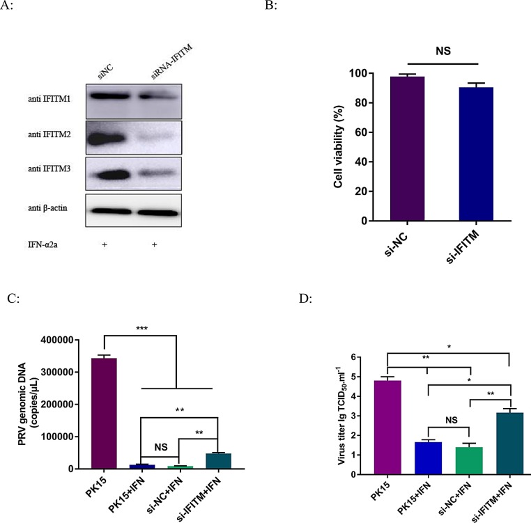
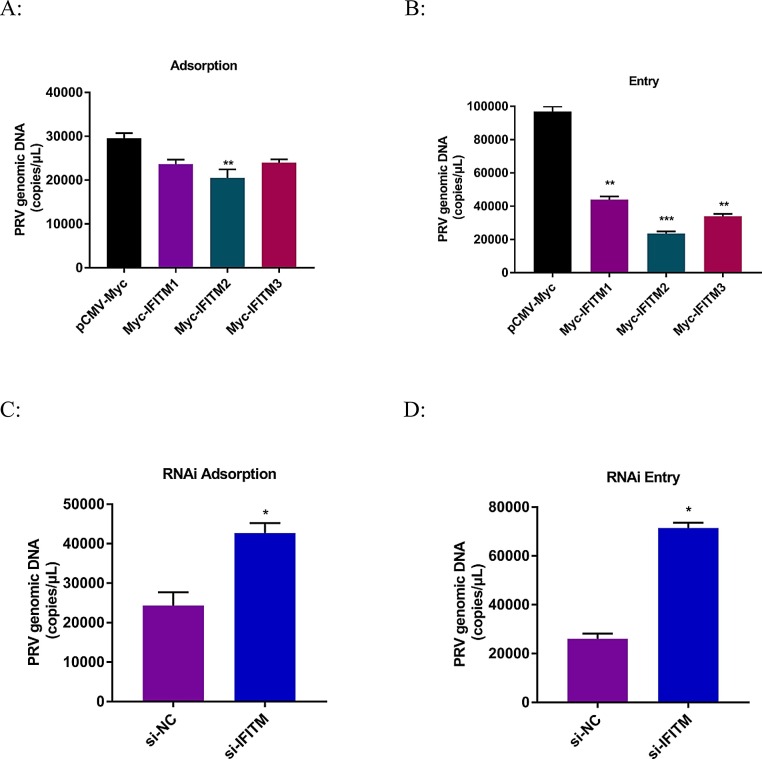
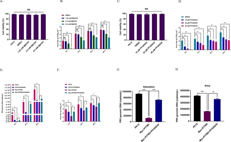
Pseudorabies virus (PRV) is one of the most destructive swine pathogens and leads to huge economic losses to the global pig industry. Type I interferons (IFNs) plays a pivotal role in the innate immune response to virus infection via induction of a series of interferon-stimulated genes (ISGs) expression. IFN-induced transmembrane (IFITM) proteins, a group of ISGs, are important host self-restriction factors, possessing a broad spectrum of antiviral effects. They are known confer resistance to a variety of RNA and DNA viruses. However, little is known about the role of IFITMs in PRV infection. In this study, we show that IFITM is crucial for controlling PRV infection and that IFITM proteins can interfere with PRV cell binding and entry. Furthermore, we showed that IFITM2-mediated inhibition of PRV entry requires the cholesterol pathway. Collectively, these results provide insight into the anti-PRV role of IFITM proteins and this inhibition possible associated with the change of cholesterol in the endosome, further underlying the importance of cholesterol in virus infection.

Keywords: Cholesterol; IFITM proteins; Pseudorabies virus.


Conflict of interest statement

The authors have no conflicts of interest to declare.

Figures

Fig. 1
Fig. 1
PRV infection and IFN-α2a treatment induces IFITM expression. (A) PRV infection induces IFITM mRNA transcription. PK15 cells were cultured in 6-well plate then infected with PRV at 100TCID50 for 12 h and 24 h, respectively. Cells were collected and extracted for RNA. 1 μg RNA was transcripted into cDNA for IFITM mRNA detection by RT-qPCR. Data were expressed as mean ± SD from three independent experiments and were measured in technical duplicate. Comparisons between groups were performed by Student’s t test. **p < 0.01, ***p < 0.001. (B and C) PRV infection induces IFN-β expression. PK15 cells were cultured in 6-well plate then infected with PRV at 100TCID50 for 12 h and 24 h, respectively. Cells were collected and extracted for RNA. 1 μg RNA was transcripted into cDNA for IFN-β mRNA detection by RT-qPCR. Culture supernatant was collected for IFN-β concentration detection by ELISA according to manufacture's instruction. Data were expressed as mean ± SD from three independent experiments and were measured in technical duplicate. Comparisons between groups were performed by Student’s t test. **p < 0.01, ***p < 0.001. (D, E, F) IFN-α2a treatment induces IFITM protein expression and inhibits PRV Bartha-61 infection in PK15 cells. The protein expression of IFITM in PK15 cells pretreated with IFN-α2a (5000 IU/mL, 10,000 IU/mL and 15,000 IU/mL) was detected by western blotting respectively. Cell samples were collected and RIPA lysis for 30min on ice. Then centrifuged for 15 min at 4°C to remove cell debris. Cell extracts were mixed with reducing loading buffer and electrophoresed by SDS-polyacrylamide (SDS-PAGE) on a 15 % polyacrylamide gel. All samples were heated at 95℃ for 5 min prior to being loaded onto an SDS-PAGE gel. After electro blotting the separated proteins onto a Polyvinylidene Fluoride (PVDF) membrane, unspecific antibody binding was blocked with phosphate-buffered saline (PBS)/Tween® 20 containing 2.5 % skimmed milk for 1h at room temperature. Then incubated blots in IFITM1, 2 or 3 specific antibodies 1: 2000 diluted at 4°C overnight. Then washed and incubated with horseradish peroxidase (HRP)-conjugated Goat anti Mouse or Rabbit secondary antibody. Blots were washed three times in PBST and developed with ECL reaction substrate. For all experiments, β-actin served as an internal reference. (G) PK15 cells were pretreated with different concentrations of IFN-α2a (5000 IU/mL, 10,000 IU/mL and 15,000 IU/mL) followed by infection with PRV Bartha-61 strain at 100TCID50. Cell culture supernatants were collected at different time points (12 h, 24 h, 36 h and 48 h). The TCID50 assay was performed to detect infectious viral particles (Reed − Muench method). Data were expressed as mean ± SD from three independent experiments.
Fig. 2
Fig. 2
Overexpression of IFITM proteins suppresses PRV infection in PK15 cells. (A) PK15/Myc-IFITM cells were cultured in a 96-well plate for 24 h, and the cell viability was determined using the CCK8 reagent. The cell viability of PK15 cells was used as the control. Data were expressed as the mean ± SD from three independent experiments. (B) PK15 cells were transfected with the expression plasmids, Myc-IFITM1, Myc-IFITM2, and Myc-IFITM3 for 24 h, following which the cells were collected for western blotting analysis. An anti-Myc antibody was used and GAPDH served as the loading control. (C) The Myc, Myc-IFITM1, Myc-IFITM2 and Myc-IFITM3 cells were infected with PRV at 100TCID50. Culture supernatant was collected at 24 h post infection. And viral DNA was extracted according to manufacture's instruction. PRV genomic DNA copies was detected using real-time TaqMan PCR. (D) Infectious progeny viral titers were determined by the TCID50 assay. Data were expressed as mean ± SD from three independent experiments and were measured in technical duplicate. Comparisons between groups were performed by Student’s t test. *p < 0.05, **p < 0.01, ***p < 0.001.
Fig. 3
Fig. 3
Knockdown of IFITM protein expression by siRNA promotes PRV replication in PK15 cells. (A) PK15 cells were pretreated with IFN-α2a 5000 IU/mL for 8 h and subsequently transfected with siRNA targeting IFITMs proteins. After 24 h, IFITM protein expression was analyzed by western blotting using specific antibodies. β-actin served as the loading control. (B) PK15/si-IFITM cells were cultured in a 96-well plate for 24 h, and the cell viability was determined using the CCK8 reagent. Cells treated with a control siRNA (si-NC) was used as the control. Data were expressed as the mean ± SD from three independent experiments. (C) Cells with knocked down IFITMs were infected with PRV at 100TCID50 for 24 h, then virus was collected and viral DNA was extracted for PRV genomic DNA copies detection by real-time TaqMan PCR. (D) Infectious progeny viral titers were determined by the TCID50 assay. Data were expressed as mean ± SD from three independent experiments and were measured in technical duplicate. Comparisons between groups were performed by Student’s t test. *p < 0.05, **p < 0.01, ***p < 0.001.
Fig. 4
Fig. 4
IFITM proteins restrict PRV entry into PK15 cells. PK15 cells overexpressing or knockdown of IFITM proteins were applied to viral adsorption and entry experiments. Cells were infected with PRV at an MOI of 1. (A,C) For the adsorption assay, cells were incubated at 4 °C for 2 h, washed 4 times with cold PBS and collected for DNA extraction. Real-time PCR was used to detect the virus copies bound to the cell surface. (B,D) For the entry assay, cells were incubated at 37 °C for another 2 h, washed 4 times with cold PBS and collected for DNA extraction. The viral entry into cells was detected by real-time TaqMan PCR. Data were expressed as mean ± SD from three independent experiments and were measured in technical duplicate. Comparisons between groups were performed by Student’s t test. *p < 0.05, **p < 0.01.
Fig. 5
Fig. 5
IFITM2 inhibit PRV entry maybe required the accumulation of cholesterol. (A,C) Chemicals were previously tested for cytotoxicity at the concentrations used. PK15 cells treated with 1.25 mM, 2.5 mM and 5.0 mM Methyl-β-cyclodextrin (MβCDX) for 45 min in 96 well plate. For cholesterol inhibitor PF429242, cells were pretreated with 10 μM, 20 μM and 40 μM PF429242 for 24 h then following other experimental operations. Cell viability was determined using the CCK8 reagent according to manufacture's instruction. Data were expressed as the mean ± SD from three independent experiments. (B,D) Infection of cells pretreated with (B) MβCDX and (D) PF429242 blocks infection with PRV Bartha-61 strain. PK15 cells were pretreated with DMSO, MβCDX or PF429242 at the indicated concentrations and hours, subsequently cells were infected with virus at 100TCID50 for 2 h and then overlaid with fresh media. All infections and incubations were carried out in the presence of indicated concentrations of PF429242, but not conclude MβCDX. For MβCDX treated cells, infection culture supernatant was collected at 6 h, 12 h, 18 h and 24 h. PF429242 treated cells supernatant was collected at 6 h, 8 h, 10 h and 12 h after PRV infection. All sampleswere used for TCID50 assay. Values were compared to viral titer of DMSO treated cells. Data were expressed as mean ± SD from three independent experiments. (E) PK15 cells infected with 100TCID50 PRV, PK15 cells pretreated with 20 μM PF429242 following 100TCID50 PRV infection, IFITM2 overexpressed cells infected with 100TCID50 PRV and IFITM2 overexpressed cells pretreated with 20 μM PF429242 following 100TCID50 PRV infection were performed and cell culture supernatant was collected at 12 h, 24 h and 48 h for viral DNA extraction. Then Real-time PCR was performed for viral DNA copies detection. PK15 cells infected with PRV alone served as control. Data were expressed as mean ± SD from three independent experiments and were measured in technical duplicate. (F) Viral titers of above virus at 12 h, 24 h and 48 h were detected by TCID50 assay. PK15 cells infected with PRV alone served as control. Data were expressed as mean ± SD from three independent experiments. (G, H) IFITM2 overexpressed cells untreated or pretreated with 20 μM PF429242 were applied to viral adsorption and entry experiments. Cells were infected with PRV at an MOI of 3. (G) For the adsorption assay, cells were incubated at 4 °C for 2 h, washed 4 times with cold PBS and collected for DNA extraction. Real-time PCR was used to detect the virus copies bound to the cell surface. (H) For the entry assay, cells were incubated at 37 °C for another 2 h, washed 4 times with cold PBS and collected for DNA extraction. The viral entry into cells was detected by real-time TaqMan PCR. Data were expressed as mean ± SD from three independent experiments and were measured in technical duplicate. Comparisons between groups were performed by Student’s t test. *p < 0.05, **p < 0.01, ***p < 0.001.

Similar articles

Cited by

References

    1. Amini-Bavil-Olyaee S., Choi Y.J., Lee J.H., Shi M., Huang I.C., Farzan M., Jung J.U. The antiviral effector IFITM3 disrupts intracellular cholesterol homeostasis to block viral entry. Cell Host Microbe. 2013;13(4):452–464. - PMC - PubMed
    1. Bailey C.C., Kondur H.R., Huang I.C., Farzan M. Interferon-induced transmembrane protein 3 is a type II transmembrane protein. J. Biol. Chem. 2013;288(45):32184–32193. - PMC - PubMed
    1. Bailey C.C., Zhong G., Huang I.C., Farzan M. IFITM-family proteins: the cell’s first line of antiviral defense. Annu. Rev. Virol. 2014;1:261–283. - PMC - PubMed
    1. Bedford J.G., O’Keeffe M., Reading P.C., Wakim L.M. Rapid interferon independent expression of IFITM3 following T cell activation protects cells from influenza virus infection. PLoS One. 2019;14(1) - PMC - PubMed
    1. Brass A.L., Huang I.C., Benita Y., John S.P., Krishnan M.N., Feeley E.M., Ryan B.J., Weyer J.L., van der Weyden L., Fikrig E., Adams D.J., Xavier R.J., Farzan M., Elledge S.J. The IFITM proteins mediate cellular resistance to influenza A H1N1 virus, West Nile virus, and dengue virus. Cell. 2009;139(7):1243–1254. - PMC - PubMed

Publication types

LinkOut - more resources