. 2020 Aug:58:102925.
doi: 10.1016/j.ebiom.2020.102925. Epub 2020 Jul 31.

Vascular occlusion by neutrophil extracellular traps in COVID-19

Affiliations

Vascular occlusion by neutrophil extracellular traps in COVID-19

Moritz Leppkes et al. EBioMedicine. 2020 Aug.

Abstract

Background: Coronavirus induced disease 2019 (COVID-19) can be complicated by severe organ damage leading to dysfunction of the lungs and other organs. The processes that trigger organ damage in COVID-19 are incompletely understood.

Methods: Samples were donated from hospitalized patients. Sera, plasma, and autopsy-derived tissue sections were examined employing flow cytometry, enzyme-linked immunosorbent assays, and immunohistochemistry.

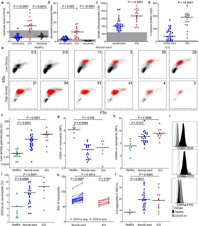
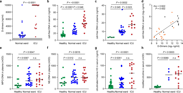
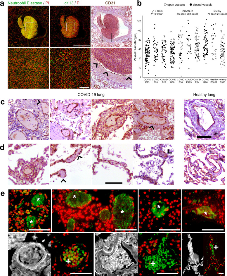
Patient findings: Here, we show that severe COVID-19 is characterized by a highly pronounced formation of neutrophil extracellular traps (NETs) inside the micro-vessels. Intravascular aggregation of NETs leads to rapid occlusion of the affected vessels, disturbed microcirculation, and organ damage. In severe COVID-19, neutrophil granulocytes are strongly activated and adopt a so-called low-density phenotype, prone to spontaneously form NETs. In accordance, markers indicating NET turnover are consistently increased in COVID-19 and linked to disease severity. Histopathology of the lungs and other organs from COVID-19 patients showed congestions of numerous micro-vessels by aggregated NETs associated with endothelial damage.

Interpretation: These data suggest that organ dysfunction in severe COVID-19 is associated with excessive NET formation and vascular damage.

Funding: Deutsche Forschungsgemeinschaft (DFG), EU, Volkswagen-Stiftung.

Keywords: Aggregated neutrophil extracellular traps; Coagulopathy; Endothelialitis; Immunothrombosis; SARS-CoV-2.


Conflict of interest statement

Declaration of Competing Interest Martin Herrmann served as adviser to Neutrolis, Cambridge, MA. The remaining authors declare no financial competing interests related to the study.

Figures

Fig. 1
Fig. 1
During acute phase responses in patients with COVID-19 circulating neutrophils display an activated, post-secretory phenotype prone to cellular aggregation. (a) Leukocyte count (Kruskal-Wallis P<0.0001) and (b) the neutrophil-lymphocyte ratio are elevated in severe COVID-19 (Kruskal-Wallis P<0.0001). Elevations of (c) lactate dehydrogenase (LDH) and (d) C-reactive protein (CRP) serum levels discriminate normal ward and ICU patients with COVID-19. The respective reference range is highlighted in grey. (e,f) Increased amounts of neutrophils are recovered from the low buoyant-density fraction after centrifugation. Numbers indicate percentage of CD16+ neutrophils of leukocytes of both densities (Kruskal-Wallis P<0.0001). (g,i upper panel) Downregulation of CD62L (Kruskal-Wallis P=0.0007) and (h,i middle panel) upregulation of CD66b in circulating neutrophils (Kruskal-Wallis P=0.001). (i lower panel, j, k) The activated phenotype of neutrophils is associated with an enhanced interaction with thrombocytes (CD41a) (Kruskal-Wallis P=0.0002) and (l) other leucocytes in whole blood precipitating cellular aggregation in patients in whole blood (WB) (Kruskal-Wallis P=0.0013) (green: healthy donors; blue: patients treated in normal wards; red: patients treated in intensive care units (ICU); black: patients recovered from COVID-19. Single dots represent individual patient samples. Kruskal-Wallis test was used for multiple group comparisons and the respective P value is depicted in the figure legend, P in the figure is deduced from post-hoc pair-wise comparison using Dunn's test) (For interpretation of the references to color in this figure legend, the reader is referred to the web version of this article).
Fig. 2
Fig. 2
Fibrin- and NET degradation products are elevated in the circulation during severe COVID-19 (a) The cross-linked fibrin degradation products (D-dimers) are elevated in hospitalized patients with COVID-19 and are strongly increased in patients requiring ICU care. The reference range is highlighted in grey. (b) Cell-free DNA was elevated in both serum (Kruskal-Wallis P<0.0001) and (c) plasma (Kruskal-Wallis P=0.0002) and (d) correlated with the D-dimer level (orange/black represents survivors/non-survivors). NET degradation products, namely (e) myeloperoxidase-DNA complexes (MPO-DNA) in citrated plasma (Kruskal-Wallis P=0.0033), (f) neutrophil elastase-DNA complexes (NE-DNA) in citrated plasma (Kruskal-Wallis P=0.0017) and (g) citrullinated histone H3 in serum were increased in the circulation of patients as compared to healthy controls (Kruskal-Wallis P=0.0001). (h) Modified protein-bound citrulline was significantly elevated in serum proteins of hospitalized patients (Kruskal-Wallis P<0.0001). (green: healthy donors; blue: patients treated in normal wards; red: patients treated in intensive care units (ICU). Single dots represent individual patient samples; Kruskal-Wallis test was used for multiple group comparisons and the respective P value is depicted in the figure legend, P in the figure is deduced from post-hoc pair-wise comparison using Dunn's test) (For interpretation of the references to color in this figure legend, the reader is referred to the web version of this article).
Fig. 3
Fig. 3
Aggregated neutrophil extracellular traps occlude pulmonary vessels. Immunohistochemistry was performed on lung tissue sections derived from autopsies of COVID-19 patients (n=8). (a) A representative picture of an occluded pulmonary blood vessel (CD31) is shown. The occlusive material is immunopositive for DNA (propidium iodide (PI) (red)), neutrophil elastase and citrullinated histone H3 (citH3) (green) typical of neutrophil extracellular traps and characterized by a high cellularity (scale bars designate 1 mm (upper panel) and 100 µm (lower panel), arrowheads indicate areas of endothelial damage). (b) Using CD31 immunohistochemistry open and occluded vessels were quantified in each lung tissue section of deceased COVID-19 patients and 2 non-COVID-19 control samples (single dots represent individual blood vessels, empty circles represent open vessels, filled circles represent occluded vessels). (c) Small and middle-sized vessels from COVID-19 sections were frequently clotted as compared to vessels in healthy control sections (Scale bars designate 100 µm, arrowheads indicate areas of endothelial damage). (d) Capillaries in dilated alveolar septa were frequently clogged with pauci-cellular material (Scale bars designate 200 µm, arrowheads indicate areas of endothelial damage). (e) Lung tissue sections of deceased COVID-19 patients were stained for expression of neutrophil elastase. The autofluorescence signal (488/525 nm) is displayed side-by-side in greyscale to identify vascular structures. Intermediate-sized pulmonary vessels from COVID-19 patients were frequently clotted by NETs (Scale bar designates 100 µm, asterisks and crosses indicate vessels clogged by pauci-cellular NETs and neutrophil-rich aggregates, respectively) (For interpretation of the references to color in this figure legend, the reader is referred to the web version of this article.).

Comment in

  • Immunothrombosis in severe COVID-19.
    Nakazawa D, Ishizu A. Nakazawa D, et al. EBioMedicine. 2020 Sep;59:102942. doi: 10.1016/j.ebiom.2020.102942. Epub 2020 Aug 15. EBioMedicine. 2020. PMID: 32810824 Free PMC article. No abstract available.

References

    1. Zhu N, Zhang D, Wang W. A Novel Coronavirus from Patients with Pneumonia in China, 2019. N Engl J Med. 2020;382(8):727–733. - PMC - PubMed
    1. WHO. Coronavirus disease 2019 (COVID-19) Situation Report –142. 10.06.2020 2020. https://www.who.int/docs/default-source/coronaviruse/situation-reports/2.... Accessed 11.06.2020 2020).
    1. Huang C, Wang Y, Li X. Clinical features of patients infected with 2019 novel coronavirus in Wuhan, China. Lancet. 2020;395(10223):497–506. - PMC - PubMed
    1. Liu Y, Yan LM, Wan L. Viral dynamics in mild and severe cases of COVID-19. Lancet Infect Dis. 2020 - PMC - PubMed
    1. Tang N, Li D, Wang X, Sun Z. Abnormal coagulation parameters are associated with poor prognosis in patients with novel coronavirus pneumonia. J Thromb Haemost. 2020;18(4):844–847. - PMC - PubMed

MeSH terms