. 2020 Aug 3;11(1):336.
doi: 10.1186/s13287-020-01852-y.

Human umbilical cord-derived mesenchymal stem cells prevent the progression of early diabetic nephropathy through inhibiting inflammation and fibrosis

Affiliations

Human umbilical cord-derived mesenchymal stem cells prevent the progression of early diabetic nephropathy through inhibiting inflammation and fibrosis

E Xiang et al. Stem Cell Res Ther. .

Abstract

Background: Diabetic nephropathy (DN) is one of the most serious complications of diabetes and the leading cause of end-stage chronic kidney disease. Currently, there are no effective drugs for treating DN. Therefore, novel and effective strategies to ameliorate DN at the early stage should be identified. This study aimed to explore the effectiveness and underlying mechanisms of human umbilical cord mesenchymal stem cells (UC-MSCs) in DN.

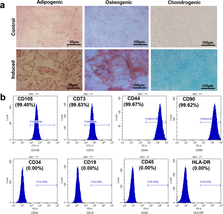
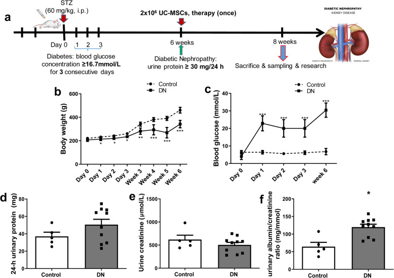
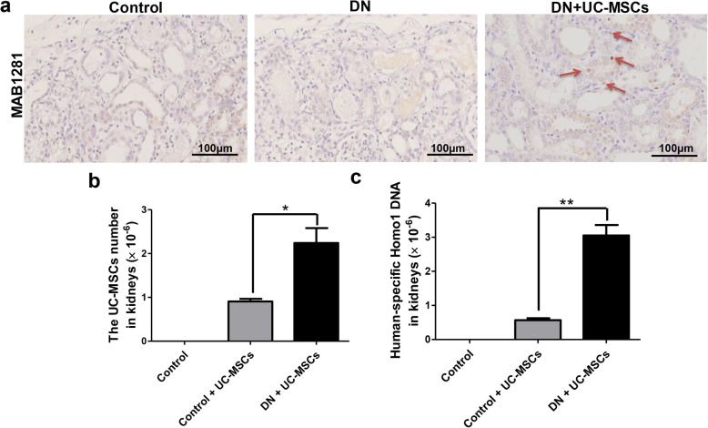
Methods: We identified the basic biological properties and examined the multilineage differentiation potential of UC-MSCs. Streptozotocin (STZ)-induced DN rats were infused with 2 × 10⁶ UC-MSCs via the tail vein at week 6. After 2 weeks, we measured blood glucose level, levels of renal function parameters in the blood and urine, and cytokine levels in the kidney and blood, and analyzed renal pathological changes after UC-MSC treatment. We also determined the colonization of UC-MSCs in the kidney with or without STZ injection. Moreover, in vitro experiments were performed to analyze cytokine levels of renal tubular epithelial cell lines (NRK-52E, HK2) and human renal glomerular endothelial cell line (hrGECs).

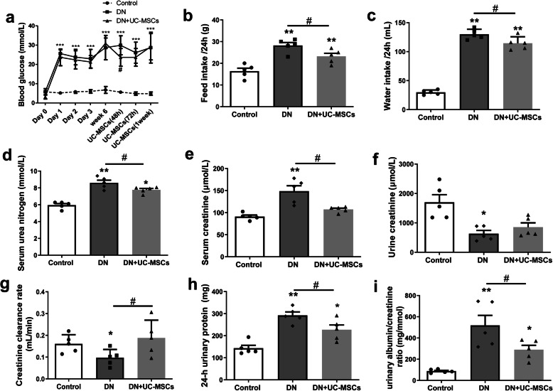
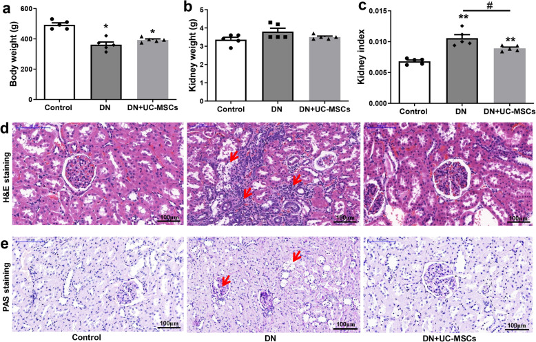
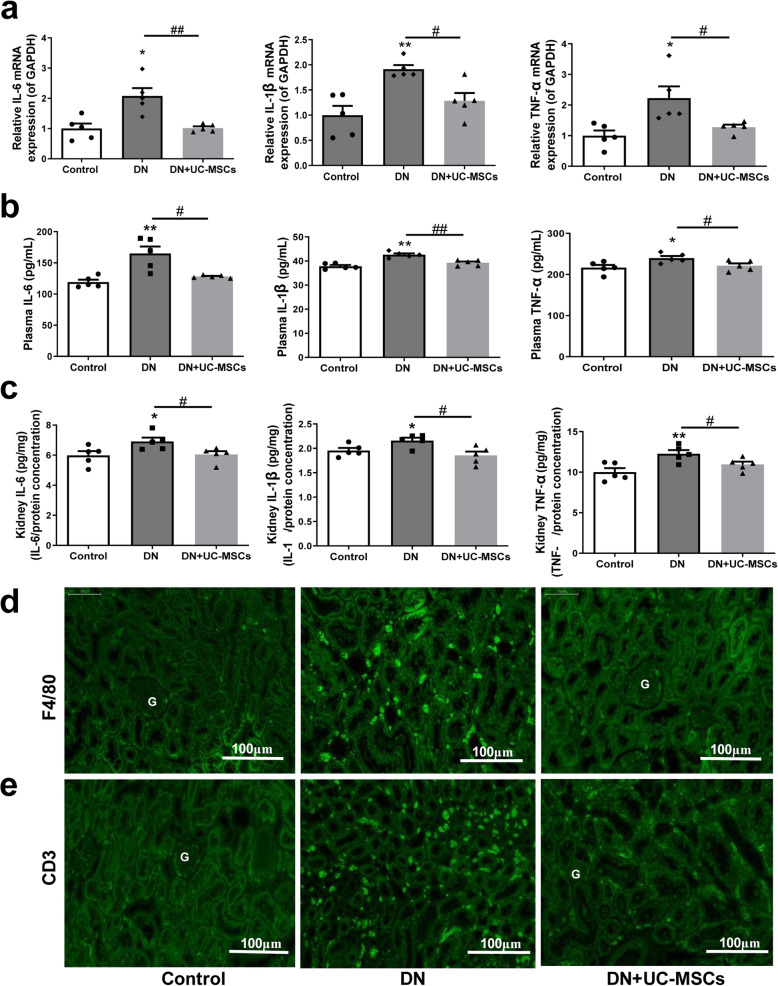
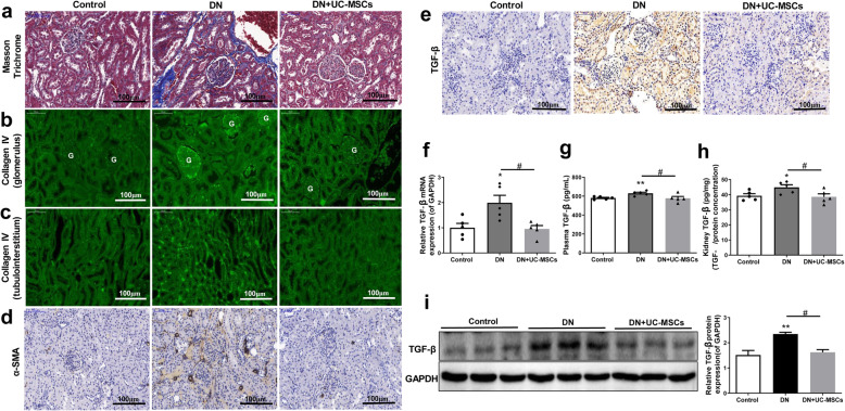
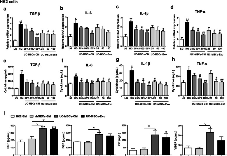
Results: UC-MSCs significantly ameliorated functional parameters, such as 24-h urinary protein, creatinine clearance rate, serum creatinine, urea nitrogen, and renal hypertrophy index. Pathological changes in the kidney were manifested by significant reductions in renal vacuole degeneration, inflammatory cell infiltration, and renal interstitial fibrosis after UC-MSC treatment. We observed that the number of UC-MSCs recruited to the injured kidneys was increased compared with the controls. UC-MSCs apparently reduced the levels of pro-inflammatory cytokines (IL-6, IL-1β, and TNF-α) and pro-fibrotic factor (TGF-β) in the kidney and blood of DN rats. In vitro experiments showed that UC-MSC conditioned medium and UC-MSC-derived exosomes decreased the production of these cytokines in high glucose-injured renal tubular epithelial cells, and renal glomerular endothelial cells. Moreover, UC-MSCs secreted large amounts of growth factors including epidermal growth factor, fibroblast growth factor, hepatocyte growth factor, and vascular endothelial growth factor.

Conclusion: UC-MSCs can effectively improve the renal function, inhibit inflammation and fibrosis, and prevent its progression in a model of diabetes-induced chronic renal injury, indicating that UC-MSCs could be a promising treatment strategy for DN.

Keywords: Diabetic nephropathy; Inflammation; Renal fibrosis; Umbilical cord mesenchymal stem cells.


Conflict of interest statement

The authors declare that they have no competing interests.

Figures

Fig. 1
Fig. 1
Induced differentiation ability and characteristic surface markers of UC-MSCs. a Differentiation abilities of cells were detected by cellular staining. The order from left to right: adipogenesis using Oil red O staining, osteogenesis using Alizarin red staining, and chondrogenesis using Alcian blue staining. b Specific surface markers of cells were examined by flow cytometry. The UC-MSCs associated with markers were positive for CD105, CD90, CD44, and CD73 and were negative for CD19, CD34, CD45, and HLA-DR
Fig. 2
Fig. 2
Timetable and flowchart of rat treatment and cell therapy as well as the identification of rat DN model. a The flowchart of rat treatment from day 0 to week 8. b The growth curve of body weight. c Blood glucose concentration curve. d 24-h urinary protein. e Urine creatinine. f Urinary albumin/creatinine ratio. N = 5 (control), N = 10 (DN), data are presented as mean ± SEM. *P < 0.05, **P < 0.01, ***P < 0.001 vs control group
Fig. 3
Fig. 3
Effects of UC-MSC treatment on biochemical indexes of DN rats. a Blood glucose concentration curve. b Feed intake/24 h. c Water inkake/24 h. d Serum urea nitrogen. e Serum creatinine. f Urine creatinine. g Creatinine clearance rate. h 24-h urinary protein. i Urinary albumin/creatinine ratio. N = 5 (per group), data are presented as mean ± SEM. *P < 0.05, **P < 0.01, ***P < 0.001 vs control. #P < 0.05 vs DN group
Fig. 4
Fig. 4
Histopathology analysis of tissues from DN rats. a Bodyweight. b Kidney weight. c Kidney index (kidney weight/bodyweight). d Hematoxylin-eosin (H&E) staining. e Periodic acid-Schiff (PAS) staining. N = 5 (per group), data are presented as mean ± SEM. *P < 0.05, **P < 0.01 vs control. #P < 0.05 vs DN group
Fig. 5
Fig. 5
The location and distribution of UC-MSCs in kidneys of DN rats at 2 weeks after intravenous infusion. a The red arrow indicates MAB1281-positive cells in the kidneys of DN rats. b The number of UC-MSCs detected in the kidneys of DN rats. c Relative expression of human-specific Homol DNA in kidneys of DN rats. N = 3–4 (per group), data are presented as mean ± SEM. *P < 0.05, **P < 0.01 vs control +UC-MSC group
Fig. 6
Fig. 6
Effects of UC-MSCs on inflammatory response in DN rats. a Relative mRNA expression of IL-6, IL-1β, and TNF-α in kidney tissues. b, c Concentration of IL-6, IL-1β, and TNF-α in plasma and kidney tissues by ELISA. d, e Immunofluorescence staining of F4/80 (d) and CD3 (e) in kidney tissues. (G: glomerulus). N = 5 (per group), data are presented as mean ± SEM. *P < 0.05, **P < 0.01 vs control. #P < 0.05, ##P < 0.01 vs DN group
Fig. 7
Fig. 7
UC-MSC treatment alleviated glomerular and tubulointerstitial fibrosis in DN rats. a Masson’s trichrome staining. b, c Immunofluorescence staining of collagen IV in glomerulus (b) and tubulointerstitium (e). d, e Immunohistochemistry staining of α-SMA (d) and TGF-β (e) in kidney tissues. f The mRNA expression of TGF-β in kidney tissues. g, h Concentration of TGF-β in plasma (g) and kidney (h) tissues. i Western blot analysis of TGF-β in kidney tissues. N = 5 (per group), data are presented as mean ± SEM. *P < 0.05, **P < 0.01 vs control. #P < 0.05 vs DN group
Fig. 8
Fig. 8
UC-MSC-CM or UC-MSC-Exo depressed cytokine expression in high glucose-injured HK2 cells. a-d mRNA expression of TGF-β (a), IL-6 (b), IL-1β (c) and TNF-α (d) in HK2 cells. e-h Concentration of TGF-β (e), IL-6 (f), IL-1β (g), and TNF-α (h) in the supernatant of HK2 cells. i Concentration of EGF, FGF, HGF, and VEGF in UC-MSC-CM and UC-MSC-Exo. N = 3 independent experiments. Data are presented as mean ± SEM. *P < 0.05, **P < 0.01 vs LG group; #P < 0.05, ##P < 0.01 vs HG group; P < 0.05, ★★P < 0.01 vs HK2-SM group, P < 0.05 vs hrGEC-SM group. UC-MSC-CM: UC-MSC conditioned medium; UC-MSC-Exo: UC-MSC-derived exosomes; LG: low glucose; HG: high glucose; SM: standard medium

References

    1. Forbes JM, Cooper ME. Mechanisms of diabetic complications. Physiol Rev. 2013;93(1):137–188. - PubMed
    1. Mathers CD, Loncar D. Projections of global mortality and burden of disease from 2002 to 2030. PLoS Med. 2006;3(11):e442. - PMC - PubMed
    1. Saran R, et al. US renal data system 2019 annual data report: epidemiology of kidney disease in the United States. Am J Kidney Dis. 2020;75(1 Suppl 1):A6–A7. - PubMed
    1. Martinez-Castelao A, et al. The concept and the epidemiology of diabetic nephropathy have changed in recent years. J Clin Med. 2015;4(6):1207–1216. - PMC - PubMed
    1. Hadjadj S, et al. Death, end-stage renal disease and renal function decline in patients with diabetic nephropathy in French cohorts of type 1 and type 2 diabetes. Diabetologia. 2016;59(1):208–216. - PubMed

Publication types

Substances