Review
. 2020 Sep 24;56(3):2002049.
doi: 10.1183/13993003.02049-2020. Print 2020 Sep.

Pathogenesis of COVID-19-induced ARDS: implications for an ageing population

Affiliations
Review

Pathogenesis of COVID-19-induced ARDS: implications for an ageing population

Manuel A Torres Acosta et al. Eur Respir J. .

Abstract

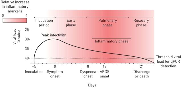
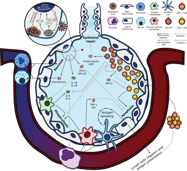
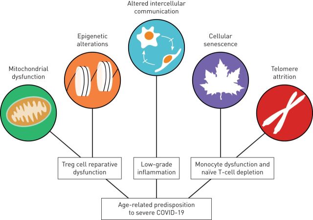
The coronavirus disease 2019 (COVID-19) pandemic has elicited a swift response by the scientific community to elucidate the pathogenesis of severe acute respiratory syndrome-coronavirus-2 (SARS-CoV-2)-induced lung injury and develop effective therapeutics. Clinical data indicate that severe COVID-19 most commonly manifests as viral pneumonia-induced acute respiratory distress syndrome (ARDS), a clinical entity mechanistically understood best in the context of influenza A virus-induced pneumonia. Similar to influenza, advanced age has emerged as the leading host risk factor for developing severe COVID-19. In this review we connect the current understanding of the SARS-CoV-2 replication cycle and host response to the clinical presentation of COVID-19, borrowing concepts from influenza A virus-induced ARDS pathogenesis and discussing how these ideas inform our evolving understanding of COVID-19-induced ARDS. We also consider important differences between COVID-19 and influenza, mainly the protean clinical presentation and associated lymphopenia of COVID-19, the contrasting role of interferon-γ in mediating the host immune response to these viruses, and the tropism for vascular endothelial cells of SARS-CoV-2, commenting on the potential limitations of influenza as a model for COVID-19. Finally, we explore hallmarks of ageing that could explain the association between advanced age and susceptibility to severe COVID-19.


Conflict of interest statement

Conflict of interest: M.A. Torres Acosta has nothing to disclose. Conflict of interest: B.D. Singer has a patent US Patent App. 15/542,380, “Compositions and Methods to Accelerate Resolution of Acute Lung Inflammation” pending.

Figures

FIGURE 1
FIGURE 1
Stereotypical clinical course of coronavirus disease 2019 complicated by acute respiratory distress syndrome (ARDS). Mean incubation period and viral load threshold cycle (Ct) values at days 0, 7, 14 and 21 according to He et al. [12]. Median days from symptom onset to dyspnoea (interquartile range 4–9 days), ARDS (interquartile range 8–15 days), and discharge or death (interquartile range 17–25 days) according to Zhou et al. [14].
FIGURE 2
FIGURE 2
Key components of the pathogenesis of influenza-induced acute respiratory distress syndrome. Influenza's replication cycle requires the presence of viral RNA in the host epithelial cell's cytoplasm, which is recognised by anti-viral pathways such as Toll-like receptors, mitochondria-associated anti-viral signalling, and the NLRP3 inflammasome. The host cell responds by upregulating nuclear factor (NF)-κB-regulated genes (NF-κBrg) and secreting type-I and -III interferons (IFN-I and IFN-III), which stimulate the transcription of interferon-stimulated genes (ISGs). Infected epithelial cell-derived IFN-I and IFN-III induce epithelial cell apoptosis, and interleukin (IL)-1β and IL-18 secretion activates alveolar macrophages, which phagocytose apoptotic cells. Alveolar macrophages also secrete IL-6 and IL-8, which recruit the central immune system, including monocytes, to the lung parenchyma. Monocytes differentiate into monocyte-derived macrophages and dendritic cells. Monocyte-derived macrophages also contribute to infected epithelial cell phagocytosis and secrete tumour necrosis factor (TNF)-α and nitric oxide (NO) to activate epithelial cell apoptosis. Dendritic cells link the innate and adaptive immune responses by sampling alveolar antigens and migrating to lymph nodes, where they present viral antigens to naïve T-cells. After undergoing antigen-specific clonal expansion, T-lymphocytes are mobilised to the lung. CD8+ T-cells exert their cytotoxicity on viral antigen-presenting cells when co-stimulated by CD4+ T-cells, which also secrete type-II interferon (IFN-II), a negative regulator of CD8+ T-cell activity. In turn, CD4+ T-cells are negatively regulated by regulatory T-cells (Treg), which also promote epithelial repair via the secretion of amphiregulin and keratinocyte growth factor (KGF), and attenuate inflammation by inhibiting alveolar macrophage-derived IL-6 and IL-8 secretion while potentiating their phagocytic activity (efferocytosis). NK: natural killer.
FIGURE 3
FIGURE 3
Examples of the contribution of hallmarks of ageing to the age-related predisposition to severe coronavirus disease 2019 (COVID-19). Age-related mitochondrial dysfunction can induce epigenetic changes in regulatory T-cells (Treg), which impair their pro-recovery functions to hinder proper resolution of inflammation and repair of lung injury. Monocytes and naïve T-lymphocytes undergo cellular senescence following telomere attrition from sustained replication, impairing the host's ability to mount an efficient immune response to a viral challenge or create a memory T-cell response to vaccines. Finally, altered intercellular communication underlies the low-grade inflammation associated with ageing, which contributes to the development of age-related comorbidities.

References

    1. Zhu N, Zhang D, Wang W, et al. . A novel coronavirus from patients with Pneumonia in China, 2019. N Engl J Med 2020; 382: 727–733. doi:10.1056/NEJMoa2001017 - DOI - PMC - PubMed
    1. World Health Organization . Coronavirus disease (COVID-19) pandemic. www.who.int/emergencies/diseases/novel-coronavirus-2019 Date last accessed: 10 July 2020.
    1. Herold S, Becker C, Ridge KM, et al. . Influenza virus-induced lung injury: pathogenesis and implications for treatment. Eur Respir J 2015; 45: 1463–1478. doi:10.1183/09031936.00186214 - DOI - PubMed
    1. Chen G, Wu D, Guo W, et al. . Clinical and immunological features of severe and moderate coronavirus disease 2019. J Clin Invest 2020; 130: 2620–2629. doi:10.1172/JCI137244 - DOI - PMC - PubMed
    1. Onder G, Rezza G, Brusaferro S. Case-fatality rate and characteristics of patients dying in relation to COVID-19 in Italy. JAMA 2020; 323: 1775–1776 - PubMed

Publication types