. 2020 Oct 2;295(40):13862-13874.
doi: 10.1074/jbc.RA120.013898. Epub 2020 Aug 3.

Extended interaction networks with HCV protease NS3-4A substrates explain the lack of adaptive capability against protease inhibitors

Affiliations

Extended interaction networks with HCV protease NS3-4A substrates explain the lack of adaptive capability against protease inhibitors

Georg Dultz et al. J Biol Chem. .

Abstract

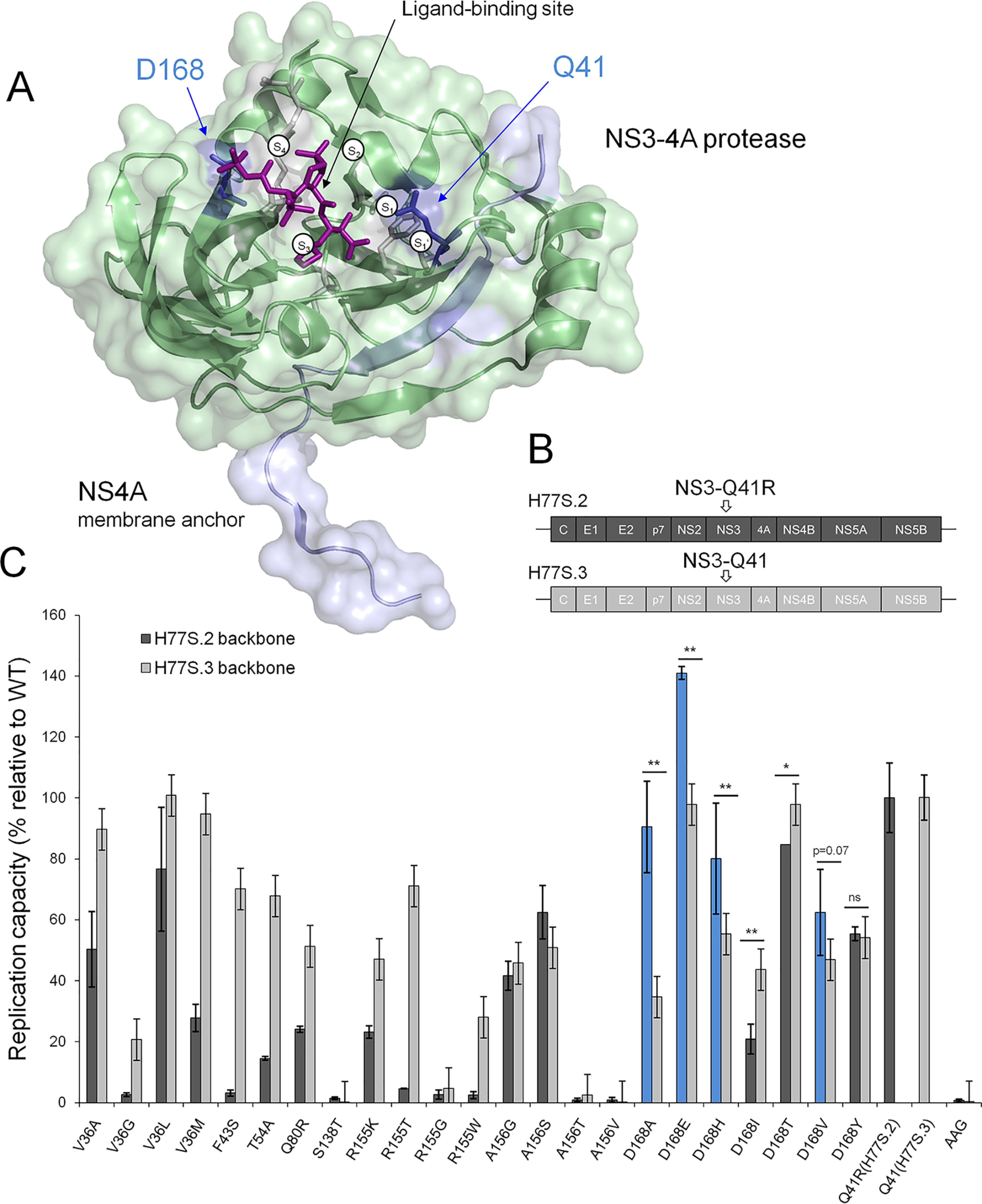
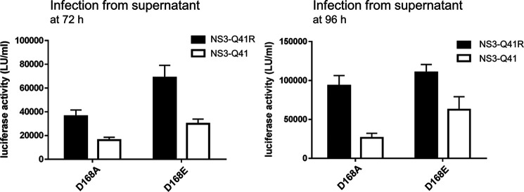
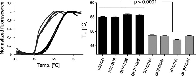
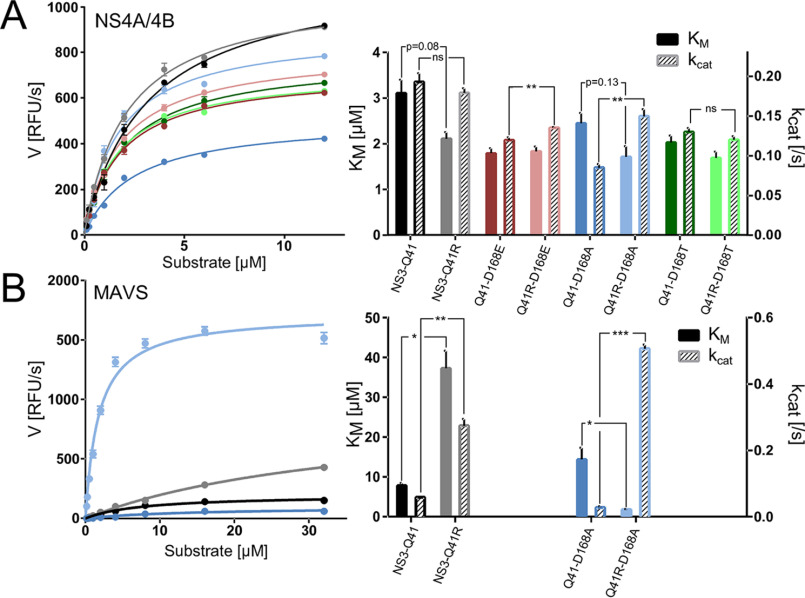
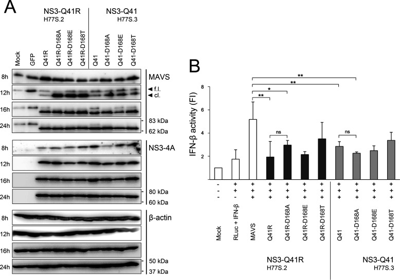
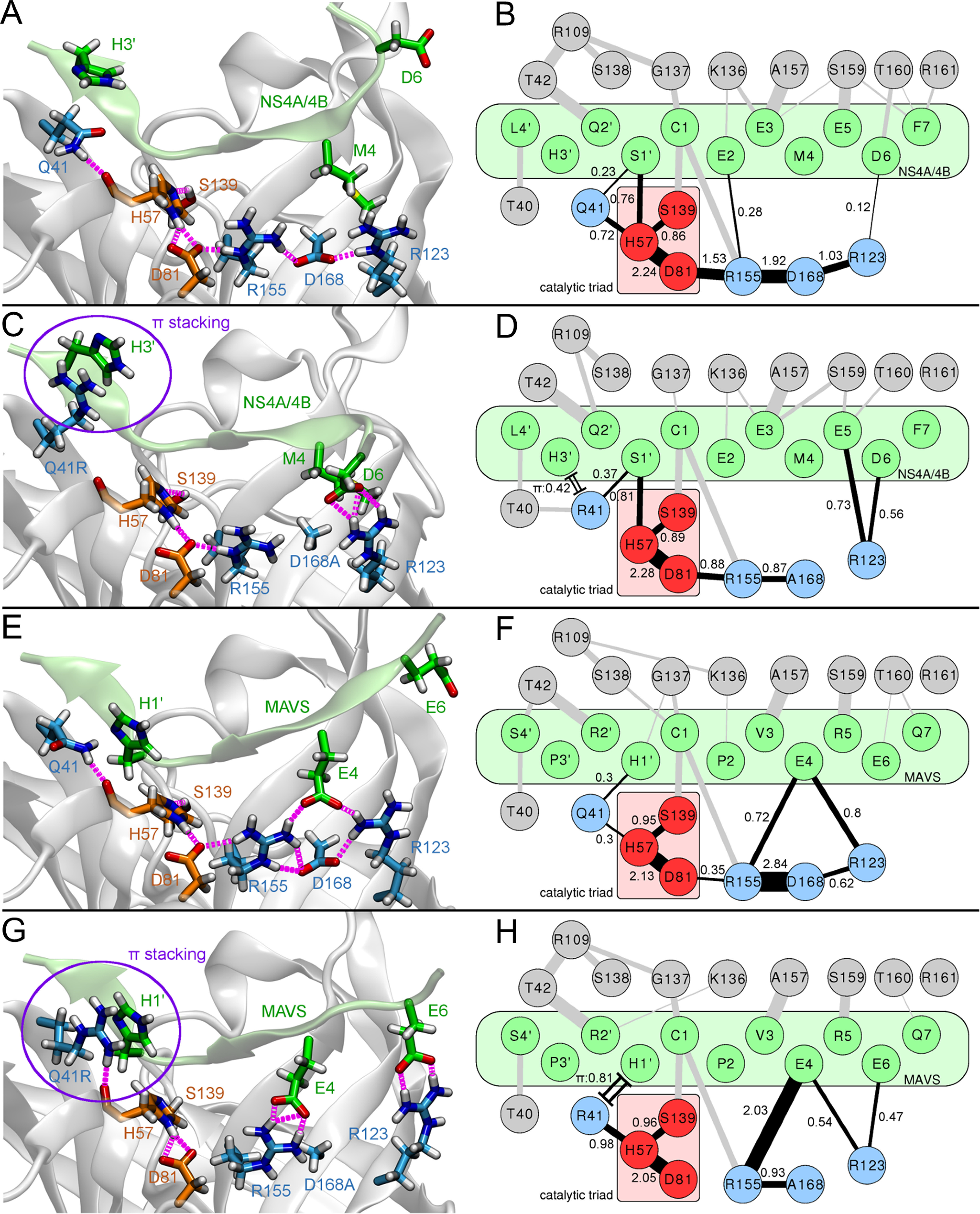
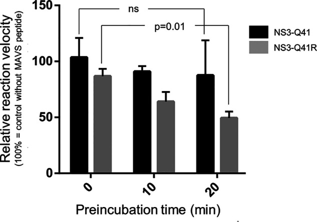
Inhibitors against the NS3-4A protease of hepatitis C virus (HCV) have proven to be useful drugs in the treatment of HCV infection. Although variants have been identified with mutations that confer resistance to these inhibitors, the mutations do not restore replicative fitness and no secondary mutations that rescue fitness have been found. To gain insight into the molecular mechanisms underlying the lack of fitness compensation, we screened known resistance mutations in infectious HCV cell culture with different genomic backgrounds. We observed that the Q41R mutation of NS3-4A efficiently rescues the replicative fitness in cell culture for virus variants containing mutations at NS3-Asp168 To understand how the Q41R mutation rescues activity, we performed protease activity assays complemented by molecular dynamics simulations, which showed that protease-peptide interactions far outside the targeted peptide cleavage sites mediate substrate recognition by NS3-4A and support protease cleavage kinetics. These interactions shed new light on the mechanisms by which NS3-4A cleaves its substrates, viral polyproteins and a prime cellular antiviral adaptor protein, the mitochondrial antiviral signaling protein MAVS. Peptide binding is mediated by an extended hydrogen-bond network in NS3-4A that was effectively optimized for protease-MAVS binding in Asp168 variants with rescued replicative fitness from NS3-Q41R. In the protease harboring NS3-Q41R, the N-terminal cleavage products of MAVS retained high affinity to the active site, rendering the protease susceptible for potential product inhibition. Our findings reveal delicately balanced protease-peptide interactions in viral replication and immune escape that likely restrict the protease adaptive capability and narrow the virus evolutionary space.

Keywords: adaptation; drug resistance; evolution; hepatitis C virus (HCV); mitochondrial antiviral signaling protein (MAVS); molecular adaptation; molecular biology; molecular dynamics; protease inhibitor; replicative fitness; resistance mutation; serine protease (NS3-4A); structure constraints.


Conflict of interest statement

Conflict of interest—The authors declare that they have no conflicts of interest with the contents of this article.

Figures

Figure 1.
Figure 1.
Replicative fitness of PI-resistant variants in NS3 ± Q41R protease genomic backgrounds. A, structural context of PI-resistance mutations depicted on the NS3-4A protease structure from PDB 2OC8 (50) with PI bound to the ligand-binding site (purple stick model); ligand-binding pockets S4 to S1′ (51). The protein surface is partially transparent; protease domain: green, NS4A: light blue. Gln41 and Asp168 are shown as blue stick models located at opposite ends of the protease substrate–binding pocket. B, the H77S.2 molecular clone contains six cell culture adaptive mutations (not shown) throughout the viral genome; the only cell culture adaptive mutation located in the NS3-4A protease domain, NS3-Q41R (depicted by arrow), is reverted back to NS3-Gln41 WT in H77S.3 (6). C, GLuc activity secreted by RNA-transfected cells normalized to pH77S.3 and pH77S.2 WT. Blue shading indicates PI-resistant variants with enhanced replicative fitness by NS3-Q41R. Data shown represent the mean ± S.D. from at least three independent experiments; *p < 0.001; **p < 0.0001; by two-sided t test. AAG, negative control.
Figure 2.
Figure 2.
Impact of NS3 ± Q41R on the infectious virus production of Asp168 variants. Comparison of infectious virus yield from D168A/E-protease mutants within NS3-Gln41 and NS3-Q41R backbones. Infectious virus production determined from supernatant fluids at 72 and 96 h infected to naïve Huh-7.5 cells as determined by GLuc activity assay. Data shown represent the mean ± S.D. from at least three independent experiments.
Figure 3.
Figure 3.
Impact of Asp168 mutants on the protein fold of the NS3-4A protease ± NS3-Q41R. Data from real-time thermal stability assay using Sypro Orange, a temperature-stable fluorophore that exhibits enhanced fluorescence upon interacting with unfolded proteins. The thermal stability of WT protease and mutants was assessed under increasing incubation temperatures. The impact of mutations on protein unfolding patterns is characterized by fluorescence emission curves (left). Melting temperatures (Tm) from purified NS3-4A protease mutants are determined by fitting the sigmoidal melt curve to the Boltzmann equation (right). Error bars represent the mean ± S.D. from at least three independent experiments; *p ≤ 0.05; **p ≤ 0.01; by two-sided t test.
Figure 4.
Figure 4.
Impact of Asp168 mutants and NS3 ± Q41R on protease enzymatic function. A, reaction velocity and Michealis-Menten kinetics as assessed from purified protein of protease WT and Asp168 mutants ± NS3-Q41R and the natural polyprotein substrate NS4A/4B and (B) assessed for the MAVS peptide (each at different peptide concentrations). Reaction constants Km and kcat were calculated after nonlinear regression curve fitting. Error bars represent the mean ± S.D. from at least three independent experiments; *p < 0.05; **p ≤ 0.01; ***p ≤ 0.001; by two-sided t test.
Figure 5.
Figure 5.
Impact of Asp168 mutants and NS3 ± Q41R on MAVS cleavage capacity and IFN-β activation. A, U-2 OS cells were left untransfected (mock), transfected with a GFP expression plasmid (control of transfection efficiency), and transfected with expression constructs harboring NS3-Gln41 or NS3-Q41R ± D168A/E/T. MAVS cleavage as assessed by Western blotting; one of five representative experiments is shown. B, U-2 OS cells co-transfected with IFN-β reporter plasmids, a full-length MAVS expression construct, and D168A/E/T mutant protease constructs ± NS3-Q41R, subsequently analyzed for IFN-β-dependent luciferase activity. Renilla luciferase (RLuc) activity determined to normalized IFN-β activity. Error bars represent the mean ± S.D. from at least three independent experiments; *p ≤ 0.05; **p ≤ 0.01; by two-sided t test; f.l., full-length; cl., cleaved form; FI, fold induction; ns, not significant.
Figure 6.
Figure 6.
Molecular characterization of protease-substrate interactions. Illustrations of representative protease binding-site conformations (left panel: A, C, E and G) and H-bond networks of protease-substrate complex simulations (right panel: B, D, F and H). (A) 3D structure and (B) residue-interaction network of the NS4A/4B substrate bound to the NS3-Gln41 protease WT or (C) 3D structure and (D) residue-interaction network for NS4A/4B bound to the Q41R-D168A protease; (E) 3D structure and (F) residue-interaction network for MAVS bound to the NS3-Gln41 protease WT or (G) 3D structure and (H) residue-interaction network for MAVS bound to the Q41R-D168A protease. Hydrogen bonds present in each time frame were extracted from MD trajectories. Edge widths (single lines) in the residue networks shown represent the average number of H-bonds during simulation time and correspond to the individual bond strength; a quantitative measure for the bond strength was provided by corresponding numbers. Only edges above a minimum occurrence limit of 0.1 are shown. Cation-π stacking interactions between arginine and histidine residues (time fraction within π stacking distance cutoff) are shown as double lines with caps. Atom colors: Substrate (green); protease catalytic triad (red); extended H-bond network of the catalytic triad (blue). H-bonds are shown in magenta (A, C, E and G) and black (B, D, F and H).
Figure 7.
Figure 7.
Inhibition of the NS3-4A protease ± Q41R by MAVS cleavage products. Relative reaction velocity for NS4A/4B cleavage as assessed for the purified NS3-Gln41 protease WT and NS3-Q41R mutant protease (measured by FRET protease assay) and different preincubation time with MAVS peptide (0, 10, and 20 min). Reference velocity (100%) is the cleavage reaction for the NS4A/4B FRET peptide without MAVS. Error bars represent the mean ± S.D. from at least three independent experiments; ns, not significant, p = 0.01; by two-sided t test.

References

    1. Sheldon J., Beach N. M., Moreno E., Gallego I., Piñeiro D., Martínez-Salas E., Gregori J., Quer J., Esteban J. I., Rice C. M., Domingo E., and Perales C. (2014) Increased replicative fitness can lead to decreased drug sensitivity of hepatitis C virus. J. Virol. 88, 12098–12111 10.1128/JVI.01860-14 - DOI - PMC - PubMed
    1. Doncheva N. T., Domingues F. S., McGivern D. R., Shimakami T., Zeuzem S., Lengauer T., Lange C. M., Albrecht M., and Welsch C. (2019) Near-neighbor interactions in the NS3-4A protease of HCV impact replicative fitness of drug-resistant viral variants. J. Mol. Biol. 431, 2354–2368 10.1016/j.jmb.2019.04.034 - DOI - PMC - PubMed
    1. Domingo E., Sheldon J., and Perales C. (2012) Viral quasispecies evolution. Microbiol. Mol. Biol. Rev. 76, 159–216 10.1128/MMBR.05023-11 - DOI - PMC - PubMed
    1. Rong L., Dahari H., Ribeiro R. M., and Perelson A. S. (2010) Rapid emergence of protease inhibitor resistance in hepatitis C virus. Sci. Transl. Med. 2, 30ra32 10.1126/scitranslmed.3000544 - DOI - PMC - PubMed
    1. Pawlotsky J. M. (2016) Hepatitis C virus resistance to direct-acting antiviral drugs in interferon-free regimens. Gastroenterology 151, 70–86 10.1053/j.gastro.2016.04.003 - DOI - PubMed

Publication types

MeSH terms

Substances

LinkOut - more resources