. 2020 Aug 18;117(33):20109-20116.
doi: 10.1073/pnas.1921315117. Epub 2020 Aug 3.

Necroptosis-based CRISPR knockout screen reveals Neuropilin-1 as a critical host factor for early stages of murine cytomegalovirus infection

Affiliations

Necroptosis-based CRISPR knockout screen reveals Neuropilin-1 as a critical host factor for early stages of murine cytomegalovirus infection

Rebecca K Lane et al. Proc Natl Acad Sci U S A. .

Abstract

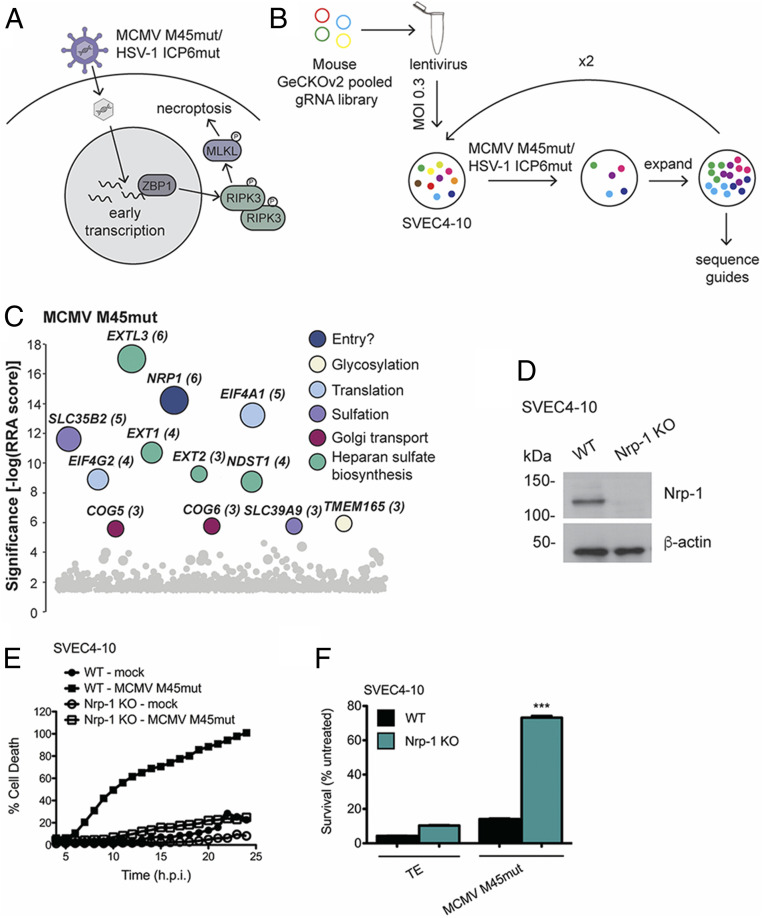
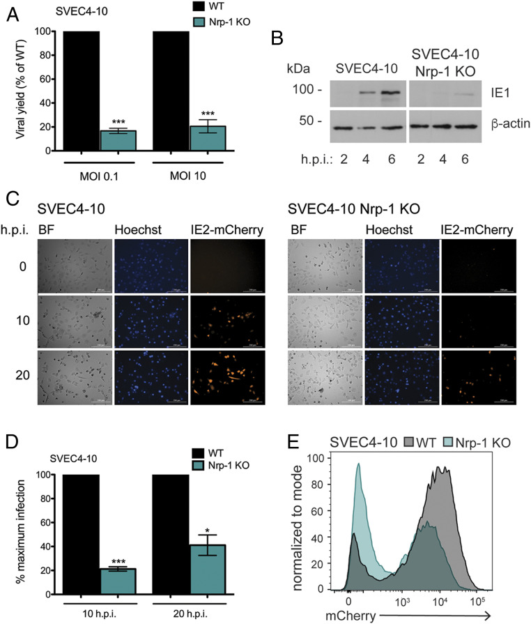
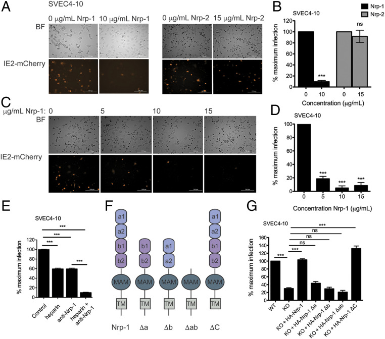
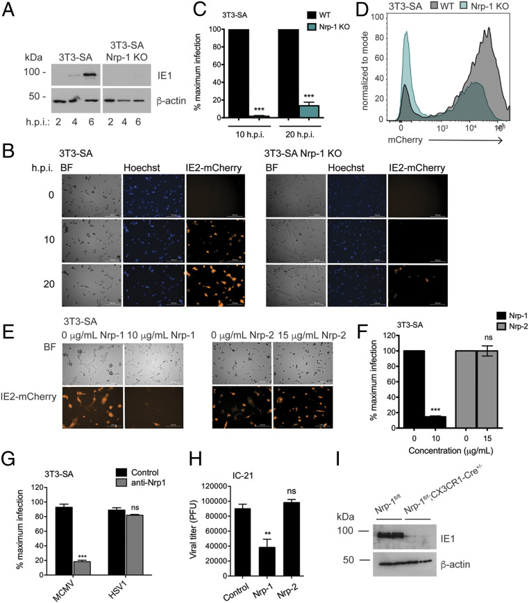
Herpesviruses are ubiquitous human pathogens that cause a wide range of health complications. Currently, there is an incomplete understanding of cellular factors that contribute to herpesvirus infection. Here, we report an antiviral necroptosis-based genetic screen to identify novel host cell factors required for infection with the β-herpesvirus murine cytomegalovirus (MCMV). Our genome-wide CRISPR-based screen harnessed the capacity of herpesvirus mutants that trigger antiviral necroptotic cell death upon early viral gene expression. Vascular endothelial growth factor (VEGF) and semaphorin-binding receptor Neuropilin-1 (Nrp-1) emerge as crucial determinants of MCMV infection. We find that elimination of Nrp-1 impairs early viral gene expression and reduces infection rates in endothelial cells, fibroblasts, and macrophages. Furthermore, preincubation of virus with soluble Nrp-1 dramatically inhibits infection by reducing virus attachment. Thus, Nrp-1 is a key determinant of the initial phase of MCMV infection.

Keywords: CRISPR; cytomegolovirus; necroptosis; neuropilin.


Conflict of interest statement

The authors declare no competing interest.

Figures

Fig. 1.
Fig. 1.
CRISPR knockout (KO) screen reveals novel candidate mediators of MCMV and HSV-1 infection. (A) Diagram of activation of death signaling during mutant herpesvirus infection. The Z-NA sensor ZBP-1 recognizes early transcriptional events in herpesvirus infection, triggering activation of necroptotic signaling. (B) Schematic of the genome-wide CRISPR screen for novel candidate mediators of MCMV infection. The mouse GeCKOv2 gRNA library was transfected into 293T cells to produce lentivirus. Library-containing lentivirus was then transduced at multiplicity of infection (MOI) 0.3 into SVEC4-10 cells. Transduced cells were selected based on puromycin resistance and infected at MOI 5 with MCMV M45mut for 1 h. After surviving cells had grown to 80% confluency, the infection process was repeated until cells were completely resistant. Cellular genomes were then sequenced to identify enriched gRNAs. (C) Screen results for MCMV. Hits are stratified along a vertical axis according to significance and arbitrarily spread along the horizontal axis. The size of the circle and the number next to the gene name both indicate the number of gRNAs for that gene that were found in the resistant population. Colors indicate gene ontology. (D) Western blot showing level of Nrp-1 KO in SVEC4-10 cells. (E) Cell death over time in wild-type (WT) and Nrp-1 KO SVEC4-10 cells infected with MCMV M45mut. Cells were infected in triplicate with MCMV M45mut (MOI 10) in media containing 50 nM Sytox green and 2 μM Hoechst and imaged every hour with a Cytation 5 Imaging Reader. Cell death was calculated as (GFP + cells/Hoechst + cells) × 100% and averaged from four images per well. (F) Cell survival after 20 h.p.i. in WT and Nrp-1 KO SVEC4-10 cells after infection with MCMV M45mut. Cells were infected with MCMV M45mut (MOI 10) or treated with 25 ng/mL TNFα + 5 μM emricasan or media alone. After 24 h, survival was quantified by Cell Titer Glo Luminescent Cell Viability Assay. Infections were performed in triplicate with three independent replicates. Error bars indicate SEM. Significance was determined using a Student’s t test (***P < 0.0001).
Fig. 2.
Fig. 2.
Loss of Nrp-1 reduces viral gene expression. (A) MCMV yield in WT and Nrp-1 KO SVEC4-10 cells. WT and Nrp-1 KO SVEC4-10 cells were infected at the indicated MOI with WT MCMV for 1 h. Cells were harvested after 48 (MOI 10) or 72 (MOI 0.1) h.p.i., and viral yield determined by plaque assay. Values shown are viral yield relative to that of WT cells. Infections were performed in triplicate. (B) Western blot for viral immediate early gene expression. (C) Fluorescence microscopy of infected WT or Nrp-1 KO SVEC4-10 cells. Cells were infected in triplicate at MOI 1 with MCMV IE2-mCherry in media containing 2 μM Hoechst and imaged with a Cytation 5 Imaging Reader every hour for 24 h. Shown are representative images. (D) Quantification of mCherry+ cells in C. Shown is the average of four images. Rates of infection in Nrp-1 KO cells were normalized to rates of infection in WT cells. (E) Flow cytometry for IE2-mCherry expression. Cells were infected as in C, and red fluorescence quantified after 20 h by flow cytometry. At least, two replicates were performed per experiment. Error bars indicate SEM. Significance was determined using a Student’s t test (***P < 0.0001, *P < 0.01).
Fig. 3.
Fig. 3.
The extracellular domains of Nrp-1 mediate MCMV infection. MCMV IE2-mCherry was incubated with soluble recombinant Nrp-1, Nrp-2, or media alone for 1 h at 37 °C before being added to WT SVEC4-10 cells at MOI 1 in triplicate. Cells were imaged 20 h postinfection with a Cytation 5 Imaging Reader (A), and mCherry+ cells quantified (B). Significance was determined using a Student’s t test. (C) Fluorescence microscopy for IE2-mCherry expression. Cells were infected in triplicate with virus pretreated as in A with varying concentrations of soluble Nrp-1 and imaged as in A. (D) Quantification of mCherry+ cells from (C). Significance was determined by using a one-way ANOVA with Dunnett’s multiple comparisons test. (E) Infection rates in SVEC4-10 cells pretreated with anti-Nrp-1 antibody or heparin. Cells were incubated for 30 min with 20 μg/mL heparin, 4 μg/mL anti-Nrp-1 antibody, or both. After 6 h of infection with WT MCMV, cells were fixed and stained for IE1 expression. Significance was determined by one-way ANOVA with Bonferroni’s multiple comparisons test. (F) Structure of Nrp-1 and domain deletion mutants. (G) Quantification of mCherry+ cells in SVEC4-10 WT or Nrp-1 KO cells reconstituted with full-length and deletion Nrp-1 mutants. Cells were infected with MCMV IE2-mCherry (MOI 1) in triplicate and imaged after 20 h.p.i. Significance was determined using a one-way ANOVA with Bonferroni’s multiple comparisons test. All microscopy data were quantified by taking the average from four images. At least, two replicates were performed per experiment. Error bars indicate SEM. (***P < 0.0001).
Fig. 4.
Fig. 4.
Nrp-1 mediates MCMV infection of multiple cell types. (A) Western blot for viral immediate early gene expression in 3T3-SA cells. (B) Fluorescence microscopy for IE2-mCherry expression. Cells were infected in triplicate at MOI 1 with MCMV IE2-mCherry in media containing 2 μM Hoechst and imaged with a Cytation 5 Imaging Reader every hour for 24 h. Shown are representative images. (C) Quantification of mCherry+ cells from B. (D) Cells were infected as in B and red fluorescence quantified after 20 h by flow cytometry. (E) Fluorescence microscopy for IE2-mCherry expression. MCMV IE2-mCherry was incubated with soluble recombinant Nrp-1, Nrp-2, or media alone for 1 h at 37 °C before being added to WT SVEC4-10 and 3T3-SA cells in triplicate at MOI 1. Cells were imaged 20 h postinfection with a Cytation 5 Imaging Reader. (F) Quantification of mCherry+ cells from E. (G) Infection rates in the presence of anti-Nrp-1 antibody. The 3T3-SA cells cultured in coverslips were preincubated with anti-Nrp1 antibody (4 μg/mL) for 30 min and then infected for 6 h with MCMV or HSV-1 (MOI 10). The percentage of infected cells was assessed by staining for IE1 (MCMV) or ICP0 (HSV-1). (H) Quantification of viral attachment at the cell surface in the presence of soluble neuropilins. MCMV was incubated with soluble recombinant Nrp-1, Nrp-2, or media alone for 1 h before being added to IC-21 cells in triplicate at MOI 1 and incubated for 30 min at 4 °C. Cells were harvested, and unbound virions washed away. The amount of adhered virus was then quantified by plaque assay. Significance was determined by using a one-way ANOVA with Dunnett’s multiple comparisons test. (I) Western blot for IE1 expression in BMDMs lacking Nrp-1 and infected with MCMV (MOI 1) for 6 h. BMDMs were generated from two mice per genotype and treated with 2 μM 4-hydroxy-tamoxifen for 7 d to induce Cre activity. All microscopy data were quantified by taking the average from four images. At least, two replicates were performed per experiment. Error bars indicate SEM. Significance was determined using a Student’s t test unless otherwise stated (***P < 0.0001, **P < 0.001).

References

    1. Brune W., Andoniou C. E., Die another day: Inhibition of cell death pathways by cytomegalovirus. Viruses 9, 249 (2017). - PMC - PubMed
    1. Upton J. W., Chan F. K., Staying alive: Cell death in antiviral immunity. Mol. Cell 54, 273–280 (2014). - PMC - PubMed
    1. Barber G. N., Host defense, viruses and apoptosis. Cell Death Differ. 8, 113–126 (2001). - PubMed
    1. Kaiser W. J., Upton J. W., Mocarski E. S., Viral modulation of programmed necrosis. Curr. Opin. Virol. 3, 296–306 (2013). - PMC - PubMed
    1. Mocarski E. S., Upton J. W., Kaiser W. J., Viral infection and the evolution of caspase 8-regulated apoptotic and necrotic death pathways. Nat. Rev. Immunol. 12, 79–88 (2011). - PMC - PubMed

Publication types