. 2020 Sep 18;88(10):e00132-20.
doi: 10.1128/IAI.00132-20. Print 2020 Sep 18.

Rab5a Promotes Cytolethal Distending Toxin B-Induced Cytotoxicity and Inflammation

Affiliations

Rab5a Promotes Cytolethal Distending Toxin B-Induced Cytotoxicity and Inflammation

Ming-Xian Chen et al. Infect Immun. .

Abstract

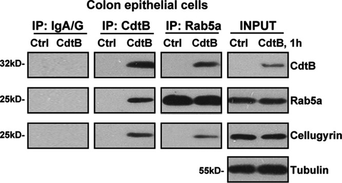
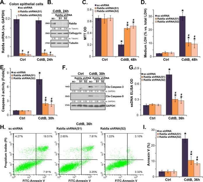
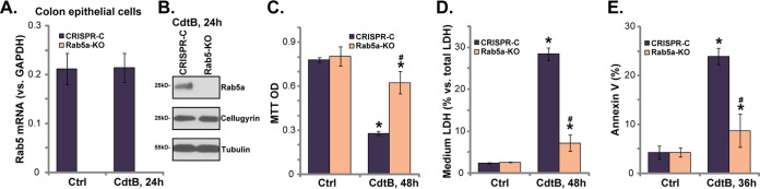
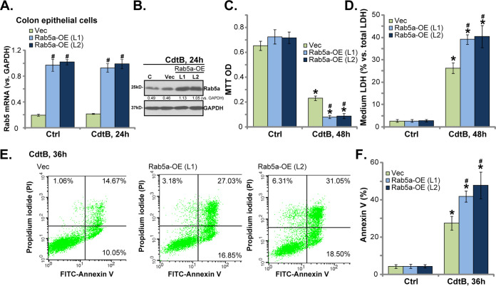
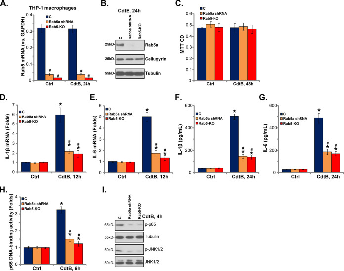
The cytolethal distending toxin B subunit (CdtB) induces significant cytotoxicity and inflammation in many cell types that are involved in the pathogenesis of postinfectious irritable bowel syndrome (PI-IBS). However, the underlying mechanisms remain unclear. This study tested the potential role of Rab small GTPase 5a (Rab5a) in the process. We tested mRNA and protein expression of proinflammatory cytokines (interleukin-1β [IL-1β] and IL-6) in THP-1 macrophages by quantitative PCR (qPCR) and enzyme-linked immunosorbent assays (ELISAs), respectively. In the primary colonic epithelial cells, Cdt treatment induced a CdtB-Rab5a-cellugyrin association. Rab5a silencing, by target small hairpin RNAs (shRNAs), largely inhibited CdtB-induced cytotoxicity and apoptosis in colon epithelial cells. CRISPR/Cas9-mediated Rab5a knockout also attenuated CdtB-induced colon epithelial cell death. Conversely, forced overexpression of Rab5a intensified CdtB-induced cytotoxicity. In THP-1 human macrophages, Rab5a shRNA or knockout significantly inhibited CdtB-induced mRNA expression and production of proinflammatory cytokines (IL-1β and IL-6). Rab5a depletion inhibited activation of nuclear factor-κB (NF-κB) and Jun N-terminal protein kinase (JNK) signaling in CdtB-treated THP-1 macrophages. Rab5a appears essential for CdtB-induced cytotoxicity in colonic epithelial cells and proinflammatory responses in THP-1 macrophages.

Keywords: Rab5a; cytolethal distending toxin B; cytotoxicity; inflammation; postinfectious irritable bowel syndrome.


Figures

FIG 1
FIG 1
Cdt holotoxin treatment induces a CdtB-Rab5a-cellugyrin association in human colonic epithelial cells. Primary human colonic epithelial cells were treated with Cdt holotoxin (labeled as “CdtB,” 2 μg/ml, same for all figures) for 1 h, and CdtB-Rab5a-cellugyrin association (“IP”) and expression (“input”) were tested by the coimmunoprecipitation (co-IP) assay and the Western blotting assay, respectively. “IP: IgA/G” is the control for the co-IP assay. The experiments shown were repeated three times, and similar results were obtained.
FIG 2
FIG 2
Rab5a shRNA inhibits CdtB-induced cytotoxicity and apoptosis in human colonic epithelial cells. Stable primary human colonic epithelial cells, with Rab5a shRNA (“S1/S2,” two different sequences) or the scrambled shRNA (sc-shRNA), were either left untreated (“Ctrl,” same for all figures) or treated with Cdt holotoxin (2 μg/ml) for the applied time, and the expression of the listed genes was tested by qPCR (A) and Western blotting (B); cell viability and cell death were tested by an MTT assay (C) and an LDH medium release assay (D), respectively. Caspase-3/-9 activation (E and F) and cell apoptosis (G) were tested by the assays mentioned in the text. Rab5a mRNA was normalized to GAPDH mRNA (same for all figures). Rab5a and cellugyrin protein expression were normalized to the loading control GAPDH/tubulin (B and F). Cell apoptosis was tested by the annexin V-FACS assay (H and I). Bars represent means ± SD (same for all figures). *, P < 0.05 versus Ctrl treatment of sc-shRNA cells. #, P < 0.05 versus CdtB treatment of sc-shRNA cells. The experiments reflected in this figure were repeated three times, and similar results were obtained.
FIG 3
FIG 3
Rab5a knockout inhibits CdtB-induced cytotoxicity and apoptosis in human colonic epithelial cells. Stable primary human colonic epithelial cells, with the lenti-CRISPR/Cas9-GFP Rab5a-KO construct (“Rab5a-KO” cells) or the empty vector (“CRISPR-C”) were treated with Cdt holotoxin (2 μg/ml) for the applied time; the expression of the listed genes was tested by qPCR (A) and Western blotting (B); cell viability and cell death were tested by an MTT assay (C) and an LDH medium release assay (D), respectively; cell apoptosis was quantitatively analyzed by the annexin V-FACS assay (E). *, P < 0.05 versus Ctrl treatment of CRISPR-C cells. #, P < 0.05 versus CdtB treatment of CRISPR-C cells. The experiments reflected in this figure were repeated three times, and similar results were obtained.
FIG 4
FIG 4
Rab5a overexpression enhances CdtB-induced cytotoxicity in human colonic epithelial cells. Stable primary human colonic epithelial cells, with Rab5a cDNA adenovirus or “Rab5-OE (L1/L2),” as well as vector control cells (“Vec”), were treated with Cdt holotoxin (2 μg/ml) for the applied time, and the expression of the listed genes was tested by qPCR (A) and Western blotting (B); cell viability and cell death were tested by an MTT assay (C) and an LDH medium release assay (D), respectively; cell apoptosis was tested by the annexin V-FACS assay (E and F). *, P < 0.05 versus Ctrl treatment of Vec cells. #, P < 0.05 versus CdtB treatment of Vec cells. The experiments reflected in this figure were repeated four times, and similar results were obtained.
FIG 5
FIG 5
Rab5a is required for CdtB-induced proinflammatory responses in THP-1 human macrophages. Stable THP-1 human macrophages, with Rab5a shRNA (“S2”) or the lenti-CRISPR/Cas9 Rab5a knockout construct (Rab5a-KO cells), as well as the parental control THP-1 macrophages (“C”), were treated with Cdt holotoxin (2 μg/ml) for the applied time; the expression of the listed genes was tested by qPCR (A, D, and E) and Western blotting (B and I); cell viability was tested by an MTT assay (C); the production of listed cytokines in the medium supernatants was tested by ELISA (F and G); p65 DNA-binding activity was tested as well (H). *, P < 0.05 versus Ctrl treatment of C cells. #, P < 0.05 versus CdtB treatment of C cells. The experiments reflected in this figure were repeated three times, and similar results were obtained.

References

    1. Downs IA, Aroniadis OC, Kelly L, Brandt LJ. 2017. Postinfection irritable bowel syndrome: the links between gastroenteritis, inflammation, the microbiome, and functional disease. J Clin Gastroenterol 51:869–877. doi: 10.1097/MCG.0000000000000924. - DOI - PubMed
    1. Dai C, Jiang M. 2012. The incidence and risk factors of post-infectious irritable bowel syndrome: a meta-analysis. Hepatogastroenterology 59:67–72. doi: 10.5754/hge10796. - DOI - PubMed
    1. Ghoshal UC, Gwee KA. 2017. Post-infectious IBS, tropical sprue and small intestinal bacterial overgrowth: the missing link. Nat Rev Gastroenterol Hepatol 14:435–441. doi: 10.1038/nrgastro.2017.37. - DOI - PubMed
    1. Schwille-Kiuntke J, Frick JS, Zanger P, Enck P. 2011. Post-infectious irritable bowel syndrome—a review of the literature. Z Gastroenterol 49:997–1003. doi: 10.1055/s-0031-1281581. - DOI - PubMed
    1. Pimentel M, Morales W, Rezaie A, Marsh E, Lembo A, Mirocha J, Leffler DA, Marsh Z, Weitsman S, Chua KS, Barlow GM, Bortey E, Forbes W, Yu A, Chang C. 2015. Development and validation of a biomarker for diarrhea-predominant irritable bowel syndrome in human subjects. PLoS One 10:e0126438. doi: 10.1371/journal.pone.0126438. - DOI - PMC - PubMed

Publication types

MeSH terms