. 2020 Aug 3;10(1):13016.
doi: 10.1038/s41598-020-69495-w.

Human pluripotent stem cell-derived cardiomyocytes as a target platform for paracrine protection by cardiac mesenchymal stromal cells

Affiliations

Human pluripotent stem cell-derived cardiomyocytes as a target platform for paracrine protection by cardiac mesenchymal stromal cells

Chrystalla Constantinou et al. Sci Rep. .

Abstract

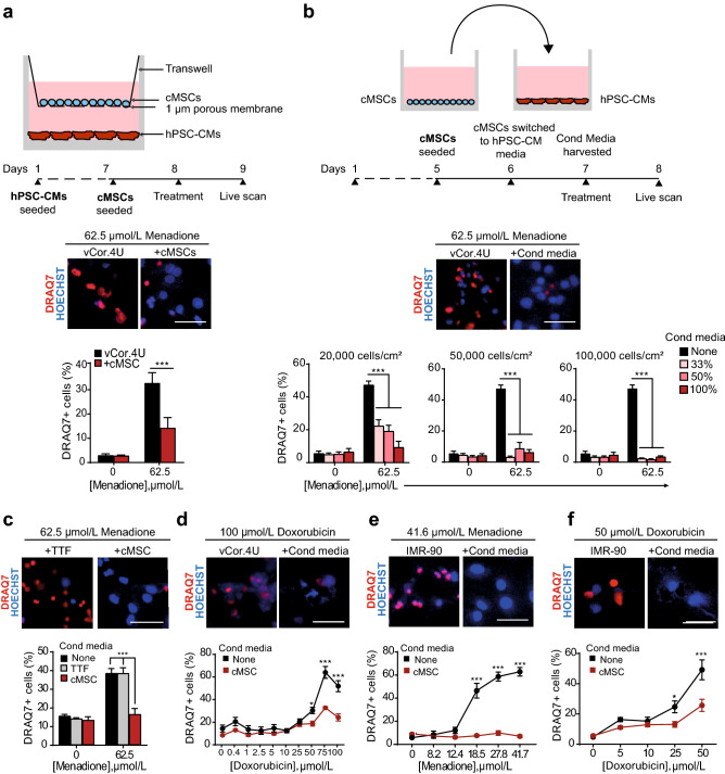
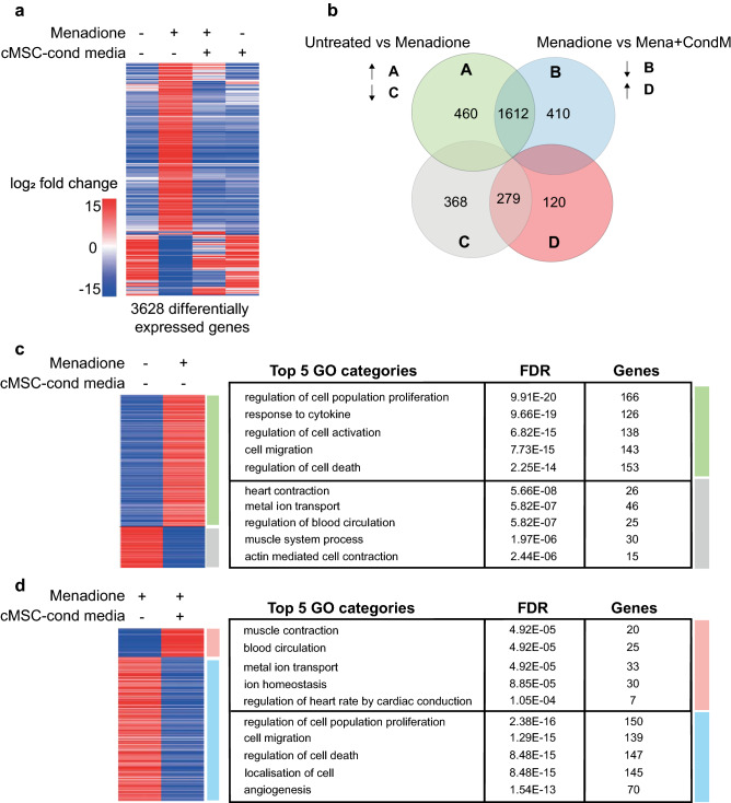
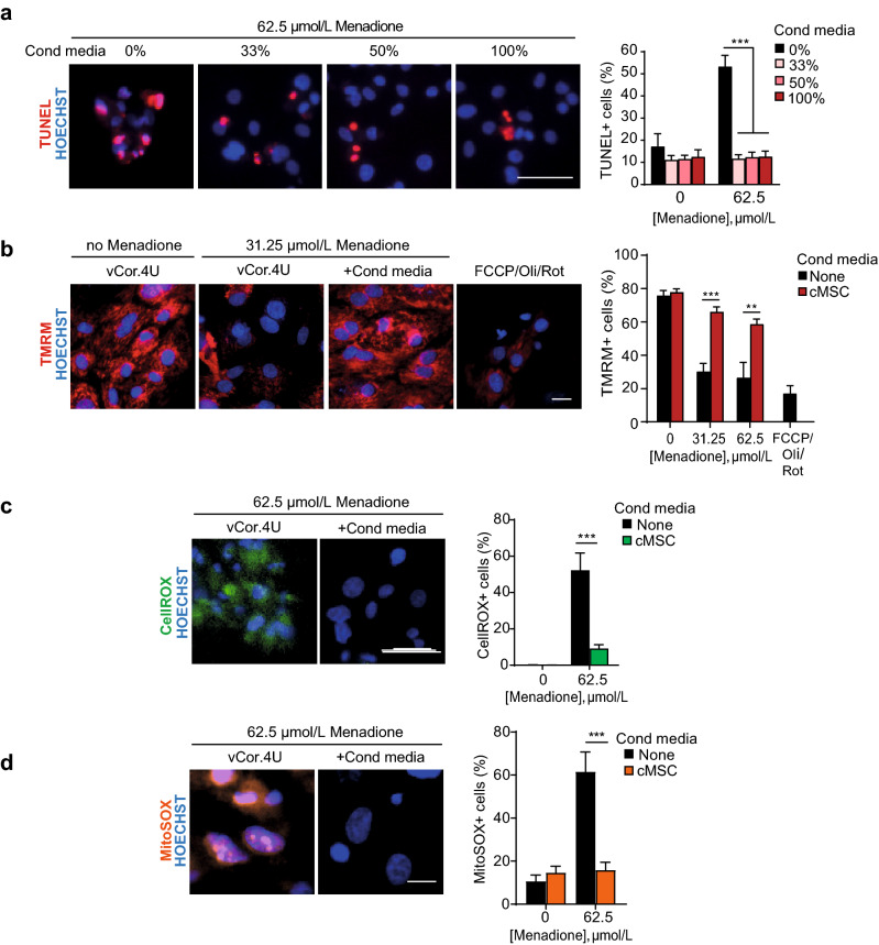
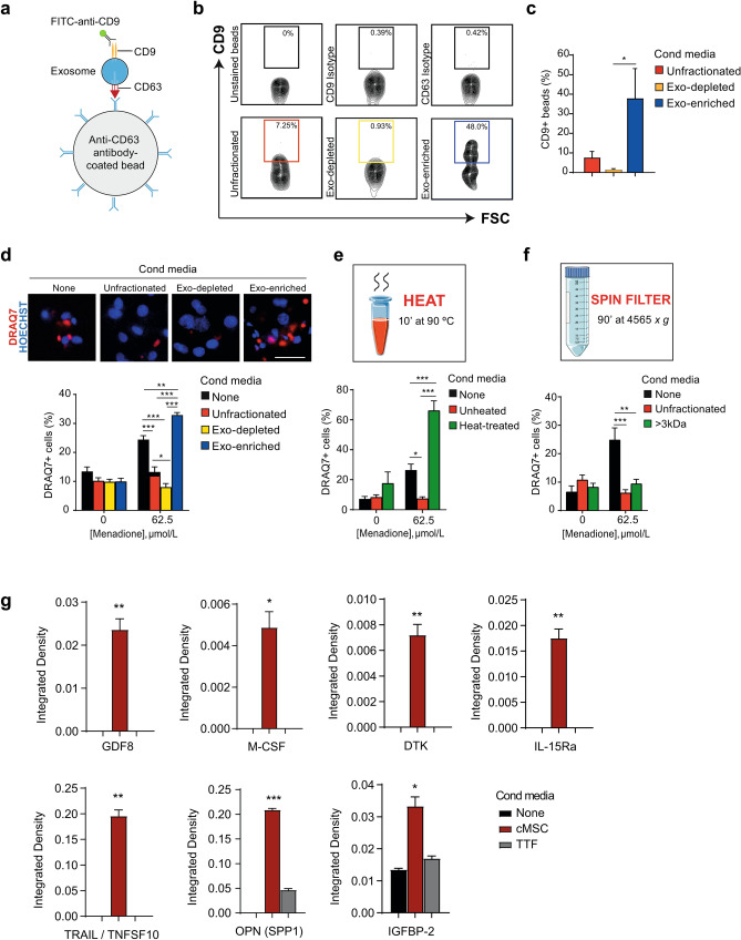
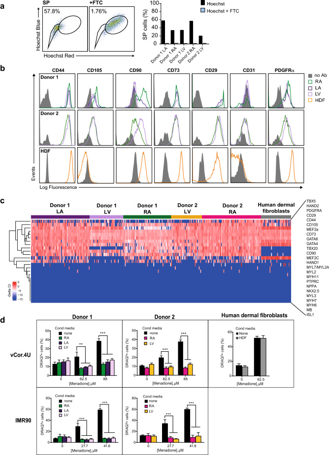
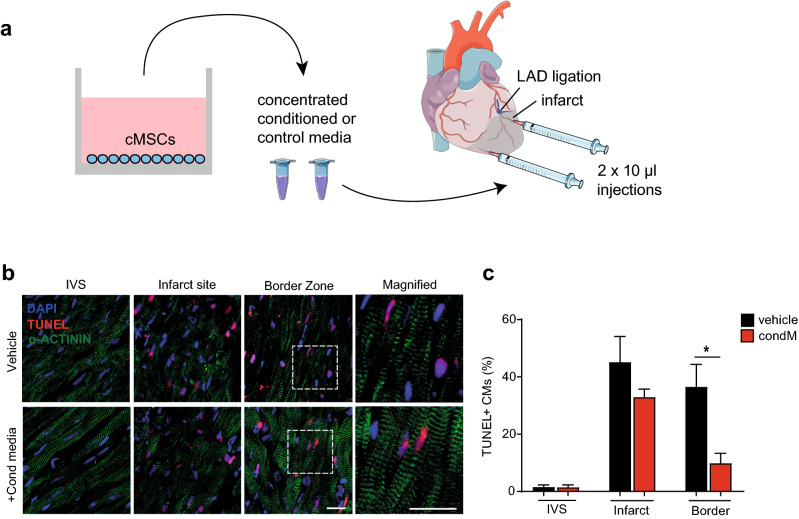
Ischemic heart disease remains the foremost cause of death globally, with survivors at risk for subsequent heart failure. Paradoxically, cell therapies to offset cardiomyocyte loss after ischemic injury improve long-term cardiac function despite a lack of durable engraftment. An evolving consensus, inferred preponderantly from non-human models, is that transplanted cells benefit the heart via early paracrine signals. Here, we tested the impact of paracrine signals on human cardiomyocytes, using human pluripotent stem cell-derived cardiomyocytes (hPSC-CMs) as the target of mouse and human cardiac mesenchymal stromal cells (cMSC) with progenitor-like features. In co-culture and conditioned medium studies, cMSCs markedly inhibited human cardiomyocyte death. Little or no protection was conferred by mouse tail tip or human skin fibroblasts. Consistent with the results of transcriptomic profiling, functional analyses showed that the cMSC secretome suppressed apoptosis and preserved cardiac mitochondrial transmembrane potential. Protection was independent of exosomes under the conditions tested. In mice, injecting cMSC-conditioned media into the infarct border zone reduced apoptotic cardiomyocytes > 70% locally. Thus, hPSC-CMs provide an auspicious, relevant human platform to investigate extracellular signals for cardiac muscle survival, substantiating human cardioprotection by cMSCs, and suggesting the cMSC secretome or its components as potential cell-free therapeutic products.


Conflict of interest statement

R.P.S. and A.M.C. were employees of Kardia Therapeutics. R.P.S. and M.D.S. were founders and stock-holders. All other authors declare no conflict of interest.

Figures

Figure 1
Figure 1
Mouse cMSC secretome suppresses cell death in human cardiomyocytes from pluripotent stem cells. (a) cMSC protect vCor.4U human ventricular myocytes from lethal oxidative stress in trans-well co-culture. Above, schematic representation and timeline. Middle, representative images of nuclear DRAQ7 staining after a menadione challenge. Below, bar graph of DRAQ7 uptake; n = 6. cMSC were prospectively sorted using Sca1 and the SP phenotype, and were 90% PDGFRα+. (b) cMSC-conditioned media protect vCor.4U human ventricular myocytes from lethal oxidative stress. Above, schematic representation and timeline. Middle, representative images as in (a). For the cultures illustrated, cMSC were seeded at 100,000 cells/cm2 and conditioned media used at a concentration of 50%. Below, bar graphs of DRAQ7 uptake, at the indicated cMSC seeding densities and media concentrations; n = 9. (c,d) Specificity and generality of protection in vCor.4U human ventricular myocytes. Above, representative images. Below, bar graphs of DRAQ7 uptake; n = 9. (c) Lack of protection from menadione by tail tip fibroblast-conditioned medium. (d) Protection from doxorubicin by cMSC-conditioned media. (e,f) cMSC-conditioned media was tested on IMR-90 hPSC-CMs, an independent human cardiomyocyte line. (e) Representative images and bar graph of DRAQ7 uptake after menadione. n = 12. (f) Representative images and bar graph of DRAQ7 uptake after doxorubicin; n = 9. For all panels: scale bar, 50 μm; data are shown as the mean ± SEM; *p < 0.05; ***p < 0.0001.
Figure 2
Figure 2
RNA-Seq analysis of IMR90-cardiomyocytes ± menadione and cMSC-conditioned medium. (a) Heatmap of the 3,628 differentially expressed genes (log2fold change > 2, p value < 0.05) across the four IMR90-CM treatment groups (n = 3), shown by unsupervised cluster analysis. Oxidative stress was induced for 24 h with 20 μM menadione with and without cMSC-conditioned medium treatment. The heatmap was created in SeqMonk using Pearson’s Correlation clustering for the genes (y-axis). (b) Venn diagram showing overlapping genes shared in the pairwise comparisons indicated. Green, up-regulated genes vs untreated; grey, down-regulated genes vs untreated; blue, up-regulated genes vs menadione-stressed; red, down-regulated genes vs menadione-stressed. (c,d) Curated heatmaps and tables of GOs for the changes induced by (c) menadione or (d) cMSC-conditioned medium. Tables include top 5 non-redundant GOs, from the ToppGene “Biological Process” database. For a full list of the generated GOs see Supplementary Fig. S7. These data were deposited on SRA public repository with accession number PRJNA629893.
Figure 3
Figure 3
Mouse cMSCs’ secretome blocks human ventricular myocyte apoptosis and the dissipation of mitochondrial potential induced by menadione. (a) Representative images and bar graph of TUNEL+ vCor.4U human ventricular myocytes, 24 h after menadione ± cMSC-conditioned media at the concentrations shown. n = 9. (b) Representative images and bar graph of mitochondrial TMRM in vCor.4U hPSC-CMs, stressed with menadione ± cMSC-conditioned media. Carbonyl cyanide 4-(trifluoromethoxy) phenylhydrazone (FCCP), oligomycin (Oli) and rotenone (Rot), uncouplers of oxidative phosphorylation, were used as controls. n = 7. (c) Representative images and bar graph of CellROX in vCor.4U hPSC-CMs 8 h after menadione ± cMSC-conditioned media. n = 12. (d) Representative images and bar graph of MitoSOX 4 h after menadione ± cMSC-conditioned media. n = 7. For all panels: scale bar 50 μm; data are shown as the mean ± SEM; *p < 0.05; ***p < 0.0001.
Figure 4
Figure 4
An exosome-independent mechanism mediates the observed protection of human cardiomyocytes by mouse cMSC. (a) Schematic of exosome capture and detection using anti-CD63-conjugated latex beads plus FITC-anti-CD9. (b,c) Bead-exosome complexes were analysed by flow cytometry. (b) Representative contour plots are shown. The gate defines the CD63+CD9+ exosomes. The top row shows controls for staining and the bottom row the proportion of exosomes in unfractionated conditioned media versus the depleted (exo-depleted) and enriched (exo-enriched) fractions. FSC forward scatter. More than 5,000 beads were scored for each condition shown. (c) Bar graph of CD9+ depletion and enrichment; n = 3. (d) DRAQ7 uptake in vCor.4U human ventricular myocytes after menadione ± cMSC-conditioned media or the indicated fractions. Above, representative images. Scale bar, 50 μm. Below, bar graph of DRAQ7 uptake; n = 10. (e) Thermostability testing of cMSC-conditioned media. Bar graph of DRAQ7 uptake; n = 9. (f) Size-fractionation of cMSC-conditioned media. Bar graph of DRAQ7 uptake; n = 6. For all panels, data are shown as the mean ± SEM. *p < 0.05; **p < 0.001; ***p < 0.0001. (g) Bar graphs of cytokine levels, using low-density membrane arrays, for the factors enriched in cMSC-conditioned medium vs medium only or TTF-conditioned medium. Results are image analysis of integrated density, normalised to the average of anti-streptavidin and anti-HRP controls for each membrane. Enlarged versions of the arrays can be found in Supplementary Fig. S9. Data are mean ± SEM; n = 2; *p < 0.05; **p < 0.01; ***p < 0.001. Unpaired one-tailed t test between cMSC and medium or cMSC and TTF. Graphics were created using Servier Medical Art website, a free medical image database with a licence under Creative Commons Attribution 3.0 Unported License (https://creativecommons.org/licenses/by/3.0/).
Figure 5
Figure 5
Human cardiac stromal cells protect human cardiomyocytes. (a) Human cardiac stromal cells are enriched for the SP phenotype. Left, representative dot plots of SP staining for the left atrium of Donor 1. FTC, ABCG2 inhibitor Fumitremorgin C. Right, bar graphs showing consistent enrichment for SP cells in five human cardiac stromal cell populations from two donors. (b) Flow cytometry comparing surface marker expression in the human cardiac stromal cell populations and human dermal fibroblasts (HDFs). Note the absence of CD105 in HDFs. (c) Single-cell qRT–PCR comparing the co-expression of selected genes in human cardiac stromal cells and HDFs. The heatmap shows expression as − ΔCt values (blue, low; red, high). Genes are ordered based on hierarchical clustering. n = 39–72 cells for each sample illustrated. See also Fig. S3 in the supplementary data. (d) Human cardiac stromal cells from the donors and chambers shown all suppress cell death induced by menadione in vCor.4U (top) and IMR-90 (bottom) human cardiomyocytes; HDFs (right) had no effect. Bar graph, mean ± SEM; vCor,4U: n = 6; IMR-90: n = 9; **p < 0.001; ***p < 0.0001. RA right atrium, LA left atrium, LV left ventricle.
Figure 6
Figure 6
Conditioned media from mouse cMSC suppress cardiomyocyte apoptosis after mouse myocardial infarction. (a) Schematic representation of cMSC-conditioned medium production, concentration and intramyocardial injection after LAD ligation. Concentrated conditioned or control media were injected into the infarct border zone, one site ~ 1 mm beneath the suture and a second site more apical. Images modified from Servier Medical Art website, a free medical image database with a licence under Creative Commons Attribution 3.0 Unported License (https://creativecommons.org/licenses/by/3.0/). (b) Representative immunohistochemistry images of TUNEL staining, 24 h after myocardial infarction. An α-actinin antibody was used to demarcate cardiomyocyte identity. Scale bars 20 μm. See also Supplementary Fig. S11. (c) Bar graph of TUNEL staining in cardiomyocytes in the remote myocardium (intraventricular septum, IVS), infarct site, and border zone. Data are shown as the mean ± SEM. n = 3; *p < 0.05.

References

    1. GBD 2016 Causes of Death Collaborators Global, regional, and national age-sex specific mortality for 264 causes of death, 1980–2016: A systematic analysis for the Global Burden of Disease Study 2016. Lancet. 2017;390:1151–1210. doi: 10.1016/S0140-6736(17)32152-9. - DOI - PMC - PubMed
    1. Mercola M, Ruiz-Lozano P, Schneider MD. Cardiac muscle regeneration: Lessons from development. Genes Dev. 2011;25:299–309. doi: 10.1101/gad.2018411. - DOI - PMC - PubMed
    1. Banerjee MN, Bolli R, Hare JM. Clinical studies of cell therapy in cardiovascular medicine: Recent developments and future directions. Circ. Res. 2018;123:266–287. doi: 10.1161/CIRCRESAHA.118.311217. - DOI - PMC - PubMed
    1. Noseda M, Abreu-Paiva M, Schneider MD. The quest for the adult cardiac stem cell. Circ. J. 2015;79:1422–1430. doi: 10.1253/circj.CJ-15-0557. - DOI - PubMed
    1. Noseda M, et al. PDGFRalpha demarcates the cardiogenic clonogenic Sca1+ stem/progenitor cell in adult murine myocardium. Nat. Commun. 2015;6:6930. doi: 10.1038/ncomms7930. - DOI - PMC - PubMed

Publication types

Substances