. 2020 Nov;16(11):1199-1207.
doi: 10.1038/s41589-020-0594-x. Epub 2020 Aug 3.

Rational discovery of molecular glue degraders via scalable chemical profiling

Affiliations

Rational discovery of molecular glue degraders via scalable chemical profiling

Cristina Mayor-Ruiz et al. Nat Chem Biol. 2020 Nov.

Erratum in

Abstract

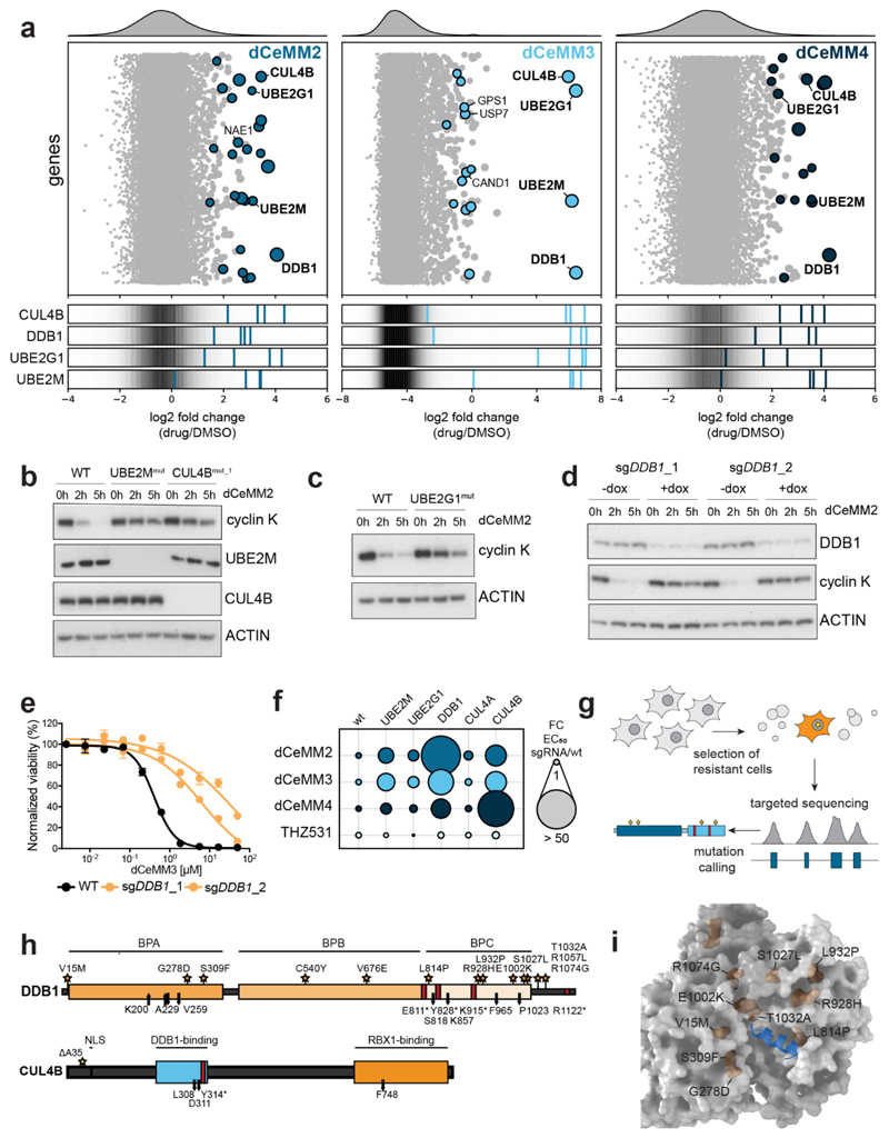
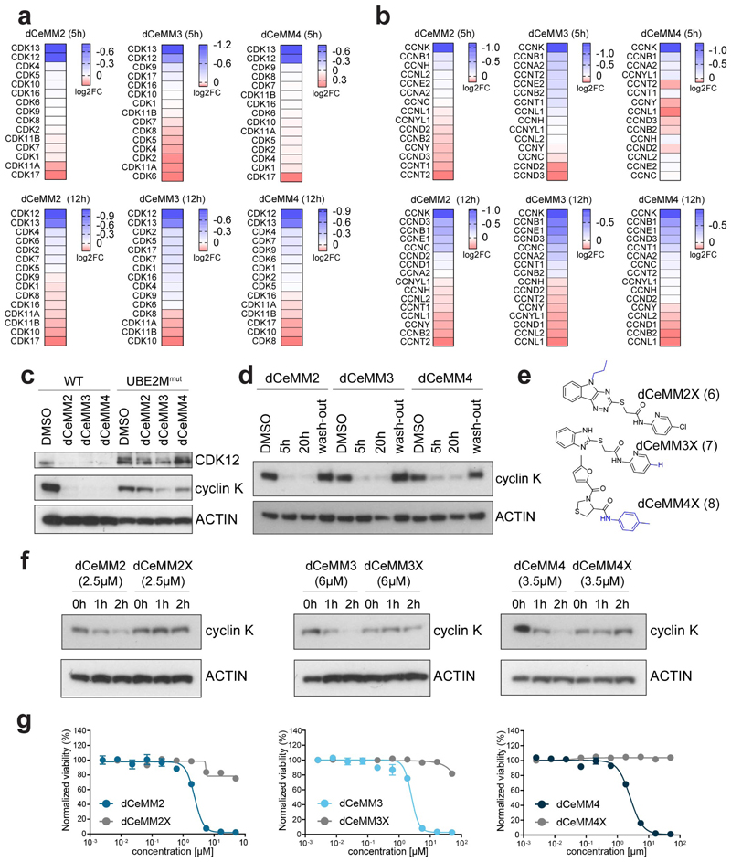
Targeted protein degradation is a new therapeutic modality based on drugs that destabilize proteins by inducing their proximity to E3 ubiquitin ligases. Of particular interest are molecular glues that can degrade otherwise unligandable proteins by orchestrating direct interactions between target and ligase. However, their discovery has so far been serendipitous, thus hampering broad translational efforts. Here, we describe a scalable strategy toward glue degrader discovery that is based on chemical screening in hyponeddylated cells coupled to a multi-omics target deconvolution campaign. This approach led us to identify compounds that induce ubiquitination and degradation of cyclin K by prompting an interaction of CDK12-cyclin K with a CRL4B ligase complex. Notably, this interaction is independent of a dedicated substrate receptor, thus functionally segregating this mechanism from all described degraders. Collectively, our data outline a versatile and broadly applicable strategy to identify degraders with nonobvious mechanisms and thus empower future drug discovery efforts.


Conflict of interest statement

Competing Interests

C.M.-R. and G.E.W. are listed as inventors of a patent application for glue discovery in neddylation-deficient cellular systems. C.M.-R., S.K. and G.E.W. are listed as inventors of patent applications covering the chemical space of dCeMM2/3/4. M.B., S.K., G.E.W and CeMM are founders and equity holders of Proxygen.

Figures

Fig.1
Fig.1. Screening in hypo-neddylated cells identifies novel molecular glue degraders rewiring the CRL4DCAF15 ligase.
a, Protein levels of UBE2M, UBE2F and neddylation status of different cullin backbones in WT, MLN4924-treated (1µM, 1h), UBE2M-deficient and UBE2M-reconstituted KBM7 cells. b, Substrate receptors expressed in KBM7 cells that associate with hypo-neddylated cullin scaffolds. c, Primary screening data comparing DMSO-normalized viability of WT and UBE2Mmut KBM7 cells treated with a (approximately) 2000 cytotoxic/cytostatic small-molecule library for 3 days. Doses tested were 10uM and 500nM. d, Chemical structure of the four prioritized chemical scaffolds (dCeMM1/2/3/4). dCeMM3-1 was structurally identical to dCeMM3 with a Br instead of the Cl. e, Dose-resolved, DMSO-normalized viability after 3-day dCeMM1/2/3/4 treatment in WT and UBE2Mmut KBM7 cells. Mean ± SEM; n=3 independent treatments. EC50s dCeMM1/2/3/4 (µM) WT=3;0,3;0.6;0.4 UBE2Mmut=8;4.2;10.7;7. f, CRL-focused CRISPR resistance screen for dCeMM1. Top: bubble plot displaying median sgRNA enrichment over DMSO, bubble size indicates significance. Bottom: sgRNAs enrichment targeting indicated genes, background indicates distribution of all sgRNAs. Results shown are the median of 2 independent screens. g, Expression proteomics after dCeMM1 treatment (25µM, 12h). h, DMSO-normalized viability after 3-day dCeMM1 treatment in WT and DCAF15mut KBM7 cells. Mean ± SEM; n=3 independent treatments. i, RBM39 levels in WT and DCAF15mut KBM7 cells after indisulam (IND) or dCeMM1 treatment.
Fig.2
Fig.2. dCeMM2/3/4 are novel and structurally different cyclin K degraders.
a, DMSO-normalized expression proteomics after 5h dCeMM2/3/4 treatment (2.5µM, 7µM, 3.5µM) in KBM7 cells. Limma statistical analysis was used. b Cyclin K levels upon dCeMM2/3/4 treatment in KBM7 cells. c, dCeMM2 (2.5µM, 5h) destabilizes cyclin K. 30min pretreatment with 1µM carfilzomib, 1µM MLN4924, 10µM TAK-243 or 1µM THZ531 rescues cyclin K destabilization. d, Recombinant kinase assays of dCeMM2/2X and THZ531 inhibition on enzymatic activity of CDK12/13/7. Mean ± SD n=2. e, dCeMM2/3/4 induce global transcriptional downregulation with phenotypic similarity to CDK12/13 inhibition by THZ531. Heatmap displays DMSO-normalized log2FC in gene expression for 27,051 transcripts, ranked by THZ531 log2FC. f, Gene set enrichment analysis of 984 genes (log2FC<-4 and adj. P-value<0.05) significantly downregulated upon THZ531 treatment in comparison to dCeMM2/3/4 (FDR<0.001). GSEA pre-ranked function was used (1000 permutations).
Fig.3
Fig.3. Induced cyclin K degradation is mediated via a CRL4B ligase complex in a SR-independent manner
a, Genome-wide CRISPR dCeMM2/3/4 resistance screens. Top: bubble plot displaying median sgRNA enrichment over DMSO, bubble size indicates significance. Bottom: sgRNA enrichment targeting indicated genes, background indicates distribution of all sgRNAs. b-d, dCeMM2-induced cyclin K degradation (2.5µM) is rescued in UBE2M-, CUL4B- (b), UBE2G1- (c) and DDB1- (d) deficient cells. e, DMSO-normalized viability in WT and 3-day doxycycline (dox) pretreated sgDDB1_1 and sgDDB1_2 dox-inducible Cas9 KBM7 cells after 3-day dCeMM3 treatment. Mean ± SEM; n=3 independent treatments. f, WT-normalized fold change in EC50 based on dose-resolved 3-day viability experiments upon exposure to the indicated drugs in the indicated genetic backgrounds. g, Targeted hybrid-capture approach coupled to next generation sequencing to identify mutations in spontaneously dCeMM2/3/4-resistant cells. h, Depiction of DDB1 and CUL4B mutations identified by hybrid capture sequencing in drug-resistant cell pools. Stars: point mutations. Red bars: premature stop codons. Arrows: frameshift mutations. i, Structure of DDB1 (gray) in complex with SV5V peptide (aa 21-39, blue), PDB: 2HYE. DDB1 point mutations located within 20Å of the peptide are highlighted in orange.
Fig.4
Fig.4. dCeMM2/3/4 induce proximity between CUL4B:DDB1 and CDK12/13:cyclin K
a, dCeMM3-NH2 chemical structure (dCeMM3 tethered analog). b, Drug affinity chromatography strategy based on probe-coupled agarose beads pulldowns after DMSO or THZ531 (competition) pretreatment in lysates. c, Cyclin K and DDB1 enrichment in dCeMM3-NH2 -based pulldowns. For quantification, eluted protein was normalized to protein available (input panels). THZ531-competed (100µM, 1h) ratios were set to 1. d, Chemical structure of dCeMM3-PAP (PAP: photo-affinity probe). e, Drug-target enrichment strategy based on cellular dCeMM3-PAP co-treatment with DMSO or THZ531 (100µM, competition) after Carfilzomib pretreatment (10µM, 30min). f, Cyclin K and DDB1 enrichment in dCeMM3-PAP -based pulldowns. For quantification, eluted protein was normalized to protein available (input panels). THZ531-competed (100µM, 1h) ratios were set to 1. g, Proximity labeling strategy to assess drug-induced dimerization in intact cells based on the biotin ligase miniTurbo (mTurbo). h, Biotin-labeled CDK12 enrichment following 1h DMSO or dCeMM2 treatment in the presence of carfilzomib (10µM) in HEKs transfected with DDB1-mTurbo fusion. i, Direct cyclin K immunoblotting of carfilzomib-pretreated KBM7 cells after 2h DMSO, dCeMM2 or THZ531+dCeMM2 (10µM) treatments. j, TR-FRET signal for CDK12-Alexa488cyclin K (0–5μM) titrated to terbiumDDB1 in DMSO or 10μM dCeMM2. “No DDB1” only contains streptavidin-terbium. Data are means ± SD (n=3). Kapparent (nM): DMSO=n.d., no DDB1 = n.d., dCeMM2 = 628.
None
None
None
None
None
None
None
None
None
None

Comment in

  • Prospecting for molecular glues.
    den Besten W, Lipford JR. den Besten W, et al. Nat Chem Biol. 2020 Nov;16(11):1157-1158. doi: 10.1038/s41589-020-0620-z. Nat Chem Biol. 2020. PMID: 32747810 No abstract available.

References

    1. Winter GE, et al. DRUG DEVELOPMENT. Phthalimide conjugation as a strategy for in vivo target protein degradation. Science. 2015;348:1376–81. - PMC - PubMed
    1. Bondeson DP, et al. Catalytic in vivo protein knockdown by small-molecule PROTACs. Nat Chem Biol. 2015;11:611–7. - PMC - PubMed
    1. Zengerle M, Chan KH, Ciulli A. Selective Small Molecule Induced Degradation of the BET Bromodomain Protein BRD4. ACS Chem Biol. 2015;10:1770–7. - PMC - PubMed
    1. Sakamoto KM, et al. Protacs: chimeric molecules that target proteins to the Skp1-Cullin-F box complex for ubiquitination and degradation. Proc Natl Acad Sci U S A. 2001;98:8554–9. - PMC - PubMed
    1. Schneekloth JS, Jr, et al. Chemical genetic control of protein levels: selective in vivo targeted degradation. J Am Chem Soc. 2004;126:3748–54. - PubMed

Publication types

MeSH terms