Review
. 2020 Oct;42(5):1257-1283.
doi: 10.1007/s11357-020-00235-8. Epub 2020 Aug 3.

Sleep, brain vascular health and ageing

Affiliations
Review

Sleep, brain vascular health and ageing

Arehally M Mahalakshmi et al. Geroscience. 2020 Oct.

Abstract

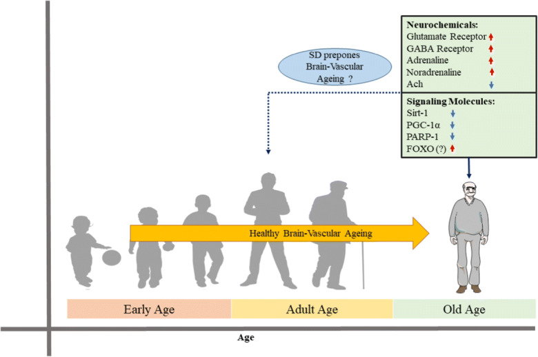
Sleep maintains the function of the entire body through homeostasis. Chronic sleep deprivation (CSD) is a prime health concern in the modern world. Previous reports have shown that CSD has profound negative effects on brain vasculature at both the cellular and molecular levels, and that this is a major cause of cognitive dysfunction and early vascular ageing. However, correlations among sleep deprivation (SD), brain vascular changes and ageing have barely been looked into. This review attempts to correlate the alterations in the levels of major neurotransmitters (acetylcholine, adrenaline, GABA and glutamate) and signalling molecules (Sirt1, PGC1α, FOXO, P66shc, PARP1) in SD and changes in brain vasculature, cognitive dysfunction and early ageing. It also aims to connect SD-induced loss in the number of dendritic spines and their effects on alterations in synaptic plasticity, cognitive disabilities and early vascular ageing based on data available in scientific literature. To the best of our knowledge, this is the first article providing a pathophysiological basis to link SD to brain vascular ageing.

Keywords: Cognition; Neurochemicals; Sleep deprivation; Synaptic plasticity; Vascular ageing.


Conflict of interest statement

The authors declare that they have no conflict of interest.

Figures

Fig. 1
Fig. 1
Sleep deprivation causes cellular respiration proteins upregulation (Ren et al. 2016) (reused as per the PLOS ONE journal’s copyright permission policy). Proteins belonging to TCA cycle and mitochondrial complexes are upregulated in sleep deprived C57BL/6 N mice brain. CG represents sleep deprivation induced by gentle handling; CL represents locomotion induced sleep deprivation. a Venn diagram showing distribution of 66 upregulated proteins in the CG group and 46 upregulated proteins in CL groups. Twenty-two shared proteins are common in both CG and CL groups. b Influence of sleep deprivation on complexes of the electron transport chain, where in complexes I–III had upregulated subunits while complexes IV and V had both up- and downregulated subunits. Upregulated proteins are shown in red boxes and downregulated proteins are shown in green. c Proteins involved in small molecule metabolism. The proteins upregulated in CG and CL groups are shown in parentheses and square brackets, respectively and presented in bold text if upregulated in both the CG and CL groups. d Preproteins synthesized in the cytosol enter mitochondria via translocases of outer membrane (TOM) and translocase of the inner membrane (TIM). Chaperones upregulated in CG or CL groups are presented in parentheses and square brackets, respectively. Chaperones upregulated in both the groups are shown in bold
Fig. 2
Fig. 2
Representative electron microscopy images show structural changes in mitochondria in locus coeruleus (Somarajan et al. 2016) (reused as per the Frontiers in Neurology journal’s copyright permission policy). One-millimetre thick brain tissue blocks of Locus Coeruleus were prepared after intracardial perfusion of rats for transmission electron microscopy to observe structural changes in mitochondria in free moving control (FMC), large platform control (LPC), REM sleep deprivation (REMSD), recovery (REC), and Prazosin treated (PRZ) groups. Control groups retain normal dumbbell-shaped mitochondria with intact cristae. Swollen mitochondria with disintegrated cristae appear in REMSD, and the REC group shows less distorted cristae. PRZ group shows marginal swelling in mitochondria with intact cristae
Fig. 3
Fig. 3
Effect of SD on structural changes in spines during LTP and LTD (Adopted with minor modifications from Chidambaram et al. 2019). SD attenuates LTP and enhances LTD, while LTP causes spine enlargement and LTD causes spine shrinkage
Fig. 4
Fig. 4
Reconstruction of dendritic segments during forced wake (EW) and sleep (S). Each row depicts the spines of a single animal from each group (de Vivo et al. 2019) (reused as per the Sleep journal’s copyright permission policy). Blocks of 1 mm2 brain tissue encompassing the primary motor cortex were prepared from mice pups. Serial images were obtained through ΣIGMA VP field emission scanning electron microscope. Spiny dendritic segments between the diameter range of 0.47–1.01 μm were selected. The results of enforced wakefulness group showed induction of shrinkage in dendritic spines compared to sleep group. Note: The red spines depict enforced wakefulness while the blue spines depict sleep state
Fig. 5
Fig. 5
Alteration in transcription and translation of various genes and proteins in sleep and wakefulness in the cortex and hippocampus of mammals (Seibt and Frank 2019) (Reused as per the Frontiers in System Neuroscience journal’s copyright permission policy). Waking data includes periods of sleep deprivation <8 h. wakefulness upregulates transcription of c-fos, Egr1, p-CREB, Arc, and BDNF while sleep deprivation has shown decreased translation of elF2a, elF4e2, elF5, Rbm3, Denr, p-mtorc1, and p-4EBP2. The methodologies employed include microarray, quantitative PCR, immunohistochemistry, Western blot, in-situ-hybridization and Rnase protection assay. The genes highlighted in black colour are active during transcription, while those highlighted in red are active during the translational phase
Fig. 6
Fig. 6
Effects of SD on neurogenesis analysed using BrdU staining. Adult male Sprague Dawley rats subjected to SD by small platform (SP) method. a, b An initial decrease in BrdU immunopositive cells in dentate gyrus of hippocampal region of SD rats. c, d Subsequent increase in granular cell proliferation and neurogenesis after 1 week of unrestricted recovery of sleep. e, f Immunofluorescence analysis indicating maturation of neuronal cells after unrestricted sleep recovery. e After 1 week of BrdU administration, BrdU positive cells showed morphological characteristics of granule cells colabelled with Tuj1, a marker for immature neurons. F indicates the BrdU positive cells colabelled with NeuN, a marker for mature neurons after 3 weeks of BrdU administration (Mirescu et al. 2006) (reused as per the “Copyright (1993-2008) National Academy of Sciences”)
Fig. 7
Fig. 7
Sketch outlining the effects of SD on the proposed accelerated brain vascular ageing. We propose that the pathological alterations in neurochemicals and signalling molecules in SD might be a risk factor for early brain-vascular ageing. These effects are not reported in healthy ageing with adequate sleep. Orange coloured arrow indicates healthy brain-vascular ageing. Red and blue colour arrows indicate the up-regulation and down-regulation of neurochemicals and signalling molecules in SD condition which could possibly contribute for the brain early vascular ageing (proposed)

Similar articles

Cited by

References

    1. Adams SK, Daly JF, Williford DN. Adolescent sleep and cellular phone use: recent trends and implications for research. Health Serv Insights. 2013;6:99–103. - PMC - PubMed
    1. Aggarwal B, Nour M, Riddhi S, Memet E, Ying W, Marie-Pierre S-O, Sanja J. Effects of inadequate sleep on blood pressure and endothelial inflammation in women: findings from the American Heart Association go red for women strategically focused research network. J Am Heart Assoc. 2018;7:e008590. - PMC - PubMed
    1. Alborch E, Torregrosa G, Terrasa JC, Estrada C. GABA receptors mediate cerebral vasodilation in the unanesthetized goat. Brain Res. 1984;321:103–110. - PubMed
    1. Alexopoulou C, Kondili E, Diamantaki E, Psarologakis C, Kokkini S, Bolaki M, Georgopoulos D. Effects of dexmedetomidine on sleep quality in critically ill patients: a pilot study. Anesthesiology. 2014;121:801–807. - PubMed
    1. Anafi RC, Kayser MS, Raizen DM. Exploring phylogeny to find the function of sleep. Nat Rev Neurosci. 2019;20:109. - PubMed

Publication types

LinkOut - more resources