. 2020 Dec;11(6):1661-1676.
doi: 10.1002/jcsm.12601. Epub 2020 Aug 3.

Satellite cell self-renewal in endurance exercise is mediated by inhibition of mitochondrial oxygen consumption

Affiliations

Satellite cell self-renewal in endurance exercise is mediated by inhibition of mitochondrial oxygen consumption

Phablo Abreu et al. J Cachexia Sarcopenia Muscle. 2020 Dec.

Abstract

Background: Skeletal muscle stem cells (satellite cells) are well known to participate in regeneration and maintenance of the tissue over time. Studies have shown increases in the number of satellite cells after exercise, but their functional role in endurance training remains unexplored.

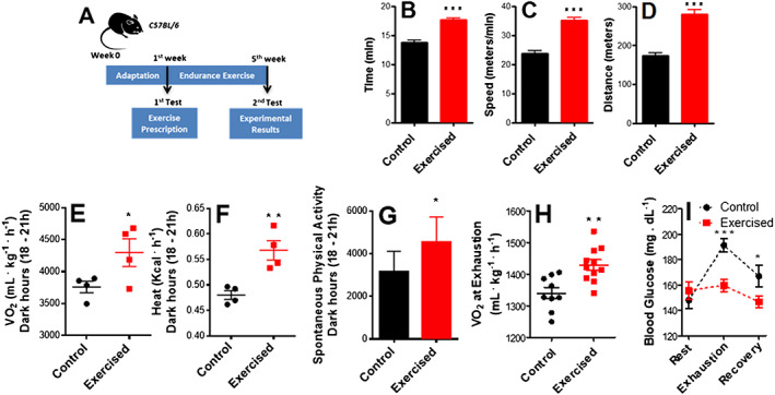
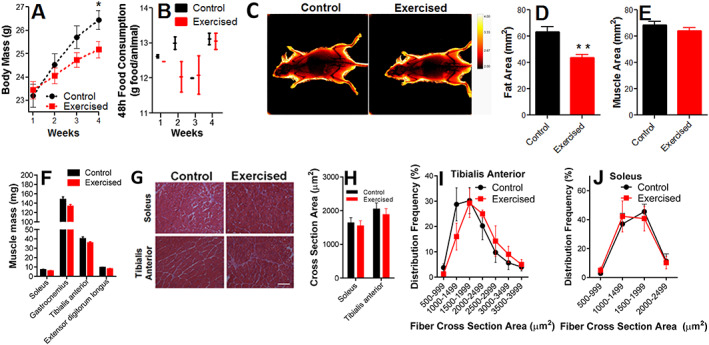
Methods: Young adult mice were submitted to endurance exercise training and the function, differentiation, and metabolic characteristics of satellite cells were investigated in vivo and in vitro.

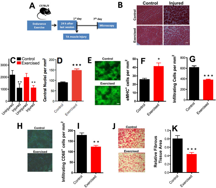
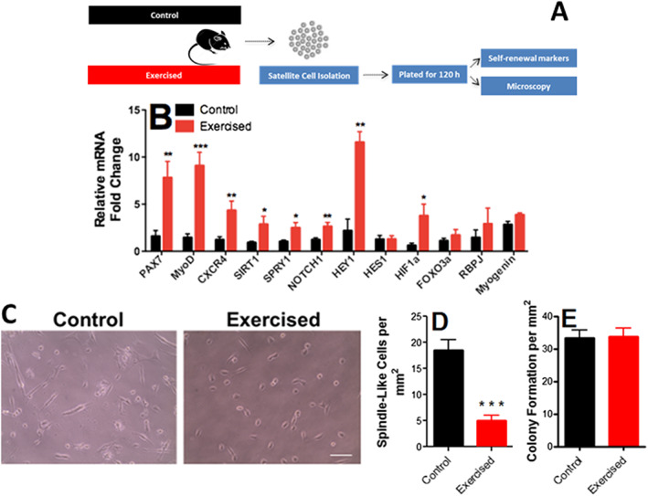
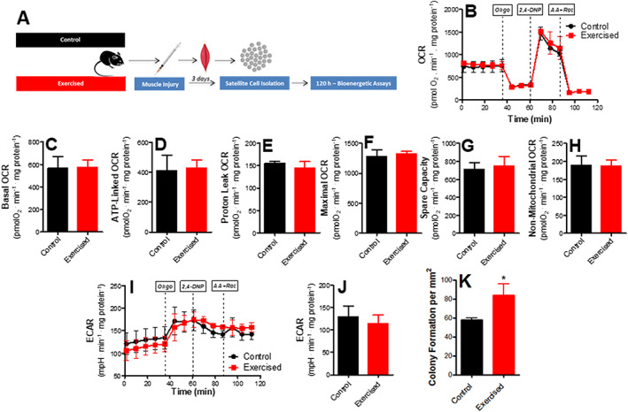
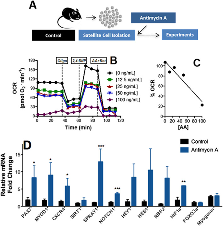
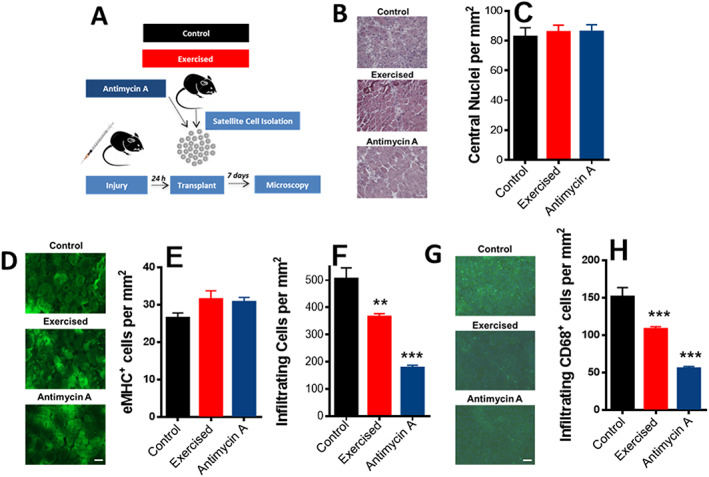
Results: We found that injured muscles from endurance-exercised mice display improved regenerative capacity, demonstrated through higher densities of newly formed myofibres compared with controls (evidenced by an increase in embryonic myosin heavy chain expression), as well as lower inflammation (evidenced by quantifying CD68-marked macrophages), and reduced fibrosis. Enhanced myogenic function was accompanied by an increased fraction of satellite cells expressing self-renewal markers, while control satellite cells had morphologies suggestive of early differentiation. The beneficial effects of endurance exercise were associated with satellite cell metabolic reprogramming, including reduced mitochondrial respiration (O2 consumption) under resting conditions (absence of muscle injury) and increased stemness. During proliferation or activated states (3 days after injury), O2 consumption was equal in control and exercised cells, while exercise enhanced myogenic colony formation. Surprisingly, inhibition of mitochondrial O2 consumption was sufficient to enhance muscle stem cell self-renewal characteristics in vitro. Moreover, transplanted muscle satellite cells from exercised mice or cells with reduced mitochondrial respiration promoted a significant reduction in inflammation compared with controls.

Conclusions: Our results indicate that endurance exercise promotes self-renewal and inhibits differentiation in satellite cells, an effect promoted by metabolic reprogramming and respiratory inhibition, which is associated with a more favourable muscular response to injury.

Keywords: Bioenergetics; Metabolism; Mitochondria; Muscle stem cells.


Conflict of interest statement

None declared.

Figures

Figure 1
Figure 1
Endurance exercise induces a performance‐dependent shift and changes energy substrate usage. (A) Schematic panel. (B) Running time. (C) Running speed. (D) Distance covered in the run‐to‐exhaustion test (n = 7, control; n = 13, exercised). (E) O2 consumption during the dark hours (18–21 h) measured by indirect calorimetry. (F) Heat production during the dark hours measured by indirect calorimetry. (G) Spontaneous physical activity during the dark hours (n = 4, control; n = 4, exercised). (H) O2 consumption at exhaustion (n = 9, control; n = 11, exercised). (I) Resting blood glucose, blood glucose during run‐to‐exhaustion, and blood glucose at 10 min rest after exhaustion (n = 7, control; n = 13, exercised). Asterisks denote statistically significant differences (* P < 0.05, ** P < 0.01, and *** P < 0.001) vs. control.
Figure 2
Figure 2
Body fat content is decreased by endurance exercise, while muscle content is preserved. (A) Body mass changes. (B) Food consumption (n = 4, control; n = 4, exercised). (C) Dual X‐ray absorptiometry scans, representative image. (D) Fat area. (E) Muscle area (n = 6, control; n = 6, exercised). (F) Muscle mass (n = 7, control; n = 9, exercised). (G) Representative images of cross‐sectional areas. (H) Cross‐sectional area quantifications. (I and J) Frequency distribution of the cross‐sectional areas of the fibres (n = 4, control; n = 4, exercised). Scale bar = 100 μm. Asterisks denote statistically significant differences (* P < 0.05; ** P < 0.01) vs. control.
Figure 3
Figure 3
Aerobic fitness enhances skeletal muscle repair and promotes anti‐inflammatory and anti‐fibrotic effects. (A) Schematic panel. (B) Illustrative images of fibre cross sections (scale bar = 100 μm). (C) Cross‐sectional area quantifications of control and exercised samples with and without injury. (D) Centrally nucleated (newly formed) myofibre counts in injured muscles. (E) Representative images of eMHC+‐stained fibres. (F) Quantification of eMHC+ fibres. (G) Infiltrating inflammatory cell counts in injured muscles. (H) Representative low magnification images, CD68+ (macrophage) staining in green and DAPI in blue. (I) Quantification of CD68+ cells (macrophages). (J) Picrosirius Red‐stained injured muscles, representative images. (K) Relative fibrous area quantification (n = 4, control; n = 4, exercised). Scale bars = 50 μm (E and H) and 100 μm (B and J). Asterisks denote statistically significant differences (* P < 0.05, ** P < 0.01, and *** P < 0.001) vs. controls.
Figure 4
Figure 4
Endurance exercise enhances self‐renewal and quiescence markers in satellite cells. (A) Schematic panel. (B) Expression of Pax7 (a satellite cell‐specific marker that increases upon activation), MyoD1 (an activation marker), CXCR4 (a satellite cell marker), Sirt1 (a self‐renewal marker), Spry1 (a return‐to‐quiescence marker), Notch1, Hey1, Hes1, and HIF1α (self‐renewal markers), FOXO3a (quiescence marker), Rbpj (involved in Notch1 signalling), and myogenin (a differentiation marker; n = 8, control; n = 8, exercised), relative to HPRT. (C) Representative images of satellite morphology. (D) Quantification of elongated, spindle‐like (more differentiated) cells. (E) Myogenic colony formation (n = 4, control; n = 4, exercised). Asterisks denote statistically significant differences (* P < 0.05, ** P < 0.01, and *** P < 0.001) vs. control.
Figure 5
Figure 5
Endurance exercise reduces mitochondrial O2 consumption in skeletal muscle stem cells. (A) Schematic panel. (B) Typical traces of real‐time O2 consumption rate (OCR) measurements in control and exercised cells determined using the Seahorse XF Analyzer. Oligomycin, 2,4‐DNP, and rotenone plus antimycin A were added where indicated. (C) Basal O2 consumption rates. (D) ATP production‐dependent OCR (basal minus oligomycin‐insensitive). (E) Proton leak (oligomycin‐insensitive) OCR. (F) Maximal OCR in the presence of 2,4‐DNP. (G) Spare respiratory capacity (maximal minus basal). (H) Non‐mitochondrial (antimycin plus rotenone‐insensitive) OCR. (I) Typical extracellular acidification rate (ECAR) measurement. (J) ECAR quantification (n = 4, control; n = 4, exercised). (K) Mitochondrial DNA/nuclear DNA ratio (mtDNA/nDNA) (n = 6, control; n = 4, exercised). (L) Expression profile of mitochondrial markers (n = 8, control; n = 8, exercised). Asterisks denote statistically significant differences (* P < 0.05, ** P < 0.01, and *** P < 0.001) vs. control.
Figure 6
Figure 6
Mitochondrial O2 consumption is unaffected in muscle stem cells from injured animals. (A) Schematic panel. (B) Typical trace. Oligomycin, 2,4‐DNP, and rotenone plus antimycin A were added where indicated. (C) Basal O2 consumption rates. (D) ATP production‐dependent oxygen consumption rate (OCR). (E) Proton leak OCR. (F) Maximal OCR. (G) Spare respiratory capacity. (H) Non‐mitochondrial OCR. (I) Typical extracellular acidification rate (ECAR) trace. (J) ECAR quantification (n = 3, control; n = 3, exercised). (K) Myogenic colony formation. Asterisks denote statistically significant differences (* P < 0.05) vs. control.
Figure 7
Figure 7
Inhibition of mitochondrial O2 consumption promotes satellite cell self‐renewal. (A) Schematic panel. (B) Typical oxygen consumption rate (OCR) trace in the presence of different concentrations of antimycin A. (B and C) Respiratory inhibition vs. antimycin A concentrations. (D) Satellite cell expression profile after 8 h in the presence of 70 ng/mL antimycin A (n = 8, control; n = 4, antimycin A), relative to HPRT. Asterisks denote statistically significant differences (* P < 0.05, ** P < 0.01, and *** P < 0.001) vs. control.
Figure 8
Figure 8
Transplant of exercised or respiratory‐inhibited satellite cells prevents inflammation. (A) Schematic panel. (B) Representative images of transplanted fibre cross sections (scale bar = 100 μm). (C) Centrally nucleated (newly formed) myofibre quantification. (D) Representative images of eMHC+‐stained fibres. (E) Quantification of eMHC+ fibres. (F) Inflammatory cell quantification. (G) Representative low magnification images, CD68+ (macrophage) staining in green and DAPI in blue. (H) Quantification of CD68+ cells (macrophages; n = 4, control; n = 4, exercised; and n = 4, antimycin A). Scale bars = 100 μm (B) and 50 μm (D and G). Asterisks denote statistically significant differences (** P < 0.01; *** P < 0.001) vs. control animals.

References

    1. Yin H, Price F, Rudnicki MA. Satellite cells and the muscle stem cell niche. Physiol Rev 2013;93:23–67. - PMC - PubMed
    1. von Maltzahn J, Jones AE, Parks RJ, Rudnicki MA. Pax7 is critical for the normal function of satellite cells in adult skeletal muscle. Proc Natl Acad Sci U S A 2013;110:16474–16479. - PMC - PubMed
    1. Bazgir B, Fathi R, Rezazadeh Valojerdi M, Mozdziak P, Asgari A. Satellite cells contribution to exercise mediated muscle hypertrophy and repair. Cell J 2017;18:473–484. - PMC - PubMed
    1. Shea KL, Xiang W, LaPorta VS, Licht JD, Keller C, Basson MA, et al. Sprouty1 regulates reversible quiescence of a self‐renewing adult muscle stem cell pool during regeneration. Cell Stem Cell 2010;6:117–129. - PMC - PubMed
    1. Chakkalakal JV, Jones KM, Basson MA, Brack AS. The aged niche disrupts muscle stem cell quiescence. Nature 2012;490:355–360. - PMC - PubMed

Publication types

LinkOut - more resources