. 2020 Aug 4:9:e57659.
doi: 10.7554/eLife.57659.

Tailored design of protein nanoparticle scaffolds for multivalent presentation of viral glycoprotein antigens

Affiliations

Tailored design of protein nanoparticle scaffolds for multivalent presentation of viral glycoprotein antigens

George Ueda et al. Elife. .

Abstract

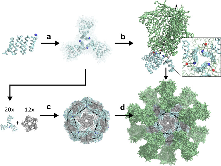
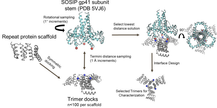
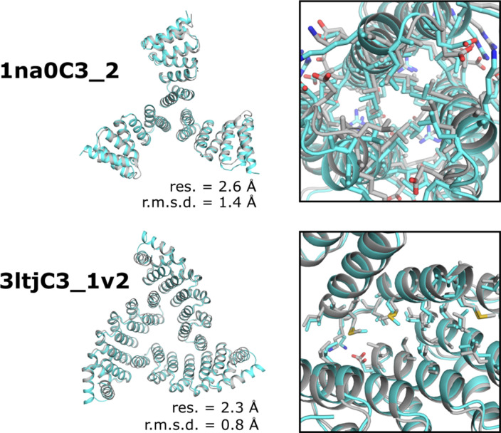
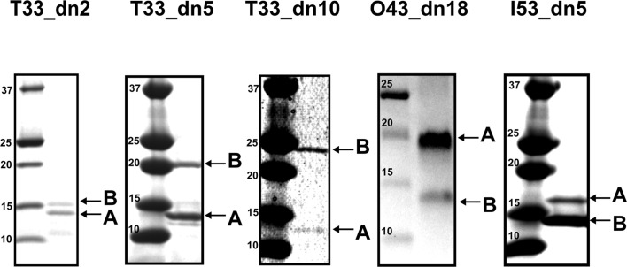
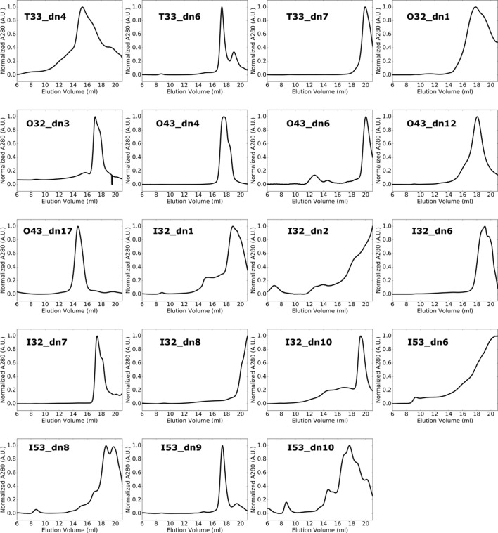
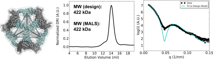
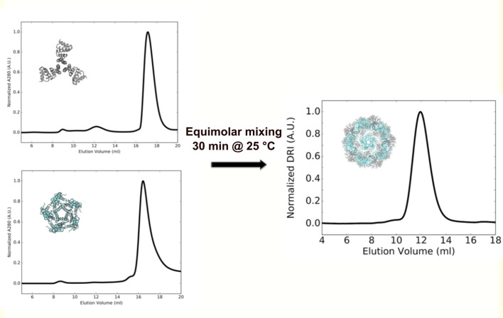
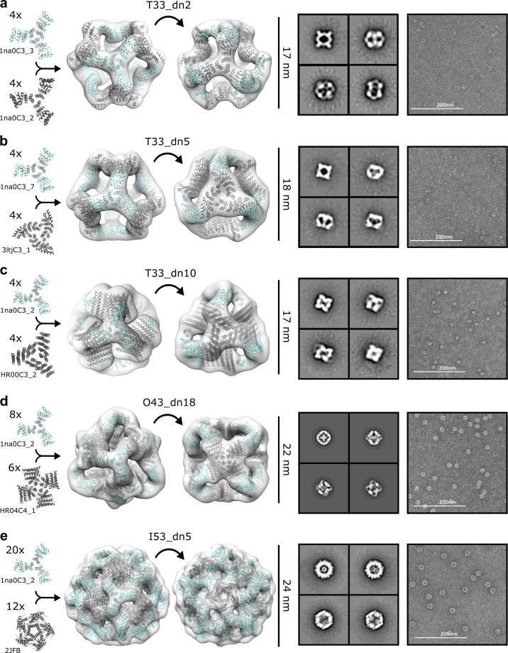
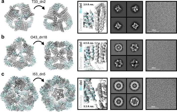
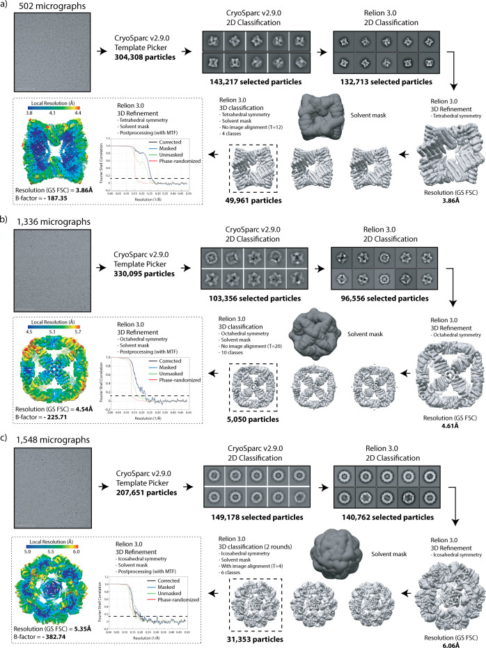
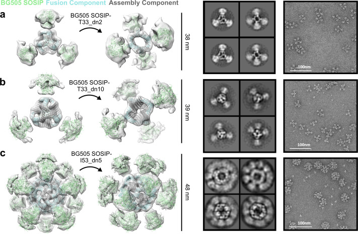
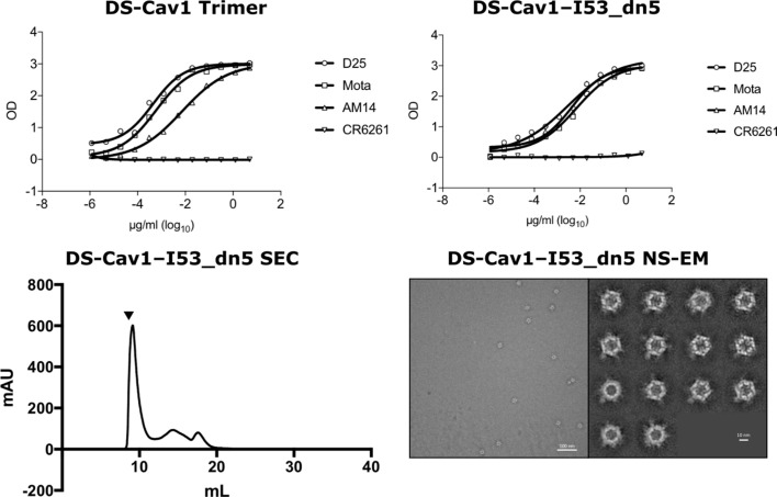
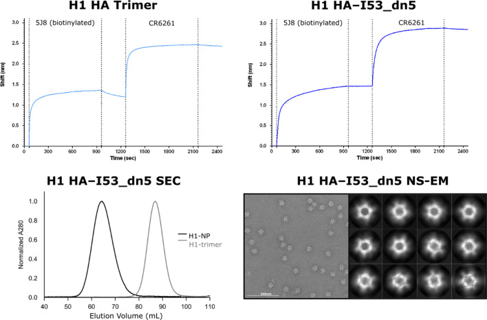
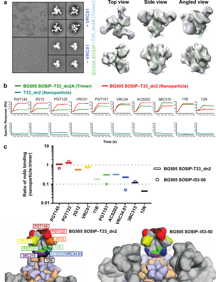
Multivalent presentation of viral glycoproteins can substantially increase the elicitation of antigen-specific antibodies. To enable a new generation of anti-viral vaccines, we designed self-assembling protein nanoparticles with geometries tailored to present the ectodomains of influenza, HIV, and RSV viral glycoprotein trimers. We first de novo designed trimers tailored for antigen fusion, featuring N-terminal helices positioned to match the C termini of the viral glycoproteins. Trimers that experimentally adopted their designed configurations were incorporated as components of tetrahedral, octahedral, and icosahedral nanoparticles, which were characterized by cryo-electron microscopy and assessed for their ability to present viral glycoproteins. Electron microscopy and antibody binding experiments demonstrated that the designed nanoparticles presented antigenically intact prefusion HIV-1 Env, influenza hemagglutinin, and RSV F trimers in the predicted geometries. This work demonstrates that antigen-displaying protein nanoparticles can be designed from scratch, and provides a systematic way to investigate the influence of antigen presentation geometry on the immune response to vaccination.

Keywords: B lymphocytes; BL21; E. coli; HEK293F; Lemo21; computational biology; human; immunology; inflammation; systems biology; virus.

Plain language summary

Vaccines train the immune system to recognize a specific virus or bacterium so that the body can be better prepared against these harmful agents. To do so, many vaccines contain viral molecules called glycoproteins, which are specific to each type of virus. Glycoproteins that sit at the surface of the virus can act as ‘keys’ that recognize and unlock the cells of certain organisms, leading to viral infection. To ensure a stronger immune response, glycoproteins in vaccines are often arranged on a protein scaffolding which can mimic the shape of the virus of interest and trigger a strong immune response. Many scaffoldings, however, are currently made from natural proteins which cannot always display viral glycoproteins. Here, Ueda, Antanasijevic et al. developed a method that allows for the design of artificial proteins which can serve as scaffolding for viral glycoproteins. This approach was tested using three viruses: influenza, HIV, and RSV – a virus responsible for bronchiolitis. The experiments showed that in each case, the relevant viral glycoproteins could attach themselves to the scaffolding. These structures could then assemble themselves into vaccine particles with predicted geometrical shapes, which mimicked the virus and maximized the response from the immune system. Designing artificial scaffolding for viral glycoproteins gives greater control over vaccine design, allowing scientists to manipulate the shape of vaccine particles and test the impact on the immune response. Ultimately, the approach developed by Ueda, Antanasijevic et al. could lead to vaccines that are more efficient and protective, including against viruses for which there is currently no suitable scaffolding.


Conflict of interest statement

GU, JF Inventor on U.S. patent application 62/422,872 titled “Computational design of self-assembling cyclic protein homo-oligomers.” Inventor on U.S. patent application 62/636,757 titled “Method of multivalent antigen presentation on designed protein nanomaterials.” Inventor on U.S. patent application PCT/US20/17216 titled “Nanoparticle-based Influenza Virus Vaccines and Uses Thereof.”, WS Inventor on U.S. patent application 62/422,872 titled “Computational design of self-assembling cyclic protein homo-oligomers.”, JC, GH, AM, AY, YT, YP, MB, BS, RG, PB, PZ, DV, RS, JM, PK, AW No competing interests declared, DE Inventor on U.S. patent application 62/636,757 titled “Method of multivalent antigen presentation on designed protein nanomaterials.” Inventor on U.S. patent application PCT/US20/17216 titled “Nanoparticle-based Influenza Virus Vaccines and Uses Thereof.”, MK Inventor on U.S. patent application PCT/US20/17216 titled “Nanoparticle-based Influenza Virus Vaccines and Uses Thereof.”, BG Inventor on U.S. patent application PCT/US20/17216 titled “Nanoparticle-based Influenza Virus Vaccines and Uses Thereof.” Member of Icosavax’s Scientific Advisory Board. NK Inventor on U.S. patent application 62/636,757 titled “Method of multivalent antigen presentation on designed protein nanomaterials.” Inventor on U.S. patent application PCT/US20/17216 titled “Nanoparticle-based Influenza Virus Vaccines and Uses Thereof.” Co-founder and shareholder of Icosavax, a company that has licensed these patent applications. Member of Icosavax’s Scientific Advisory Board. DB Inventor on U.S. patent application 62/422,872 titled “Computational design of self-assembling cyclic protein homo-oligomers.” Inventor on U.S. patent application 62/636,757 titled “Method of multivalent antigen presentation on designed protein nanomaterials.” Inventor on U.S. patent application PCT/US20/17216 titled “Nanoparticle-based Influenza Virus Vaccines and Uses Thereof.” Co-founder and shareholder of Icosavax, a company that has licensed these patent applications. Member of Icosavax’s Scientific Advisory Board.

Figures

Figure 1.
Figure 1.. De novo design of protein nanoparticles tailored for multivalent antigen presentation.
(a) Computational docking of monomeric repeat proteins into C3-symmetric trimers using the RPX method. (b) Selection of trimers for design based on close geometric match between their N termini (blue spheres) and C termini (red spheres) of a viral antigen (green, BG505 SOSIP shown for illustration). (c) Design of two-component nanoparticles incorporating a fusion component (cyan) and assembly component (gray). (d) Nanoparticle assembled with antigen-fused trimeric component yields multivalent antigen-displaying nanoparticle.
Figure 1—figure supplement 1.
Figure 1—figure supplement 1.. Computational docking and design of trimers for fusion to a specific viral glycoprotein.
Design models for C3-symmetric trimers (gray) with labeled N termini (blue spheres) screened against the BG505 SOSIP trimeric glycoprotein subunit gp41 (cyan) with C-terminal residues labeled (red spheres).
Figure 2.
Figure 2.. Biophysical characterization of antigen-tailored trimers and nanoparticles.
Top rows, design models. Middle rows, SEC chromatograms and calculated molecular weights from SEC-MALS. Bottom rows, comparisons between experimental SAXS data and scattering profiles calculated from design models. (a) 1na0C3_2. (b) 3ltjC3_1v2. (c) 3ltjC3_11. (d) HR04C3_5v2. (e) T33_dn2. (f) T33_dn10. (g) O43_dn18. (h) I53_dn5.
Figure 2—figure supplement 1.
Figure 2—figure supplement 1.. SEC-MALS chromatograms for designed trimers occupying an off-target oligomeric state.
Predominant oligomeric species for each design were collected by fractionation from initial SEC purification, and fourteen chromatograms are presented here from a subsequent round of high-performance SEC-MALS using a Superdex 200 column. *Chromatogram obtained using a Superdex 75 10/300 GL column.
Figure 2—figure supplement 2.
Figure 2—figure supplement 2.. SEC chromatograms for designed trimers with off-target retention volumes after Ni2+ IMAC.
Primary SEC chromatograms obtained from a Superdex 200 column for soluble proteins after purification by Ni2+ IMAC. Designs presented here formed off-target or polydisperse assemblies based on retention volume.
Figure 2—figure supplement 3.
Figure 2—figure supplement 3.. Comparison between the experimentally determined crystal structures and corresponding models of two designed trimers.
Left, design models (gray) and crystal structures (gray) superposed indicating resolution of structure (res.) and backbone r.m.s.d. between structure and design model. Right, magnified view of the de novo designed interface and side chain packing.
Figure 2—figure supplement 4.
Figure 2—figure supplement 4.. SDS-PAGE of bicistronically-expressed designed nanoparticles eluted from Ni2+ IMAC.
For each designed nanoparticle: Left - protein standard (Precision Plus Dual Xtra, Bio-Rad). Right - labeled bands corresponding to the expected size of each component.
Figure 2—figure supplement 5.
Figure 2—figure supplement 5.. SEC profiles for two-component nanoparticles with off-target retention volumes after Ni2+ IMAC.
Primary SEC chromatograms obtained from a Superose six column for soluble proteins directly after purification by Ni2+ IMAC. Designs presented here formed off-target or polydisperse assemblies based on retention volume.
Figure 2—figure supplement 6.
Figure 2—figure supplement 6.. Biophysical characterization of T33_dn5.
Left - designed model. Middle - SEC chromatograms and calculated molecular weights from SEC-MALS. Right - comparisons between experimental SAXS data and scattering profile computed from the design model.
Figure 2—figure supplement 7.
Figure 2—figure supplement 7.. In vitro assembly of I53_dn5.
SEC chromatograms of individual components I53_dn5A (pentameric assembly component, cyan) and I53_dn5B (trimeric fusion component, gray), and nanoparticle purified from equimolar assembly run on a Superose 6 10/300 GL column.
Figure 3.
Figure 3.. NS-EM analysis of antigen-tailored nanoparticles.
From left to right: designed trimers incorporated in each designed nanoparticle, nanoparticle design models fit into NS-EM density (views shown down each component axis of symmetry), designed nanoparticle 2D class-averages, raw electron micrographs of designed nanoparticles. (a) T33_dn2. (b) T33_dn5. (c) T33_dn10. (d) O43_dn18. (e) I53_dn5.
Figure 4.
Figure 4.. Cryo-EM analysis of antigen-tailored nanoparticles.
From left to right: cryo-EM maps with refined nanoparticle design models fit into electron density, view of designed nanoparticle interface region fit into cryo-EM density with indicated resolution (res.), designed nanoparticle 2D class-averages, raw cryo-EM micrographs of designed nanoparticles. (a) T33_dn10. (b) O43_dn18. (c) I53_dn5.
Figure 4—figure supplement 1.
Figure 4—figure supplement 1.. Cryo-EM data processing workflow.
Statistics are presented for (a) T33_dn10, (b) O43_dn18, and (c) I53_dn5.
Figure 5.
Figure 5.. NS-EM analysis of BG505 SOSIP-displaying nanoparticles.
From left to right: BG505 SOSIP-displaying nanoparticle models fit into NS-EM density, 2D class-averages, raw NS-EM micrographs of assembled BG505 SOSIP-displaying nanoparticles. (a) BG505 SOSIP–T33_dn2. (b) BG505 SOSIP–T33_dn10. (c) BG505 SOSIP–I53_dn5.
Figure 5—figure supplement 1.
Figure 5—figure supplement 1.. Structural and antigenic characterization of DS-Cav1–I53_dn5.
Top panel: ELISA using anti-DS-Cav1 antibodies D25, Motavizumab (Mota), AM14, or negative control CR6261 added to DS-Cav1 trimer with foldon or antigen-displaying nanoparticle DS-Cav1–I53_dn5. Bottom panel: SEC chromatogram of DS-Cav1–I53_dn5 on a Superose six column and NS-EM field image and 2D class averages for DS-Cav1–I53_dn5.
Figure 5—figure supplement 2.
Figure 5—figure supplement 2.. Structural and antigenic characterization of HA–I53_dn5.
Top panel: Octet bio-layer interferometry using plate-coated head-directed mAb 5J8 for antigen capture, and subsequent stem-directed mAb CR6261 addition to both antigen-fused trimeric component HA–I53_dn5B and antigen-displaying nanoparticle HA–I53_dn5. Bottom panel: SEC chromatogram for nanoparticle HA–I53_dn5 compared to trimer HA–I53_dn5B on a Sephacryl S-500 column, and NS-EM field image and 2D class averages for HA–I53_dn5.
Figure 6.
Figure 6.. BG505 SOSIP epitope accessibility compared between tetrahedral and icosahedral presentation geometries.
(a) NS-EM micrographs of BG505 SOSIP–T33_dn2A with and without VRC01 Fab bound, 2D class averages, and models fit into NS-EM density. (b) Representative sensorgrams of indicated proteins binding to anti-Env mAbs. (c) Relative accessibility of epitopes on BG505 SOSIP–T33_dn2 nanoparticles and BG505 SOSIP–I53-50 nanoparticles as determined by mAb binding (top). Ratio of moles of macromolecules are means of 2–4 experimental replicates. Epitopes mapped onto BG505 SOSIP are presented on models of T33_dn2 and I53-50 (bottom). Wheat, antigen-fused trimeric component; purple, assembly component; gray, neighboring BG505 SOSIP trimers on the nanoparticle surface.

References

    1. Abbott RK, Lee JH, Menis S, Skog P, Rossi M, Ota T, Kulp DW, Bhullar D, Kalyuzhniy O, Havenar-Daughton C, Schief WR, Nemazee D, Crotty S. Precursor frequency and affinity determine B cell competitive fitness in germinal centers, tested with Germline-Targeting HIV vaccine immunogens. Immunity. 2018;48:133–146. doi: 10.1016/j.immuni.2017.11.023. - DOI - PMC - PubMed
    1. Adams PD. PHENIX: a comprehensive Python-based system for macromolecular structure solution. International Tables for Crystallography. 2012;66:213–221. doi: 10.1107/S0907444909052925. - DOI - PMC - PubMed
    1. Afonine PV, Grosse-Kunstleve RW, Echols N, Headd JJ, Moriarty NW, Mustyakimov M, Terwilliger TC, Urzhumtsev A, Zwart PH, Adams PD. Towards automated crystallographic structure refinement with phenix.refine. Acta Crystallographica Section D Biological Crystallography. 2012;68:352–367. doi: 10.1107/S0907444912001308. - DOI - PMC - PubMed
    1. Antanasijevic A, Ueda U, Brouwer JMP, Copps J. Structural and functional evaluation of de novo-designed two-component nanoparticle carriers for HIV env trimer immunogens. bioRxiv. 2020 doi: 10.1101/2020.01.31.929273. - DOI - PMC - PubMed
    1. Bale JB, Gonen S, Liu Y, Sheffler W, Ellis D, Thomas C, Cascio D, Yeates TO, Gonen T, King NP, Baker D. Accurate design of megadalton-scale two-component icosahedral protein complexes. Science. 2016;353:389–394. doi: 10.1126/science.aaf8818. - DOI - PMC - PubMed

Publication types