. 2020 Sep 1;130(9):4704-4709.
doi: 10.1172/JCI136760.

Pancreatic ductal adenocarcinoma progression is restrained by stromal matrix

Affiliations

Pancreatic ductal adenocarcinoma progression is restrained by stromal matrix

Honglin Jiang et al. J Clin Invest. .

Abstract

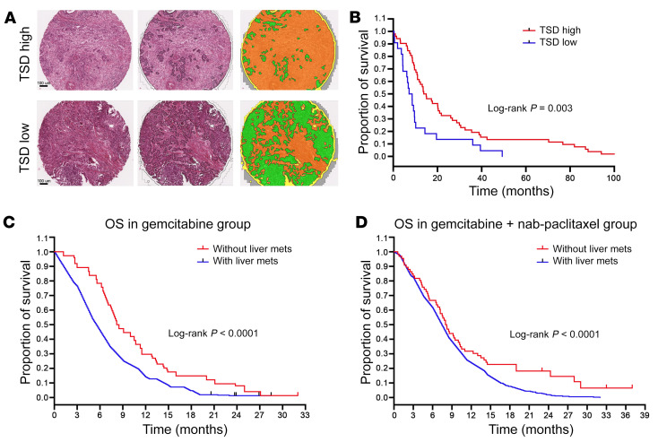
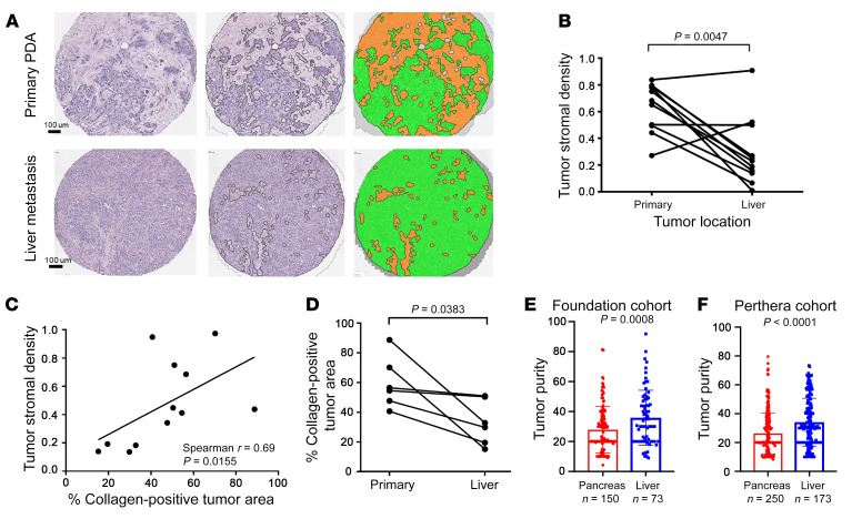
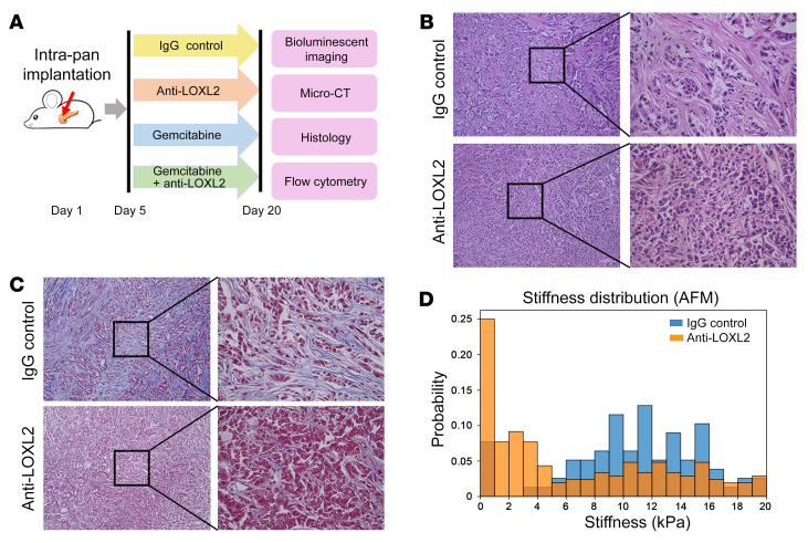
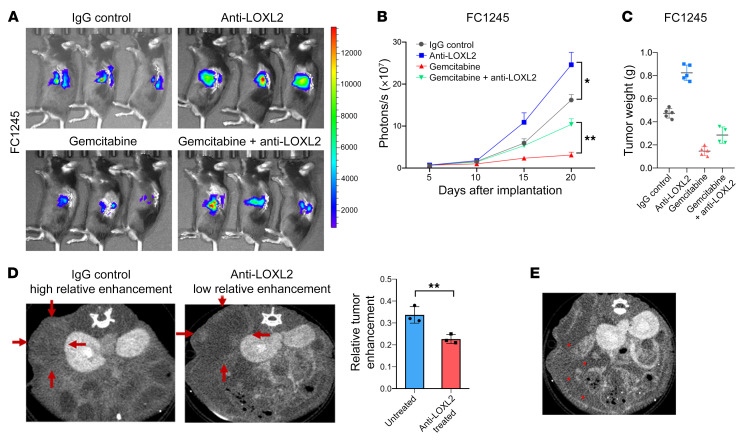
Desmoplasia describes the deposition of extensive extracellular matrix and defines primary pancreatic ductal adenocarcinoma (PDA). The acellular component of this stroma has been implicated in PDA pathogenesis and is being targeted therapeutically in clinical trials. By analyzing the stromal content of PDA samples from numerous annotated PDA data sets and correlating stromal content with both anatomic site and clinical outcome, we found PDA metastases in the liver, the primary cause of mortality to have less stroma, have higher tumor cellularity than primary tumors. Experimentally manipulating stromal matrix with an anti-lysyl oxidase like-2 (anti-LOXL2) antibody in syngeneic orthotopic PDA mouse models significantly decreased matrix content, led to lower tissue stiffness, lower contrast retention on computed tomography, and accelerated tumor growth, resulting in diminished overall survival. These studies suggest an important protective role of stroma in PDA and urge caution in clinically deploying stromal depletion strategies.

Keywords: Cancer; Extracellular matrix; Oncology.


Conflict of interest statement

Conflict of interest: KM is an employee of Foundation Medicine. MJP serves as a consultant for Perthera and holds stock in the company. EB is an employee of Perthera. BL is an employee of Bristol-Myers Squibb. ML is an employee and shareholder of Bristol-Myers Squibb. ZJW serves as a consultant for GE Healthcare. EC has Research Funding from Astra Zeneca, Ferro Therapeutics, Senti Biosciences, Merck KgA, and Bayer to UCSF; stock ownership in Tatara Therapeutics, Clara Health, BloodQ, Guardant Health, Illumina, Pacific Biosciences, and Exact Biosciences; and has received consulting income from Takeda and Merck.

Figures

Figure 1
Figure 1. Low tumor stromal content correlates with worse patient survival.
(A) Resected PDA specimens stained with H&E were segmented into tumor epithelium (green) and tumor stroma (yellow). Scale bars: 100 μm. (B) Kaplan-Meier curves of overall survival (OS) for high versus low tumor stromal density (TSD) in resected pancreatic adenocarcinoma (PDA) specimens from the Berlin patient cohort. Stratified by TSD from primary tumor, patients with high TSD had significantly longer OS than those with low TSD. (C and D) Kaplan-Meier survival analysis of patients with/without liver metastases (mets) from the MPACT study. Shorter OS was observed in patients with liver metastases in both the gemcitabine alone and gemcitabine plus nab-paclitaxel groups.
Figure 2
Figure 2. Stromal content varies in primary PDA tumors and liver metastases.
(A) Representative images following automated tumor epithelial and stromal segmentation of H&E-stained primary PDA and liver metastasis (original magnification, ×10). Left: Unsegmented H&E sections. Middle and right: Segmented H&E sections (green = tumor epithelium; orange = stroma). (B) Tumor stromal density (TSD) was quantified in both primary PDA and paired liver metastases using automated tissue segmentation (P = 0.0047 by paired t test). (C) Correlation of automated TSD quantification and collagen immunofluorescence staining (percentage of tumor area) in primary PDA tissue and matched liver metastasis tissues (P value and correlation coefficient from Spearman’s correlation paired t test, P = 0.0383). (D) Collagen content was quantified in both primary PDA and paired liver metastases using immunofluorescence staining. (E and F) Higher tumor cellularity in liver metastases compared with primary tumors was found in both the Foundation cohort (P = 0.0008 by unpaired t test) and the Know Your Tumor (Perthera) cohort (P < 0.0001 by unpaired t test).
Figure 3
Figure 3. Anti-LOXL2 treatment abolished extracellular matrix in murine PDA model.
(A) Schematic of the animal study. (B and C) H&E and trichrome staining revealed that intratumoral collagen fibers were notably reduced after anti-LOXL2 mAb. Original magnification, ×10 (left) and ×40 (right). (D) Anti-LOXL2–treated tumors had a stiffer extracellular matrix in the periductal region, as measured by atomic force microscopy (AFM).
Figure 4
Figure 4. Reduction of fibrosis augments murine PDA progression.
(A) Representative bioluminescence images of FC1245-fLuc orthotopic pancreatic tumor xenografts receiving IgG, anti-LOXL2 mAb, gemcitabine, or the combination of anti-LOXL2 mAb and gemcitabine. (B) Bioluminescent imaging signal changes (mean ± SEM) of FC1245 xenografts, n = 5 mice/group. Data were analyzed by 1-way ANOVA. *P < 0.05; **P < 0.01. (C) Tumor weight from B was measured at the endpoint. (D) Murine PDA tissues treated with anti-LOXL2 mAb showed relatively low enhancement compared with untreated tissue (P = 0.01). Red arrows indicate the region of interest. (E) Anti-LOXL2–treated mouse developed ascites, indicated with red asterisks.

References

    1. Kleeff J, et al. Pancreatic cancer. Nat Rev Dis Primers. 2016;2:16022. - PubMed
    1. Ho CK, Rahib L, Liao JC, Sriram G, Dipple KM. Mathematical modeling of the insulin signal transduction pathway for prediction of insulin sensitivity from expression data. Mol Genet Metab. 2015;114(1):66–72. doi: 10.1016/j.ymgme.2014.11.003. - DOI - PMC - PubMed
    1. Collisson EA, et al. Subtypes of pancreatic ductal adenocarcinoma and their differing responses to therapy. Nat Med. 2011;17(4):500–503. doi: 10.1038/nm.2344. - DOI - PMC - PubMed
    1. Moffitt RA, et al. Virtual microdissection identifies distinct tumor- and stroma-specific subtypes of pancreatic ductal adenocarcinoma. Nat Genet. 2015;47(10):1168–1178. doi: 10.1038/ng.3398. - DOI - PMC - PubMed
    1. Bailey P, et al. Genomic analyses identify molecular subtypes of pancreatic cancer. Nature. 2016;531(7592):47–52. doi: 10.1038/nature16965. - DOI - PubMed

Publication types

MeSH terms