Multicenter Study
. 2020 Sep;50(10):1354-1368.
doi: 10.1007/s00247-020-04747-5. Epub 2020 Aug 4.

Thoracic imaging of coronavirus disease 2019 (COVID-19) in children: a series of 91 cases

Collaborators, Affiliations
Multicenter Study

Thoracic imaging of coronavirus disease 2019 (COVID-19) in children: a series of 91 cases

Pablo Caro-Dominguez et al. Pediatr Radiol. 2020 Sep.

Abstract

Background: Pulmonary infection with SARS-CoV-2 virus (severe acute respiratory syndrome coronavirus 2; COVID-19) has rapidly spread worldwide to become a global pandemic.

Objective: To collect paediatric COVID-19 cases worldwide and to summarize both clinical and imaging findings in children who tested positive on polymerase chain reaction testing for SARS-CoV-2.

Materials and methods: Data were collected by completion of a standardised case report form submitted to the office of the European Society of Paediatric Radiology from March 12 to April 8, 2020. Chest imaging findings in children younger than 18 years old who tested positive on polymerase chain reaction testing for SARS-CoV-2 were included. Representative imaging studies were evaluated by multiple senior paediatric radiologists from this group with expertise in paediatric chest imaging.

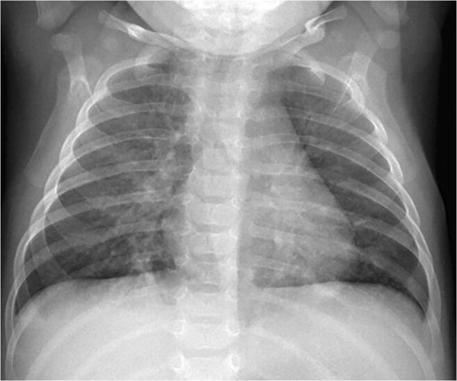
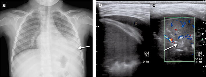
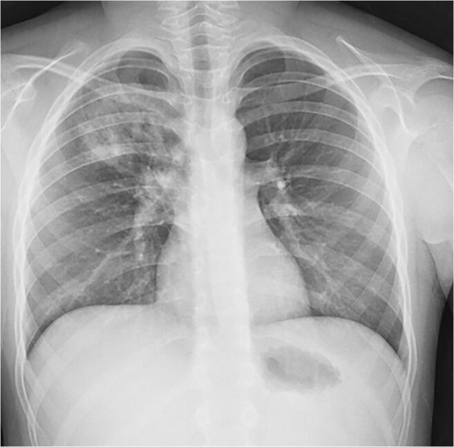
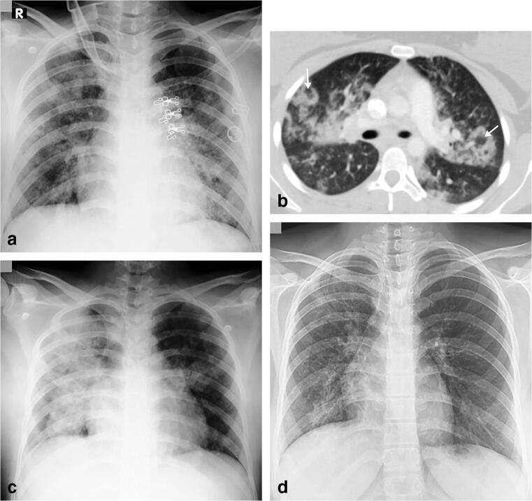
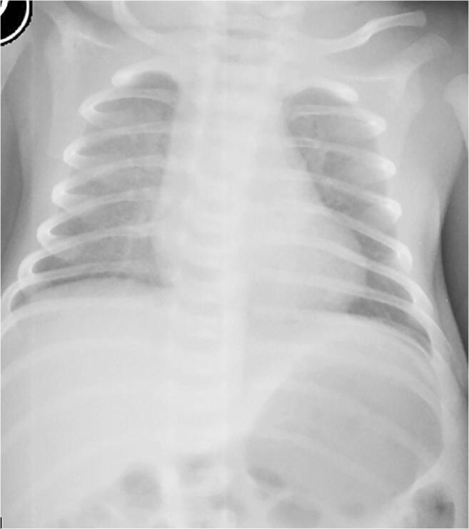
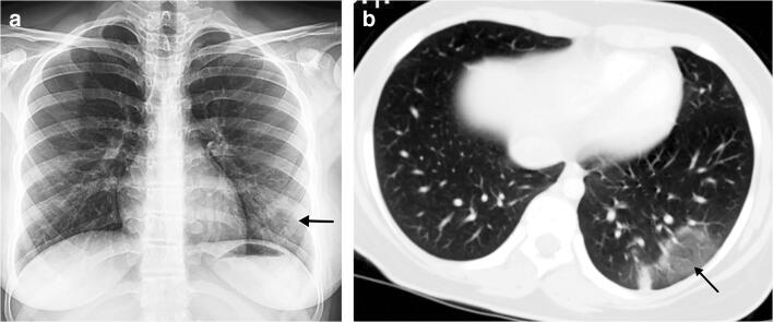
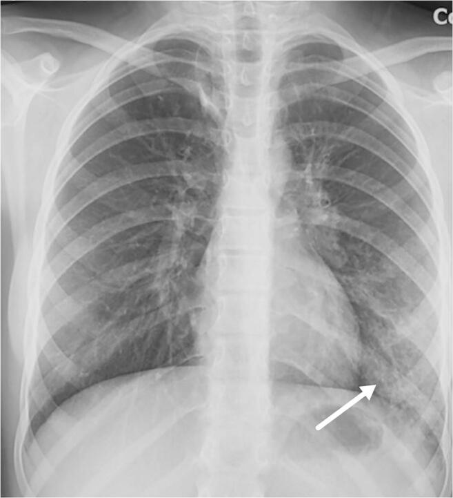
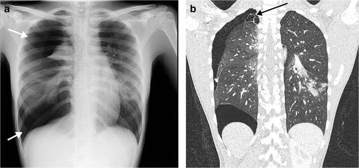
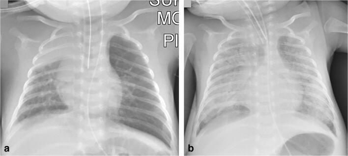
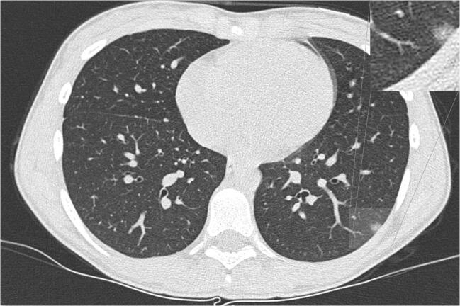
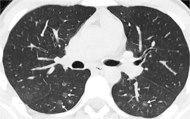
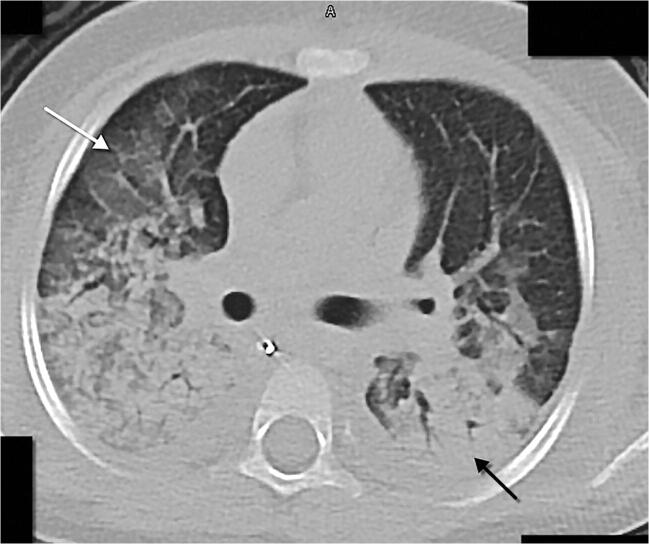
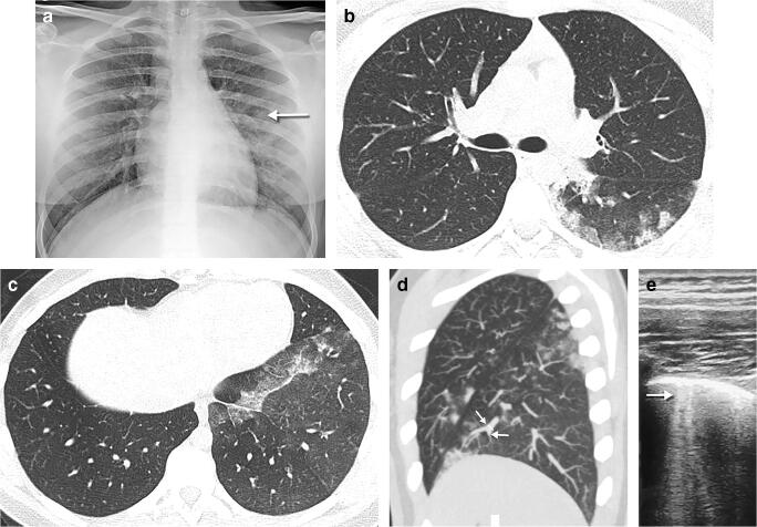
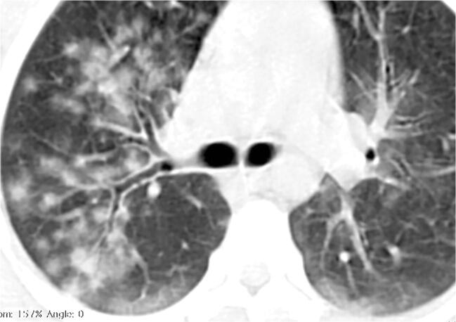
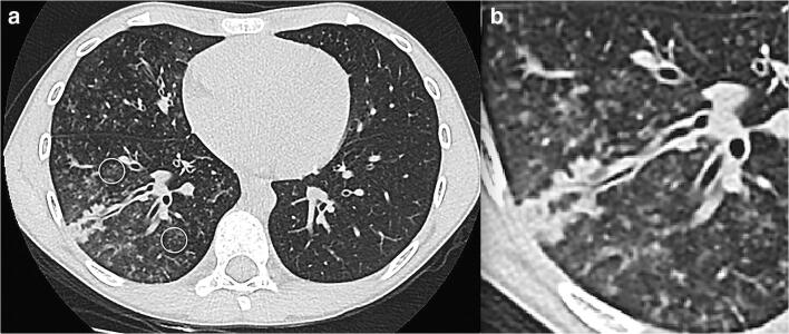
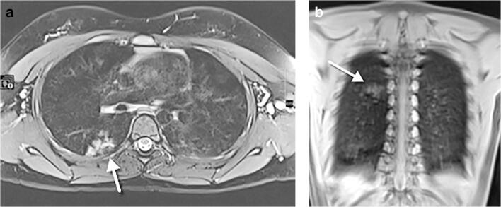
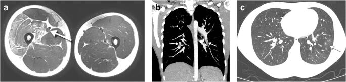
Results: Ninety-one children were included (49 males; median age: 6.1 years, interquartile range: 1.0 to 13.0 years, range: 9 days-17 years). Most had mild symptoms, mostly fever and cough, and one-third had coexisting medical conditions. Eleven percent of children presented with severe symptoms and required intensive unit care. Chest radiographs were available in 89% of patients and 10% of them were normal. Abnormal chest radiographs showed mainly perihilar bronchial wall thickening (58%) and/or airspace consolidation (35%). Computed tomography (CT) scans were available in 26% of cases, with the most common abnormality being ground glass opacities (88%) and/or airspace consolidation (58%). Tree in bud opacities were seen in 6 of 24 CTs (25%). Lung ultrasound and chest magnetic resonance imaging were rarely utilized.

Conclusion: It seems unnecessary to perform chest imaging in children to diagnose COVID-19. Chest radiography can be used in symptomatic children to assess airway infection or pneumonia. CT should be reserved for when there is clinical concern to assess for possible complications, especially in children with coexisting medical conditions.

Keywords: COVID-19; Children; Computed tomography; Coronavirus; Imaging; Lower respiratory tract infection; Pneumonitis; Radiography.


Figures

Fig. 1
Fig. 1
A 1-month-old boy presented with fever and cough. Anteroposterior chest radiograph shows bilateral perihilar peribronchial wall thickening along with subsegmental atelectasis in the lower lobes
Fig. 2
Fig. 2
A 4-year-old girl presented after 2 days of fever and cough. a Anteroposterior chest radiograph shows bilateral perihilar peribronchial thickening along with left upper and lower lobe focal airspace consolidations and moderate left pleural effusion (arrow). b, c Coronal lung ultrasound image (b) and coronal colour Doppler image (c) show extensive subpleural consolidation within the posterobasal area of the left lung, as well as a simple pleural effusion (E). Within the subpleural consolidation there were air bronchograms (arrow) and normal flow on colour Doppler
Fig. 3
Fig. 3
A 10-year-old boy presented with fever, dyspnoea and cough. Anteroposterior chest radiograph shows a focal airspace consolidation in the right upper lobe. This finding resolved on follow-up radiograph 3 days later (not shown)
Fig. 4
Fig. 4
A 16-year-old girl presented with fever, cough, sputum, nasal discharge and dyspnoea. a Anteroposterior chest radiograph, on the day of admission, shows multifocal airspace consolidations, ground glass opacities and reticular opacities in both lungs. b Axial contrast-enhanced chest CT scan performed the same day demonstrates focal bilateral patchy rounded ground glass opacities (arrows) surrounded by a more or less complete ring-like consolidation (reverse halo sign) suggesting organizing pneumonia, which was a sign of severe extent of the disease. c Anteroposterior chest radiograph, 3 days later, showed increased right and improved left airspace opacities. d Posteroanterior chest radiograph, 17 days later, showed improvement in bilateral airspace opacities with residual reticular and ground glass opacities in the right upper, right lower and left lower lobes
Fig. 5
Fig. 5
A 9-day-old girl presented with tachypnoea, after being asymptomatic following normal delivery at term. Anteroposterior chest radiograph shows bilateral diffuse ground glass opacity. The parents of the neonate were confirmed COVID-19 cases. This is the youngest PCR positive patient in our case series
Fig. 6
Fig. 6
A 15-year-old girl presented with fever, dyspnoea, cough and sputum. a Posteroanterior chest radiograph shows reticular and focal ground glass opacities (arrow) in the left lower lobe. b Axial contrast-enhanced chest CT, performed 3 days later, confirms the left lower lobe focal ground glass opacity (arrow)
Fig. 7
Fig. 7
A 12-year-old girl presented with fever and cough. Anteroposterior chest radiograph shows subtle reticulonodular pattern and ground glass opacities (arrow) in the left lower lobe
Fig. 8
Fig. 8
A 15-year-old girl presented with dyspnoea. a Posteroanterior chest radiograph shows a large right-side pneumothorax (arrows) with atelectasis of the right upper lobe and multifocal airspace opacities in the right lower and left lower lobes. There was a slight shift of the mediastinum to the left suggestive of a tension pnemothorax. b Coronal contrast-enhanced chest CT, performed 4 days later, demonstrates a small apical bullae (arrow) at the apex of the right upper lobe, along with persistence of the pneumothorax
Fig. 9
Fig. 9
A 5-week-old boy born at 32 weeks’ gestation with a history of intubation was discharged at 37 weeks’ corrected gestational age. On the following day, he presented with dyspnoea, apnoea, cough and nasal discharge and was admitted to the paediatric intensive care unit. a Anteroposterior chest radiograph demonstrates right upper lobe atelectasis and left lung hyperaeration (probably related to the low position of the endotracheal tube), multifocal airspace and ground glass opacities in the right middle and bilateral lower lobes and bilateral parahilar reticular opacities. b Anteroposterior chest radiograph, 6 days later, shows bibasilar pneumothoraces, which had increased in size compared to Day 3 (not shown) and increased bilateral airspace opacities with air bronchograms
Fig. 10
Fig. 10
A 13-year-old girl with fever and cough. An axial non-contrast CT, 8 days after the onset of symptoms, shows a small rounded subpleural ground glass opacity in the left lower lobe. A magnification of the left lower lobe finding is inset
Fig. 11
Fig. 11
A 16-year-old boy following 1 day of fever. An axial non-enhanced CT shows small rounded multifocal ground glass opacities bilaterally in the lower lobes associated with small vessels (arrows)
Fig. 12
Fig. 12
A 3-year-old girl with a previous history of polyartrhitis treated with methotrexate and prednisone who presented with fever and dyspnoea. An axial chest CT show areas of ground glass opacity in the upper lobes and middle lobe (white arrow), with confluent peribronchial consolidations (in keeping with the known COVID-19 histological association with diffuse alveolar damage), mostly in the lower lobes (black arrow), and mild bilateral pleural effusion. Despite resuscitation, this patient died
Fig. 13
Fig. 13
A 15-year-old girl in close contact with a relative with COVID-19 presented with 3 days of fever and dyspnoea. a A posteroanterior chest radiograph shows small opacities in the middle fields of the left lung (arrow). b An axial chest CT performed the same day demonstrates multifocal areas of rounded ground glass opacities, with a predominantly peripheral, subpleural location in the posterobasal segment of the left lower lobe. Intralobular reticulations can be seen superimposed on the ground glass opacities, resulting in a crazy paving pattern. c Axial images of the lung bases in the same CT show focal unilateral band of ground glass in the left lower lobe around the pleural reflection overlying the phrenic nerve. d Axial thin maximum intensity sagittal reconstruction in the same CT demonstrates focal vascular engorgement (arrows) in the anteromedial segment of the left lower lobe, compared with the upper lobe. e An axial lung ultrasound image obtained 2 days later as a follow-up diagnostic procedure shows B lines (arrow) within the lower posterior and lateral lung areas of the left lung, corresponding to the opacities seen on radiography, as well as pleural thickening
Fig. 14
Fig. 14
A 14-year-old boy unresponsive to wide-spectrum antibiotics. An axial non-contrast chest CT shows diffuse opacities with a rounded morphology and visible halo sign with both central and peripheral distribution and with relative subpleural sparing. Bilateral subpleural intralobular reticulations can be seen superimposed on the ground glass opacities, resulting in a crazy paving pattern of the posterobasal segments of the lower lobes
Fig. 15
Fig. 15
An 8-year-old boy with fever and dyspnoea. a An axial non-contrast chest CT shows peripheral tree in bud mostly in the right lower lobe (circles) and middle lobe either representing vasculitis from direct damage to the pulmonary vascular endothelium or exudative bronchiolitis due to hypersecretion/bacterial superimposed infection. b Magnification view of the right lower lobe findings
Fig. 16
Fig. 16
An 11-year-old girl, asymptomatic, who underwent whole-body MRI for Ollier disease. a An axial T2-weighted turbo spin echo fat-supressed image. b A coronal T1-weighted ultrashort echo time image. Both images demonstrate hyperintense focal infiltration (arrows) within the superior segment of the right lower lobe, as an incidental finding. Following a radiologist’s suggestion of COVID-19, the girl tested positive. A frontal chest radiograph performed 24 h after the MRI did not show abnormalities (not shown)
Fig. 17
Fig. 17
A 16-year-old boy with right leg pain due to deep vein thrombosis and pulmonary embolism, without respiratory symptoms, whose father had COVID-19. a An axial T2-weighted MR image shows a swollen right thigh, with oedema of the quadriceps muscle. Thrombus in the superficial femoral vein is seen as a distended vessel with absent flow void and increased signal intensity (arrow). b A coronal contrast-enhanced chest CT, performed due to tachycardia, demonstrates multiple emboli (arrows) within the segmental arteries of the lower lobes. c An axial contrast-enhanced CT in lung window shows small foci of ground glass opacity (arrow) with mosaic attenuation

References

    1. Dong Y, Mo X, Hu Y et al (2020) Epidemiological characteristics of 2143 pediatric patients with 2019 coronavirus disease in China. Pediatrics. 10.1542/peds.2020-0702
    1. Zu ZY, Jiang MD, Xu PP et al (2020) Coronavirus disease 2019 (COVID-19): a perspective from China. Radiology 200490 - PMC - PubMed
    1. Poggiali E, Dacrema A, Bastoni D, et al. Can lung US help critical care clinicians in the early diagnosis of novel coronavirus (COVID-19) pneumonia? Radiology. 2020;295:E6. doi: 10.1148/radiol.2020200847. - DOI - PMC - PubMed
    1. Xie X, Zhong Z, Zhao W et al (2020) Chest CT for typical 2019-nCoV pneumonia: relationship to negative RT-PCR testing. Radiology. 10.1148/radiol.2020200343:200343 - PMC - PubMed
    1. Maturu VN, Agarwal R. Reversed halo sign: a systematic review. Respir Care. 2014;59:1440–1449. doi: 10.4187/respcare.03020. - DOI - PubMed

Publication types