. 2020 Jul 29;9(8):1805.
doi: 10.3390/cells9081805.

PKHhigh/CD133+/CD24- Renal Stem-Like Cells Isolated from Human Nephrospheres Exhibit In Vitro Multipotency

Affiliations

PKHhigh/CD133+/CD24- Renal Stem-Like Cells Isolated from Human Nephrospheres Exhibit In Vitro Multipotency

Silvia Bombelli et al. Cells. .

Abstract

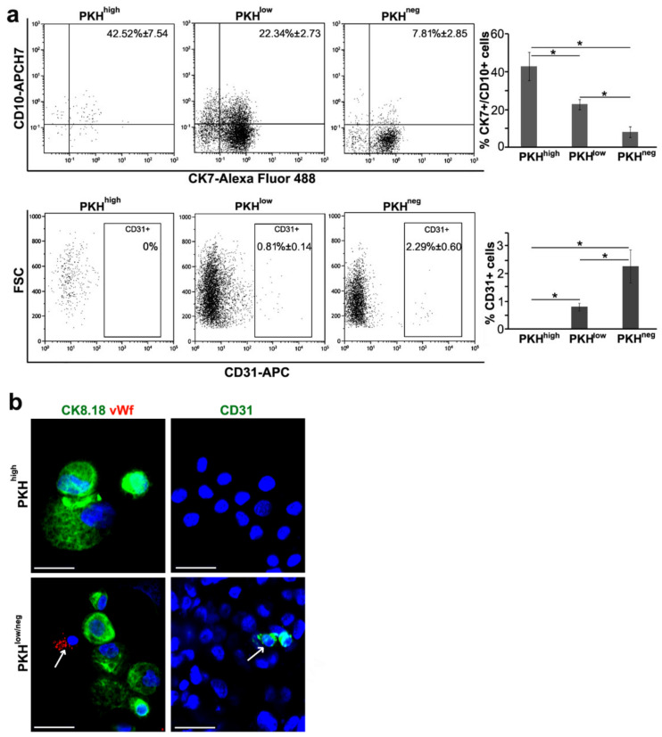
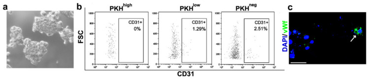
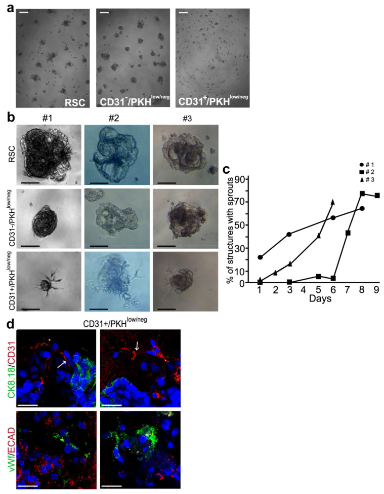
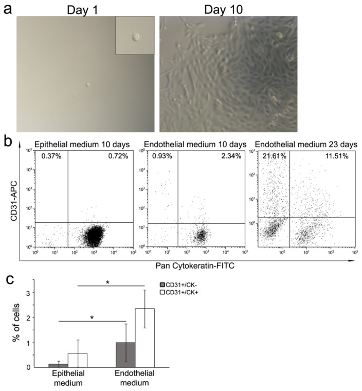
The mechanism upon which human kidneys undergo regeneration is debated, though different lineage-tracing mouse models have tried to explain the cellular types and the mechanisms involved. Different sources of human renal progenitors have been proposed, but it is difficult to argue whether these populations have the same capacities that have been described in mice. Using the nephrosphere (NS) model, we isolated the quiescent population of adult human renal stem-like PKHhigh/CD133+/CD24- cells (RSC). The aim of this study was to deepen the RSC in vitro multipotency capacity. RSC, not expressing endothelial markers, generated secondary nephrospheres containing CD31+/vWf+ cells and cytokeratin positive cells, indicating the coexistence of endothelial and epithelial commitment. RSC cultured on decellularized human renal scaffolds generated endothelial structures together with the proximal and distal tubular structures. CD31+ endothelial committed progenitors sorted from nephrospheres generated spheroids with endothelial-like sprouts in Matrigel. We also demonstrated the double commitment toward endothelial and epithelial lineages of single RSC. The ability of the plastic RSC population to recapitulate the development of tubular epithelial and endothelial renal lineages makes these cells a good tool for the creation of organoids with translational relevance for studying the parenchymal and endothelial cell interactions and developing new therapeutic strategies.

Keywords: endothelium; human adult stem cell; kidney; multipotency; nephrosphere; scaffold.


Conflict of interest statement

The authors declare no conflicts of interest.

Figures

Figure 1
Figure 1
Histological characterization of the decellularized scaffolds repopulated with adult renal stem-like PKHhigh/CD133+/CD24− cells (RSC). (a) Representative Z-stack 3D reconstruction of images after DAPI (blue) and phalloidin (purple) immunofluorescence staining of the scaffolds at 30 days after RSC seeding. Total thicknesses of the shown structure was 31.2-µm original magnification, ×400; (bc) five independent experiments of human kidney scaffold repopulation for 30 days with RSC in presence of basal medium were performed; (b) representative Hematoxylin and Eosin (H&E) staining of formalin fixed, paraffin embedded (FFPE) repopulated scaffold sections. Scale bars, 100 µm; (c) representative sequential IF analysis of the FFPE-repopulated scaffolds with the antibodies against the indicated markers that recognize specific tubular or vascular phenotypes. The different antibodies combinations identify proximal tubules (top), distal tubules (middle) and endothelium (bottom). Scale bars, 50 µm. CK—Cytokeratin; AQP—Aquaporin; vWf—Von Willebrand Factor; blue—DAPI.
Figure 2
Figure 2
Immunophenotypical characterization of nephrosphere cell subpopulations. (a) FACS analysis of nephrospheres (NS) cell subpopulations PKHhigh, PKHlow, PKHneg gated on PKH26 fluorescence. PKHhigh cells were the brightest PKH-positive cells and gated as 0.8–1% of the total population. PKHlow cells, with intermediate fluorescence, were gated as 15–20% of the total cell population and the PKHneg cells, without fluorescence, as 60–70% of the total cell population. The antibodies against the indicated markers were used. For each subpopulation, the overall weighted-mean percentages ± SD referred to three (top) and four (bottom) independent experiments, are reported in the dot plots. CK—cytokeratin; FSC—forward scatter. Chi-squared test, *p < 0.001; (b) immunofluorescence (IF) analysis of PKHhigh, PKHlow/neg cells sorted from dissociated NS and then cytospinned. The indicated markers were evaluated. Blue—DAPI. Results are representative of at least three independent experiments. Original magnification 400×. Scale bars, 50 µm. Arrows: vWf+ (left panel) and CD31+ (right panel) cells.
Figure 3
Figure 3
Characterization of secondary NS generated from FACS-sorted CD31- RSC. (a) Contrast-phase image of secondary NS generated from CD31- RSC FACS sorted from primary NS; (b) FACS analysis of CD31 expression in PKHhigh, PKHlow, PKHneg cell subpopulations of secondary NS cells gated on PKH26 fluorescence; (c) IF analysis of cytospinned cells obtained from secondary NS dissociation. Antibody against vWf was used. Blue—DAPI. Original magnification 400×. Scale bar, 50 µm. Arrow: vWf+ cells.
Figure 4
Figure 4
Morphogenesis 3D assay. (a) Contrast-phase images of RSC (left), CD31−/PKHlow/neg (middle) and CD31+/PKHlow/neg (right) at 6 h of culture. Scale bars, 100 µm; (b) contrast-phase images of structures obtained in Matrigel at 6–7 days of culture from the 3 different samples #1, #2, #3 of RSC (top), CD31−/PKHlow/neg (middle), CD31+/PKHlow/neg (bottom). Scale bars, 100 µm; (c) percentage of structures with sprouts in the three different samples of CD31+/PKHlow/neg at different time points; (d) 3D IF staining of the structures obtained from CD31+/PKHlow/neg cells with the antibodies against the indicated markers. Two different fields for each staining are shown. CK: cytokeratin; ECAD: E-cadherin; vWf: von Willebrand Factor; Blue—DAPI. Original magnification 400×. Scale bars, 50 µm. Arrows: CD31+ cells in the sprout-like extensions.
Figure 5
Figure 5
Single cell differentiation. (a) Contrast-phase images of a representative single-sorted RSC (left panel) and the representative clone generated at day 10. Original magnification: 100×. Insert: 2X digital zoom; (b) FACS analysis of two representative clones obtained from single RSC at ten days of culture in epithelial (left panel) and endothelial (middle panel) media. FACS analysis at 23 days of culture of one representative clone in endothelial medium (right panel). The CD31 and cytokeratin markers were evaluated; (c) graphic representation of CD31+/CK- and CD31+/CK+ cell percentage within the clones obtained after 10 days of culture in specific media. Weighted mean ± SD is referred to four independent experiments of 10 clones grown in epithelial medium and 6 in endothelial medium. Chi-squared test, *p < 0.01.

References

    1. Prescott L.F. The normal urinary excretion rates of renal tubular cells, leucocytes and red blood cells. Clin. Sci. 1966;31:425–435. - PubMed
    1. Lameire N.H., Bagga A., Cruz D., de Maesneer J., Endre Z., Kellum J.A., Liu K.D., Mehta R.L., Pannu N., van Biesen W., et al. Acute kidney injury: An increasing global concern. Lancet. 2013;382:170–179. doi: 10.1016/S0140-6736(13)60647-9. - DOI - PubMed
    1. Humphreys B.D., Valerius M.T., Kobayashi A., Mugford J.W., Soeung S., Duffield J.S., McMahon A.P., Bonventre J.V. Intrinsic epithelial cells repair the kidney after injury. Cell Stem Cell. 2008;2:284–291. doi: 10.1016/j.stem.2008.01.014. - DOI - PubMed
    1. Rinkevich Y., Montoro D.T., Contreras-Trujillo H., Harari-Steinberg O., Newman A.M., Tsai J.M., Lim X., Van-Amerongen R., Bowman A., Januszyk M., et al. In vivo clonal analysis reveals lineage-restricted progenitor characteristics in mammalian kidney development, maintenance, and regeneration. Cell Rep. 2014;7:1270–1283. doi: 10.1016/j.celrep.2014.04.018. - DOI - PMC - PubMed
    1. Johnson H.A., Vera-Roman J.M. Compensatory renal enlargement. Hypertrophy versus hyperplasia. Am. J. Pathol. 1996;49:1–13. - PMC - PubMed

Publication types

MeSH terms