. 2020 Jul 30;10(8):1303.
doi: 10.3390/ani10081303.

Septic Tenosynovitis of the Digital Flexor Tendon Sheath in 83 Cattle

Affiliations

Septic Tenosynovitis of the Digital Flexor Tendon Sheath in 83 Cattle

Alexandra Hund et al. Animals (Basel). .

Abstract

Septic tenosynovitis of the digital flexor tendon sheath (DFTS) is the second most prevalent infection of deeper structures of the distal limb in cattle, after septic arthritis of the distal interphalangeal (DIP) joint. Depending on the type of infection and the involvement of adjacent anatomical structures, various surgical techniques may be used for therapy: Incising the DFTS to resect one or both digital flexor tendons (RDFT), additional resection of the DIP joint (RDIP) or additional digital amputation (RAMP). Our goal was to describe clinical findings and outcome in cattle patients (euthanasia vs. treatment) and the success of surgical methods including improvement of locomotion and postoperative survival time (POST). Data of eighty-three cattle with a mean age of 4.3 years were reviewed in this retrospective study. Overall, 57.7% of tenosynovitis cases were in the lateral DFTS of a hind limb. Fifty-five cattle were treated surgically; the remaining 28 cattle were euthanized following diagnosis. The median cumulative POST was 17.3, 83.1, and 11.9 months for RDFT, RDIP, and RAMP, respectively. Fatal postoperative complications occurred in three cattle. We conclude that the applied methods were successful and allowed the animals to almost reach the average life expectancy of an Austrian dairy cow.

Keywords: cattle; digital flexor tendon sheath; postoperative survival time; tendon resection; tenosynovitis.


Conflict of interest statement

The authors declare no conflict of interest.

Figures

Figure 1
Figure 1
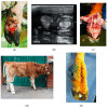
(a) Right hind limb of a five-year-old Fleckvieh cow with a four-day old penetrating wound (arrow), which only became apparent after clipping the hair; (b) Transversal sonogram (7.5 MHz linear probe) of the lateral DFTS approximately 2 cm proximal to the dewclaw with almost homogenously hypoechoic contents in both severely distended inner and outer compartments of the DFTS and no flow phenomena, indicating a fibrinous effusion. SDFT: superficial digital flexor tendon, DDFT: deep digital flexor tendon, BSLS: branch of the suspensory ligament to SDFT, SL: one of the suspensory ligament branches, OC: outer proximal compartment, IC: inner proximal compartment; (c) Intraoperative situation after incision of the lateral DFTS: the lumen is filled entirely with clotted fibrin; (d) Intraoperative situation after resection of both DFT, debridement and removal of all fibrin. The dorsal wall of the DFTS in severely hyperemic; (e) Use of sterile polyurethane soft foam as a drain; (f) Status three days after surgery during the first bandage change with a marked reduction in swelling and formation of granulation tissue; (g) Status at 12 days postsurgery with the wound almost entirely filled with healthy granulation tissue. Note that no part of the wound was sutured. After removal of the drain seven days post-surgery, the bandage facilitated satisfactory reduction of the width of the wound.
Figure 2
Figure 2
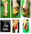
(a): Right hind limb of a seven-month-old Murbodner Blondvieh cross heifer with an eight-day old contused laceration across both DFTS with marked hypergranulation; (b) Transversal sonogram (7.5 MHz linear probe) of both DFTS approximately 1 cm proximal to the dewclaws showing marked distension, heterogeneous hypoechoic effusion with flow phenomena in the lateral DFTS indicating a purulent effusion and a mainly anechoic effusion with flow phenomena in medial DFTS indicating a serofibrinous effusion. SDFT: superficial digital flexor tendon, DDFT: deep digital flexor tendon, BSLS: branch of the suspensory ligament to the SDFT, SL: one of the suspensory ligament branches, OC: outer proximal compartment, IC: inner proximal compartment; (c) Intraoperative situation after opening of the lateral and medial DFTS, resection of both DFT of the lateral digit. The DDFT of the medial digit is visible where partial resection of tendon tissue was necessary. The entire medial DFTS was curetted and irrigated thoroughly; (d) The heifer eight days after surgery with a supportive bandage made of two PVC pipes; (e) Status at 14 days postsurgery with the wound almost entirely filled with healthy granulation tissue.
Figure 3
Figure 3
(a) Left hind limb of a three-year old Fleckvieh cow with the leg restrained for surgery; (b), (c) Lateral and plantar view of a 14-day-old wound above the lateral DFTS with marked hypergranulation and purulent exudate. Fibrinous-purulent tenosynovitis of the left lateral DFTS was diagnosed, as well as purulent tendinitis of both DFT and a purulent arthritis of the PIP joint; (d) Transversal sonogram (7.5 MHz linear probe) of the lateral DFTS approximately 2 cm proximal to the dewclaws showing heterogeneous hypoechoic effusion without flow phenomena and severe circumscribed loss of echogenicity of the DDFT as well as diffuse loss of echogenicity of the SDFT (CL: “core lesion“ caused by purulent disintegration of the tissue of the DDFT). There is marked effusion of the DFTS. SL: one of the suspensory ligament branches, MT: surface of the metatarsal bone. (e), (f) Status at 4 and 8 days postsurgery, respectively.

References

    1. Anderson D.E., Allen D., St-Jean G., Parks A.H. Use of a multifenestrated indwelling lavage system for treatment of septic digital teno-synovitis in cattle. Aust. Vet. J. 1997;75:796–799. doi: 10.1111/j.1751-0813.1997.tb15654.x. - DOI - PubMed
    1. Anderson D.E., Desrochers A., van Amstel S.R. Surgical procedures of the distal limb for treatment of sepsis in cattle. Vet. Clin. North Am. Food Anim. Pract. 2017;33:329–350. doi: 10.1016/j.cvfa.2017.02.011. - DOI - PubMed
    1. Kofler J. Neue Möglichkeiten der Diagnostik der septischen Tendovaginitis der Fesselbeugesehnenscheide des Rindes mittels Sonographie – Therapie und Langzeitergebnisse. Dtsch. Tierärztl. Wschr. 1994;101:215–222. - PubMed
    1. Bertagnoli A., Räber M., Morandi N., Mortellaro C.M., Steiner A. Tenovaginoscopic approach to the common digital flexor tendon sheath of adult cattle: Technique, normal findings and preliminary results in four clinical cases. Vet.J. 2012;191:121–127. doi: 10.1016/j.tvjl.2010.12.009. - DOI - PubMed
    1. Müller M., Gehringer S. Pathologie komplizierter Klauenerkrankungen. In: Fiedler A., Maierl J., Nuss K., editors. Erkrankungen der Klauen und Zehen des Rindes. 2nd ed. Thieme; Stuttgart: 2019. pp. 209–217.

LinkOut - more resources