. 2020 Jul 30;21(15):5445.
doi: 10.3390/ijms21155445.

Proteomic Analysis of Peri-Wounding Tissue Expressions in Extracorporeal Shock Wave Enhanced Diabetic Wound Healing in a Streptozotocin-Induced Diabetes Model

Affiliations

Proteomic Analysis of Peri-Wounding Tissue Expressions in Extracorporeal Shock Wave Enhanced Diabetic Wound Healing in a Streptozotocin-Induced Diabetes Model

Rong-Fu Chen et al. Int J Mol Sci. .

Abstract

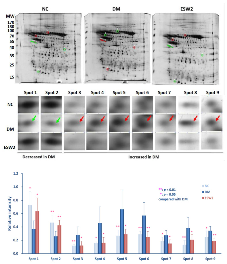
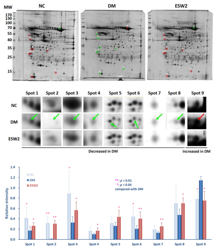
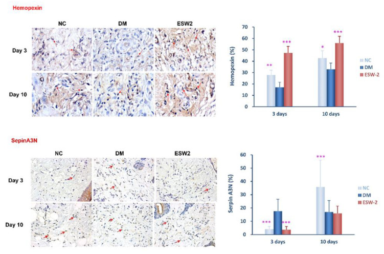
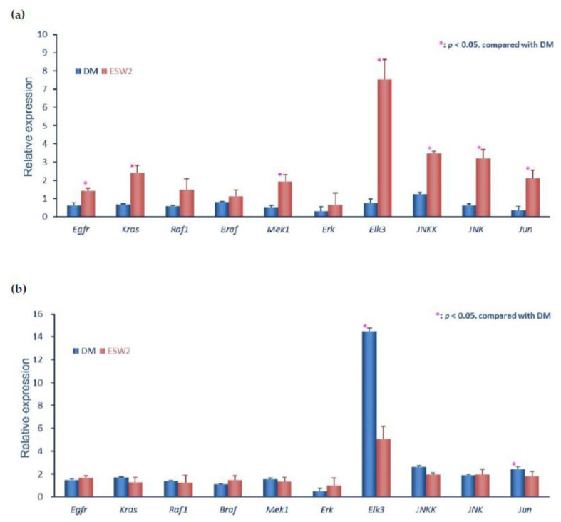
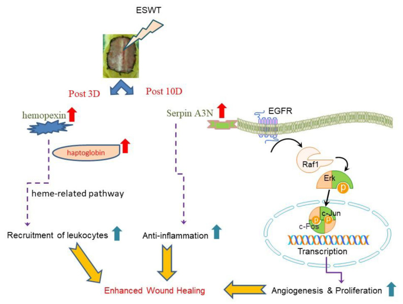
Our former studies have demonstrated that extracorporeal shock wave therapy (ESWT) could enhance diabetic wound healing but the bio-mechanisms remain elusive. This study investigated the changes of topical peri-wounding tissue expressions after ESWT in a rodent streptozotocin-induced diabetic wounding model by using the proteomic analysis and elucidated the molecular mechanism. Diabetic rats receiving ESWT, normal control, and diabetic rats receiving no therapy were analyzed. The spots of interest in proteome analysis were subjected to mass spectrometry to elucidate the peptide mass fingerprints. Protein expression was validated using immunohistochemical staining and related expression of genes were analyzed using real-time RT-PCR. The proteomic data showed a significantly higher abundance of hemopexin at day 3 of therapy but down-regulation at day 10 as compared to diabetic control. In contrast, the level of serine proteinase inhibitor (serpin) A3N expression was significantly decreased at day 3 therapy but expression was upregulated at day 10. Using real-time RT-PCR revealed that serpin-related EGFR-MAPK pathway was involved in ESWT enhanced diabetic wound healing. In summary, proteome analyses demonstrated the expression change of hemopexin and serpin with related MAPK signaling involved in ESWT-enhanced diabetic wound healing. Modulation of hemopexin and serpin related pathways are good strategies to promote wound healing.

Keywords: diabetic wound healing; hemopexin; proteomics; serpin; shock wave.


Conflict of interest statement

We declare that we have no conflict of interest.

Figures

Figure 1
Figure 1
Protein spots of taking into consideration in 2D gel electrophoretograms of peri-wounding skin tissue proteins acquired from and a diabetic rodent on day 3 after two sessions of ESWT (ESW2), and that long period of normal control (NC) and diabetic control (DM). (Above) Representative 2D gel electrophoretograms of peri-wounding skin tissue proteins. The protein samples (150 µg) were exposed to isoelectric focusing (pH 4 to 7), SDS-PAGE, and silver staining. The numbers on the left demonstrate the molecular weight (MW), in kilodaltons. (Center) Enlarged fields of the 9 spots of interest in the sliver-stained SDS-PAGE gels. The spots in the gels of NC, DM, and ESW2 were coordinated utilizing Bio-Rad Proteoweaver 2-D Analysis Software Version 4.0. The arrows prevail upon the spots of interest. (Below) Relative densities of the emphatically distinguished proteins. Relative intensity was determined by separating the thickness of coordinated spots by the thickness of all the coordinated spots in the individual gel. Abbreviation: ESWT, extracorporeal shock wave therapy.
Figure 2
Figure 2
Protein spots of taking into consideration in 2D gel electrophoretograms of peri-wounding skin tissue proteins acquired from day 10 after two sessions of ESWT (ESW2), normal controls (NC), a diabetes controls (DM) at the same days. (Above) Representative 2D gel electrophoretograms of peri-wounding skin tissue proteins. The skin tissue protein samples (150 µg) were exposed to isoelectric focusing (pH 4 to 7), SDS-PAGE, and silver staining. The numbers on the left demonstrate the molecular weight (MW), in kilodaltons. (Center) Enlarged fields of the 9 spots of interest in the sliver-stained SDS-PAGE gels. The spots in the gels of NC, DM, and ESW2 were coordinated utilizing Bio-Rad Proteoweaver 2-D Analysis Software Version 4.0. The arrows prevail upon the spots of interest. (Below) Relative densities of the emphatically distinguished proteins. Relative intensity was determined by separating the thickness of coordinated spots by the thickness of all the coordinated spots in the individual gel. Abbreviation: ESWT, extracorporeal shock wave therapy.
Figure 3
Figure 3
Hemopexin and serine protease inhibitor (SERPIN) A3N expression of peri-wounding tissue samples were analyzed by using immunohistochemical staining at day 3 among extracorporeal shock wave therapy (ESWT), normal controls (NC), and diabetic control (DM) groups. The original magnification is 400× and representative microscopic fields are shown. (Above) Cells positively stained for hemopexin antibody are also shown. *** p < 0.001. (Below) Cells positively stained for serine protease inhibitor A3N antibody are also shown. * p < 0.05. ** p < 0.005. *** p < 0.001.
Figure 4
Figure 4
ESWT enhanced wound healing is associated with early activation of the epidermal growth factor receptor (EGFR) pathway. The expression levels of the genes of the EGFR signaling cascade by real-time quantitative RT-PCR were performed on mRNA samples extracted from biopsy test acquired from the transitional zone of the wound edge at day 3 (a) and day 10 (b) after two sessions of ESWT (ESW2), normal control (NC), control diabetic (DM) rats.
Figure 5
Figure 5
The purposed bio-mechanisms of ESWT improve wound healing are through modulation of hemopexin and Serpin with EGFR-MAPK pathways to increase cell proliferation and anti-inflammatory response. Abbreviation: ESWT, extracorporeal shock wave therapy; EGFR, epidermal growth factor receptor; Erk, extracellular-signal-regulated kinase; Serpin, serine protease inhibitor.

References

    1. Cavanagh P.R., Lipsky B.A., Bradbury A.W., Botek G. Treatment for diabetic foot ulcers. Lancet. 2005;366:1725–1735. doi: 10.1016/S0140-6736(05)67699-4. - DOI - PubMed
    1. Orneholm H., Apelqvist J., Larsson J., Eneroth M. Heel ulcers do heal in patients with diabetes. Int. Wound J. 2017;14:629–635. doi: 10.1111/iwj.12654. - DOI - PMC - PubMed
    1. Dalton S.J., Whiting C.V., Bailey J.R., Mitchell D.C., Tarlton J.F. Mechanisms of chronic skin ulceration linking lactate, transforming growth factor-beta, vascular endothelial growth factor, collagen remodeling, collagen stability, and defective angiogenesis. J. Investig. Dermatol. 2007;127:958–968. doi: 10.1038/sj.jid.5700651. - DOI - PubMed
    1. Antonic V., Mittermayr R., Schaden W., Stojadinovic A. Evidence supporting extracorporeal shock wave therapy for acute and chronic soft tissue wounds. Wounds. 2011;23:204–215. - PubMed
    1. Tinazzi E., Amelio E., Marangoni E., Guerra C., Puccetti A., Codella O.M., Simeoni S., Cavalieri E., Montagnana M., Adani R., et al. Effects of shock wave therapy in the skin of patients with progressive systemic sclerosis: A pilot study. Rheumatol. Int. 2011;31:651–656. doi: 10.1007/s00296-009-1339-z. - DOI - PubMed