Review
. 2020 Jul 30;12(8):835.
doi: 10.3390/v12080835.

Nucleocapsid Structure of Negative Strand RNA Virus

Affiliations
Review

Nucleocapsid Structure of Negative Strand RNA Virus

Ming Luo et al. Viruses. .

Abstract

Negative strand RNA viruses (NSVs) include many important human pathogens, such as influenza virus, Ebola virus, and rabies virus. One of the unique characteristics that NSVs share is the assembly of the nucleocapsid and its role in viral RNA synthesis. In NSVs, the single strand RNA genome is encapsidated in the linear nucleocapsid throughout the viral replication cycle. Subunits of the nucleocapsid protein are parallelly aligned along the RNA genome that is sandwiched between two domains composed of conserved helix motifs. The viral RNA-dependent-RNA polymerase (vRdRp) must recognize the protein-RNA complex of the nucleocapsid and unveil the protected genomic RNA in order to initiate viral RNA synthesis. In addition, vRdRp must continuously translocate along the protein-RNA complex during elongation in viral RNA synthesis. This unique mechanism of viral RNA synthesis suggests that the nucleocapsid may play a regulatory role during NSV replication.

Keywords: 5H+3H; capsid protein motif; cofactor; cross subunit interactions; negative strand RNA virus; nucleocapsid; viral RNA-dependent RNA polymerase.


Conflict of interest statement

The authors declare no conflict of interest.

Figures
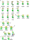
Figure 1
Figure 1
(A) A ribbon drawing of VSV nucleocapsid protein (VSVN) in complex with 9 nucleotides (sticks) encapsidated. The 5H in the N-lobe is colored green, and 3H in the C-lobe is colored yellow. Termini are labeled as N and C, respectively. The linker between the two lobes is colored red. The same ribbon drawing without RNA is shown to the right. Ribbons drawings in this and the following figures are prepared with PyMol [40]. A cartoon is drawn to illustrate the 5H+3H motif. Ten other related N structures in Table 2 are also shown using the same color scheme. (B) Eleven N structures in Polyploviricotina are shown, with those from BUNV (BUNVN), RVFV (RVFVN), and HTNV (HTNVN) in complex with encapsidated nucleotides (sticks). The N-domain in the N core is colored green and the C-domain in the N core is colored yellow. Structures of closely relayed N proteins according to Table 3 and Table 5 are grouped together. The structure from CCHFV (CCHFVN) is colored rainbow from the N-terminus (blue) to the C-terminus (red) because no clear N- and C-domains could be identified. (C) Two structures from the +ss RNA viruses are shown, with that from PepMV (PepMVN) in complex with encapsidated RNA. The color scheme is the same as in (B).
Figure 2
Figure 2
Linear assembly of NSV nucleocapsids. (A) Assembly of VSV nucleocapsid represented by five subunits (colored respectively) [14]. The encapsidated RNA (45 nucleotides) is represented by an orange ribbon and blue sticks. The cross subunit interactions between the red N-terminus and the green C_lobe (named contact I); between the red C_loop and the blue C_lobe (named contact II); and between the green C_loop and the blue N-terminus (named contact III) are labeled with orange letters. (B) Assembly of LACV nucleocapsid represented by four subunits (colored respectively) [21]. The encapsidated RNA (44 nucleotides) is represented by an orange ribbon and blue sticks. The cross subunit interactions between the red N-terminus and the green subunit (named contact A); and between the red C-terminus and the blue subunit (named contact B) are labeled with red letters.
Figure 3
Figure 3
Monomeric N proteins. (A) A N subunit in the NLP of PIV5 (left) is compared with that of N–P complex (right). The two structures are superimposed by use of the C-lobes. The RNA is represented by an orange ribbon and blue sticks. The blue and red regions represent the N- and C-termini, respectively as noted by N and C, in the N-RNA complex. The green polypeptide in the N–P complex corresponds to residues 7–25 in the P protein. Its N- and C-termini are noted by N and C, respectively. The P polypeptide occupies the site where the blue N-terminus of the neighboring N subunit is located in the nucleocapsid, even though it has an opposite orientation. (B) A N subunit in the NLP of RVFV (left) is compared with that of RNA free N (right). The blue region represent the N-terminal residues 1–34. The RNA is shown in the same color scheme.
Figure 4
Figure 4
Mechanism of viral RNA synthesis by NSV. (A) Recognition of the nucleocapsid template by the cofactor P of VSV viral RNA-dependent-RNA polymerase (vRdRp) [78]. Five N subunits are colored yellow, green, red, blue and gray, respectively. The nucleocapsid binding domain of the P protein is colored magenta or orange. One magenta P subunit sits at the top between the C-lobes of the green and red N subunits. (B) A cartoon for a model of the VSV viral RNA replication. The L (light blue) and P subunits (yellow) in VSV vRdRp form an activity bubble by opening N subunits (blue, red and green) to reveal the genomic RNA. The newly synthesized viral RNA exits the L subunit and is encapsidated concomitantly by incoming N subunits. The activity bubble is translocated from the 3′ end to the 5′ end, opening the N subunits (brown and purple) and leaving the N subunits (blue and red) behind to close on the genomic RNA.

References

    1. Knipe D., Howley P., Fields B.N., Griffin D.E. Fields’ Virilogy: Principles of Virus Structure. Lippincott Williams and Wilkins; Philadelphia, PA, USA: 2001. pp. 53–86.
    1. Lefkowitz E.J., Dempsey D.M., Hendrickson R.C., Orton R.J., Siddell S.G., Smith D.B. Virus taxonomy: The database of the International Committee on Taxonomy of Viruses (ICTV) Nucleic Acids Res. 2018;46:D708–D717. doi: 10.1093/nar/gkx932. - DOI - PMC - PubMed
    1. Amarasinghe G.K., Ayllón M.A., Bào Y., Basler C.F., Bavari S., Blasdell K.R., Briese T., Brown P.A., Bukreyev A., Balkema-Buschmann A., et al. Taxonomy of the order Mononegavirales: Update 2019. Arch. Virol. 2019;164:1967–1980. doi: 10.1007/s00705-019-04247-4. - DOI - PMC - PubMed
    1. Amarasinghe G.K., Ayllón M.A., Bào Y., Basler C.F., Bavari S., Blasdell K.R., Briese T., Brown P.A., Bukreyev A., Balkema-Buschmann A., et al. Crystal structure of the borna disease virus nucleoprotein. Structure. 2003;11:1219–1226. doi: 10.1016/j.str.2003.08.011. - DOI - PubMed
    1. Dong S., Yang P., Li G., Liu B., Wang W., Liu X., Xia B., Yang C., Lou Z., Guo Y., et al. Insight into the Ebola virus nucleocapsid assembly mechanism: Crystal structure of Ebola virus nucleoprotein core domain at 1.8 A resolution. Protein Cell. 2015;6:351–362. doi: 10.1007/s13238-015-0163-3. - DOI - PMC - PubMed

Publication types

MeSH terms

LinkOut - more resources