Title: Pachydrusen in Fellow Eyes Predict Response to Aflibercept Monotherapy in Patients with Polypoidal Choroidal Vasculopathy
- PMID: 32752023
- PMCID: PMC7463500
- DOI: 10.3390/jcm9082459
Title: Pachydrusen in Fellow Eyes Predict Response to Aflibercept Monotherapy in Patients with Polypoidal Choroidal Vasculopathy
Abstract
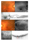
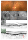
We investigated whether responses to as-needed intravitreal aflibercept injections (IAIs) for polypoidal choroidal vasculopathy (PCV) differed among patients based upon drusen characteristics in fellow eyes. 110 eyes from 110 patients with PCV received 3 monthly IAI and thereafter Pro re nata (PRN) IAI over 12 months. Patients were classified into 4 groups depending on fellow eye findings. Group 1 (n = 16): pachydrusen; Group 2 (n = 45): no drusen; Group 3 (n = 35): soft drusen; Group4 (n = 14) PCV/scarring. Best-corrected visual acuity improved at 12 months in all groups, but not significantly in Group 1 and Group 4; however, visual improvement was similar among the groups after adjusting baseline confounders. Group 1 had a significantly lower percentage of eyes needing retreatment (all p < 0.001; Group 1: 16.7%; Group 2: 50.8%; Group 3: 80%; Group 4: 85.7%). The mean number of retreatments was least in Group 1 among the groups (all p-value < 0.003; Group 1: 0.50 ± 1.32; Group 2: 1.73 ± 2.08; Group 3:2.71 ± 1.99; Group 3: 2.71 ± 2.16). Patients with pachydrusen in fellow eyes were less likely to require additional IAI following the loading dose and may be ideal candidates for aflibercept monotherapy in their first year.
Keywords: as-needed aflibercept therapy; pachydrusen; polypoidal choroidal vasculopathy; treatment burden.
Conflict of interest statement
The authors declare no conflict of interest.
Figures


Similar articles
-
Clinical Characteristics of Unilateral Macular Neovascularization Patients with Pachydrusen in the Fellow Eye.J Clin Med. 2024 Jun 27;13(13):3757. doi: 10.3390/jcm13133757. J Clin Med. 2024. PMID: 38999321 Free PMC article.
-
Response to photodynamic therapy combined with intravitreal aflibercept for polypoidal choroidal vasculopathy depending on fellow-eye condition:2-year results.PLoS One. 2020 Aug 11;15(8):e0237330. doi: 10.1371/journal.pone.0237330. eCollection 2020. PLoS One. 2020. PMID: 32780752 Free PMC article.
-
Pachydrusen in polypoidal choroidal vasculopathy in an Indian cohort.Indian J Ophthalmol. 2019 Jul;67(7):1121-1126. doi: 10.4103/ijo.IJO_1757_18. Indian J Ophthalmol. 2019. PMID: 31238425 Free PMC article.
-
INTRAVITREAL INJECTION OF AFLIBERCEPT IN PATIENTS WITH POLYPOIDAL CHOROIDAL VASCULOPATHY: A 3-YEAR FOLLOW-UP.Retina. 2018 Oct;38(10):2001-2009. doi: 10.1097/IAE.0000000000001818. Retina. 2018. PMID: 28816730
-
[Polypoidal choroidal vasculopathy].Nippon Ganka Gakkai Zasshi. 2012 Mar;116(3):200-31; discussion 232. doi: 10.4264/numa.71.282. Nippon Ganka Gakkai Zasshi. 2012. PMID: 22568102 Review. Japanese.
Cited by
-
Comparison of Outcomes between 3 Monthly Brolucizumab and Aflibercept Injections for Polypoidal Choroidal Vasculopathy.Biomedicines. 2021 Sep 5;9(9):1164. doi: 10.3390/biomedicines9091164. Biomedicines. 2021. PMID: 34572350 Free PMC article.
-
Comparative Efficacy of Brolucizumab and Aflibercept in Polypoidal Choroidal Vasculopathy: A Systematic Review and Meta-Analysis.Cureus. 2025 Jan 7;17(1):e77073. doi: 10.7759/cureus.77073. eCollection 2025 Jan. Cureus. 2025. PMID: 39917144 Free PMC article. Review.
-
Association of Fundus Autofluorescence Abnormalities and Pachydrusen in Central Serous Chorioretinopathy and Polypoidal Choroidal Vasculopathy.J Clin Med. 2022 Sep 11;11(18):5340. doi: 10.3390/jcm11185340. J Clin Med. 2022. PMID: 36142987 Free PMC article.
-
Association of Polyp Regression after Loading Phase with 12-Month Outcomes of Eyes with Polypoidal Choroidal Vasculopathy.Pharmaceuticals (Basel). 2024 May 27;17(6):687. doi: 10.3390/ph17060687. Pharmaceuticals (Basel). 2024. PMID: 38931354 Free PMC article.
-
Clinical Characteristics of Unilateral Macular Neovascularization Patients with Pachydrusen in the Fellow Eye.J Clin Med. 2024 Jun 27;13(13):3757. doi: 10.3390/jcm13133757. J Clin Med. 2024. PMID: 38999321 Free PMC article.
References
-
- Sakurada Y., Yoneyama S., Sugiyama A., Tanabe N., Kikushima W., Mabuchi F., Kume A., Kubota T., Iijima H. Prevalence and Genetic Characteristics of Geographic Atrophy among Elderly Japanese with Age-Related Macular Degeneration. PLoS ONE. 2016;11:e0149978. doi: 10.1371/journal.pone.0149978. - DOI - PMC - PubMed
-
- Kang S.W., Lee H., Bae K., Shin J.Y., Kim S.J., Kim J.M. Korean Age-related Maculopathy Study (KARMS) Group Investigation of precursor lesions of polypoidal choroidal vasculopathy using contralateral eye findings. Graefe’s Arch. Clin. Exp. Ophthalmol. 2016;255:281–291. doi: 10.1007/s00417-016-3452-5. - DOI - PMC - PubMed
LinkOut - more resources
Full Text Sources

