. 2020 Jul 31;13(15):3399.
doi: 10.3390/ma13153399.

Xeno-Free In Vitro Cultivation and Osteogenic Differentiation of hAD-MSCs on Resorbable 3D Printed RESOMER®

Affiliations

Xeno-Free In Vitro Cultivation and Osteogenic Differentiation of hAD-MSCs on Resorbable 3D Printed RESOMER®

Marline Kirsch et al. Materials (Basel). .

Abstract

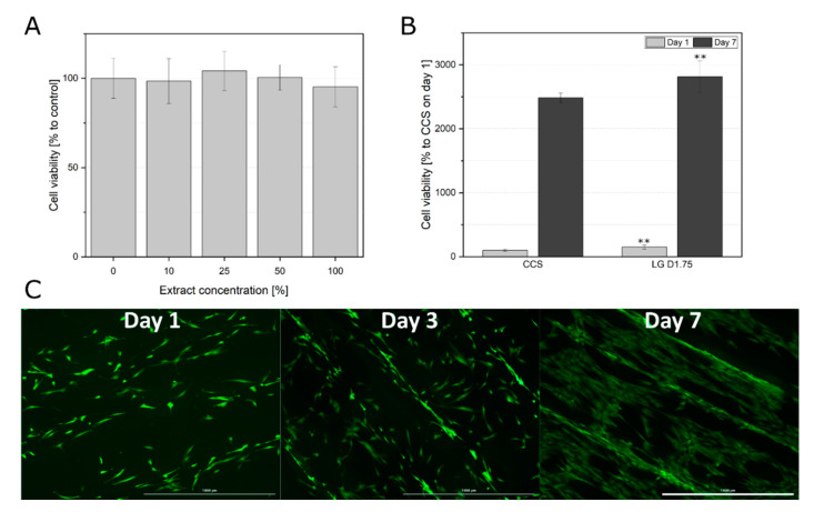
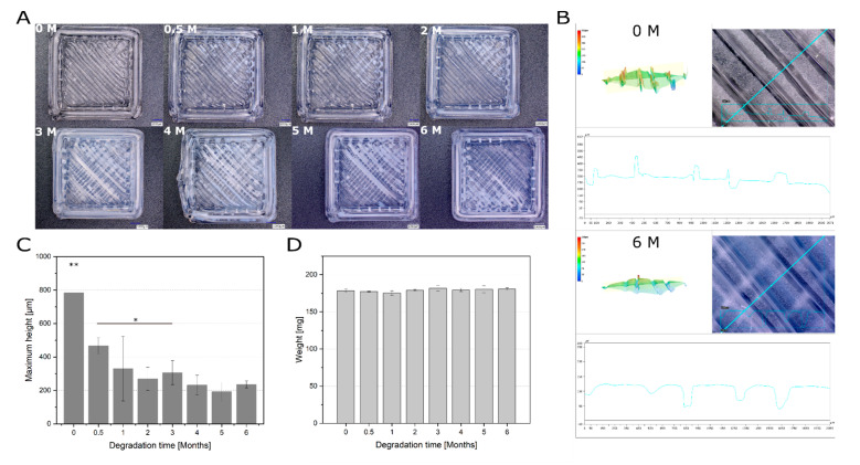
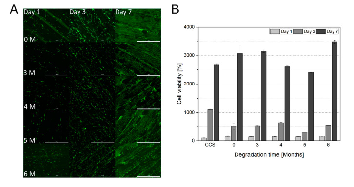
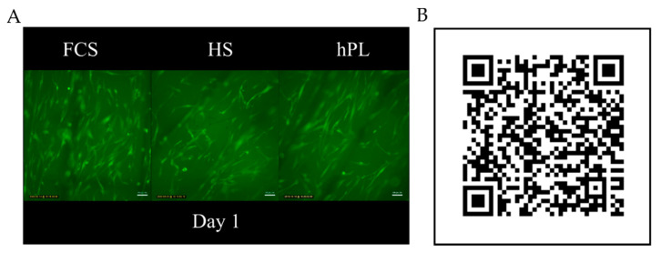
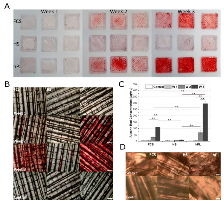
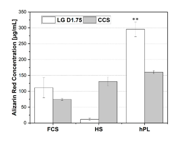
The development of alloplastic resorbable materials can revolutionize the field of implantation technology in regenerative medicine. Additional opportunities to colonize the three-dimensionally (3D) printed constructs with the patient's own cells prior to implantation can improve the regeneration process but requires optimization of cultivation protocols. Human platelet lysate (hPL) has already proven to be a suitable replacement for fetal calf serum (FCS) in 2D and 3D cell cultures. In this study, we investigated the in vitro biocompatibility of the printed RESOMER® Filament LG D1.75 materials as well as the osteogenic differentiation of human mesenchymal stem cells (hMSCs) cultivated on 3D printed constructs under the influence of different medium supplements (FCS, human serum (HS) and hPL). Additionally, the in vitro degradation of the material was studied over six months. We demonstrated that LG D1.75 is biocompatible and has no in vitro cytotoxic effects on hMSCs. Furthermore, hMSCs grown on the constructs could be differentiated into osteoblasts, especially supported by supplementation with hPL. Over six months under physiological in vitro conditions, a distinct degradation was observed, which, however, had no influence on the biocompatibility of the material. Thus, the overall suitability of the material LG D1.75 to produce 3D printed, resorbable bone implants and the promising use of hPL in the xeno-free cultivation of human MSCs on such implants for autologous transplantation have been demonstrated.

Keywords: 3D printing; RESOMER®; adipose tissue-derived mesenchymal stem cells (hAD-MSCs); fetal calve serum; human platelet lysate; human serum; in vitro biocompatibility; in vitro degradation; osteogenic differentiation; resorbable polymers.


Conflict of interest statement

The authors declare no conflict of interest.

Figures

Figure 1
Figure 1
(A) Cell viability of hAD-MSCs after 24 h incubation with extraction medium (0, 10, 25, 50, and 100% extraction medium). Data represent the mean ± SD for an eight to ten-fold determination. (B) Comparison of cell viabilities of hAD-MSCs grown on the construct surface (LG D1.75) and cell culture surface (CCS) after one and seven days of proliferation. Cell viability determined with the help of CTB assay, CTB fluorescence signal of the cells grown on the CCS on day one served as 100% control. Data represent the mean ± SD for a three to six-fold determination. ** p < 0.01. (C) Morphological examination of hAD-MSCs growing on the construct surface. After cultivation of 1, 3, and 7 days, the cells were stained with calcein-AM; 4× objective, scale bar 1000 µm.
Figure 2
Figure 2
Degradation of printed RESOMER® Filament LG D1.75 over six months. (A) Surface of 3D printed constructs before and after different time points of degradation under physiological conditions. Scale bar: 1000 µm. (B) Representative 3D profiles of the printed constructs which were used to determine the surface roughness as a threefold determination. One construct without degradation (0 M) and one after six months of degradation (6 M) is shown. (C) Maximum height (Sz) as an indicator for the surface roughness of the construct surface during the degradation (200x magnification). Data represent the mean ± SD for a threefold determination. *p < 0.05, **p < 0.01. (D) Weight change of printed RESOMER® Filament LG D1.75 during the degradation. Data represent the mean ± SD for a three-fold determination.
Figure 3
Figure 3
(A) Morphological examination of hAD-MSCs growing on degraded RESOMER® PLGA scaffolds (0, 3, 4, 5, and 6 months). After cultivation of 1, 3, and 7 days, the cells were stained with calcein-AM; 4 × objective, scale bar 1000 µm. (B) Comparison of cell viabilities of hAD-MSCs grown on degraded RESOMER® PLGA (0, 3, 4, 5, and 6 months) after one, three, and seven days of proliferation. Cell viability determined with the help of CTB assay, CTB fluorescence signal of the cells grown on the CCS on day one served as 100% control. Data represent the mean ± SD for a three-fold determination.
Figure 4
Figure 4
Time-lapse microscope video of hAD-MSCs cultivated on RESOMER® PLGA in medium supplemented with FCS, HS and hPL. (A) Single image of the time-lapse video of hAD-MSCs cultivated in medium supplemented with FCS, HS, and hPL on day 1 of cultivation. (B) QR code linking to a high speed time-lapse video of hAD-MSCs migrating on RESOMER® PLGA (11270 × speed).
Figure 5
Figure 5
(A) Overview over alizarin red stained hAD-MSCs grown on LG D1.75 constructs after 7, 14, and 21 days after induction of the osteogenic differentiation under influence of FCS, HS, and hPL. (B) Osteogenic differentiation of hAD-MSCs cultivated on printed RESOMER® Filament LG D1.75 and stained with alizarin red 7, 14, and 21 days after induction of the osteogenic differentiation under the influence of FCS, HS, and hPL. Proliferation control after week 3; 2x objective, scale bar 500 µm. (C) Quantification of osteogenic differentiation hAD-MSCs grown on printed RESOMER® Filament LG D1.75 cultivated in medium supplemented with FCS, HS, or hPL. The quantification was performed after day 7, 14, and 21 of differentiation. RESOMER® PLGA in proliferation medium was used as a control. Data represent the mean ± SD for a three-fold (control) and nine-fold (differentiation) determination.** p < 0.01. (D) Alkaline phosphatase staining of differentiated hAD-MSCs cultivated and for seven days differentiated on printed RESOMER® Filament LG D1.75. Proliferation control after seven days, 4x objective, scale bar 200 µm.
Figure 6
Figure 6
Comparison of alizarin red stained hAD-MSCs grown and differentiated on the printed RESOMER® Filament LG D1.75 construct and on cell culture surface (CCS) for three weeks after induction of osteogenic differentiation under the influence of FCS, HS, and hPL. Data represent the mean ± SD for a three-fold (cell culture surface) and nine-fold (construct surface) determination. ** p < 0.01.

References

    1. Hull C.W. Apparatus for Production of Three-Dimensional Objects by Stereolithography. No. 638905. U.S. Patent. 1984 Aug 8;
    1. Keyhan S.O., Fallahi H., Jahangirnia A., Amirzade-Iranaq M.T., Amirzade-Iranaq M.H. Application of 3-D Printing for Tissue Regeneration in Oral and Maxillofacial Surgery: What is Upcoming. In: Dobrzański L.A., editor. Biomaterials in Regenerative Medicine. InTech; London, UK: 2018.
    1. Dawood A., Marti Marti B., Sauret-Jackson V., Darwood A. 3D printing in dentistry. Br. Dent. J. 2015;219:521–529. doi: 10.1038/sj.bdj.2015.914. - DOI - PubMed
    1. Cunningham L.L., Madsen M.J., Peterson G. Stereolithographic modeling technology applied to tumor resection. J. Oral Maxillofac. Surg. 2005;63:873–878. doi: 10.1016/j.joms.2005.02.027. - DOI - PubMed
    1. Chae M.P., Rozen W.M., McMenamin P.G., Findlay M.W., Spychal R.T., Hunter-Smith D.J. Emerging Applications of Bedside 3D Printing in Plastic Surgery. Front. Surg. 2015;2:25. doi: 10.3389/fsurg.2015.00025. - DOI - PMC - PubMed