. 2020 Jul 31;25(15):3510.
doi: 10.3390/molecules25153510.

In Silico Study Identified Methotrexate Analog as Potential Inhibitor of Drug Resistant Human Dihydrofolate Reductase for Cancer Therapeutics

Affiliations

In Silico Study Identified Methotrexate Analog as Potential Inhibitor of Drug Resistant Human Dihydrofolate Reductase for Cancer Therapeutics

Rabia Mukhtar Rana et al. Molecules. .

Abstract

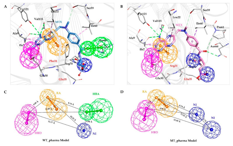
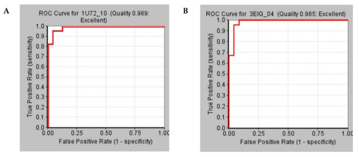
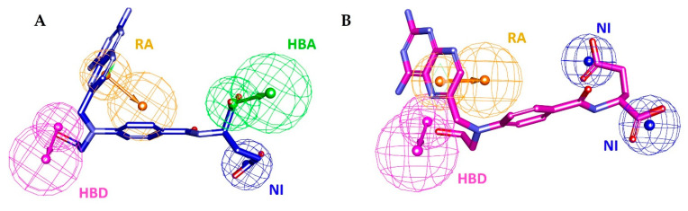
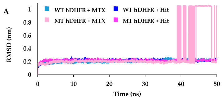
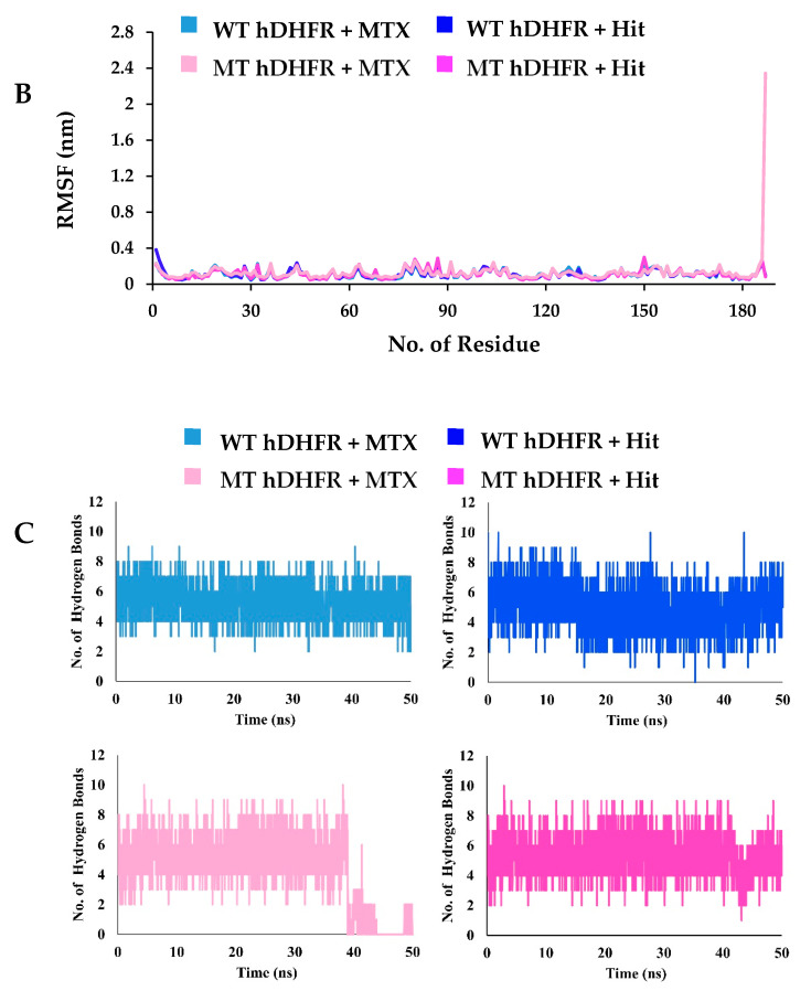
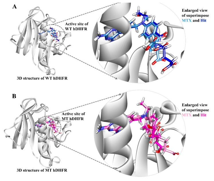
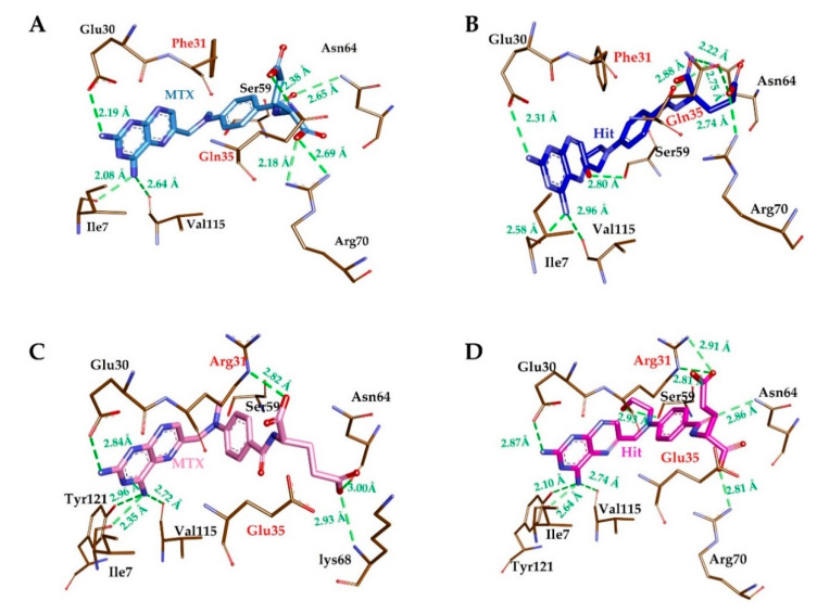
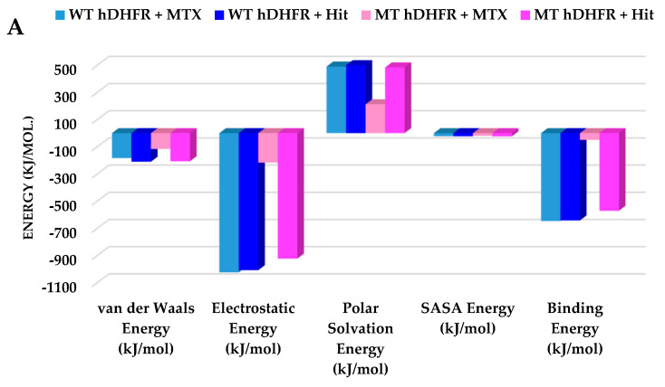
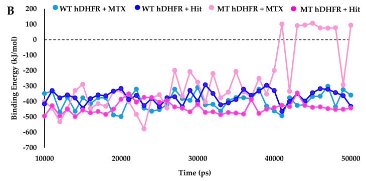
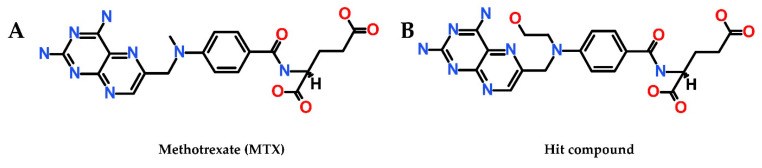
Drug resistance is a core issue in cancer chemotherapy. A known folate antagonist, methotrexate (MTX) inhibits human dihydrofolate reductase (hDHFR), the enzyme responsible for the catalysis of 7,8-dihydrofolate reduction to 5,6,7,8-tetrahydrofolate, in biosynthesis and cell proliferation. Structural change in the DHFR enzyme is a significant cause of resistance and the subsequent loss of MTX. In the current study, wild type hDHFR and double mutant (engineered variant) F31R/Q35E (PDB ID: 3EIG) were subject to computational study. Structure-based pharmacophore modeling was carried out for wild type (WT) and mutant (MT) (variant F31R/Q35E) hDHFR structures by generating ten models for each. Two pharmacophore models, WT-pharma and MT-pharma, were selected for further computations, and showed excellent ROC curve quality. Additionally, the selected pharmacophore models were validated by the Guner-Henry decoy test method, which yielded high goodness of fit for WT-hDHFR and MT-hDHFR. Using a SMILES string of MTX in ZINC15 with the selections of 'clean', in vitro and in vivo options, 32 MTX-analogs were obtained. Eight analogs were filtered out due to their drug-like properties by applying absorption, distribution, metabolism, excretion, and toxicity (ADMET) assessment tests and Lipinski's Rule of five. WT-pharma and MT-pharma were further employed as a 3D query in virtual screening with drug-like MTX analogs. Subsequently, seven screening hits along with a reference compound (MTX) were subjected to molecular docking in the active site of WT- and MT-hDHFR. Through a clustering analysis and examination of protein-ligand interactions, one compound was found with a ChemPLP fitness score greater than that of MTX (reference compound). Finally, a simulation of molecular dynamics (MD) identified an MTX analog which exhibited strong affinity for WT- and MT-hDHFR, with stable RMSD, hydrogen bonds (H-bonds) in the binding site and the lowest MM/PBSA binding free energy. In conclusion, we report on an MTX analog which is capable of inhibiting hDHFR in wild type form, as well as in cases where the enzyme acquires resistance to drugs during chemotherapy treatment.

Keywords: Drug resistance; human dihydrofolate reductase; methotrexate; molecular docking; molecular dynamics simulation.; pharmacophore modeling; virtual screening.


Conflict of interest statement

The authors declare no competing interests. The funders had no role in the design of the study; in the collection, analyses, or interpretation of data; in the writing of the manuscript, or in the decision to publish the results.

Figures

Figure 1
Figure 1
Structure-based pharmacophore generation. (A) Residues of wild type hDHFR active site complementing pharmacophoric features are shown as a thin stick. Bound inhibitor (MTX) is shown as a light blue colored thick stick model. HBA, HBD, RA, and NI are colored as green, magenta, orange and blue respectively. (B) Residues of mutant hDHFR active site complementing pharmacophoric features are shown as a thin stick. Bound inhibitor (MTX) is shown as a pink-colored thick stick model. HBA, HBD, RA, and NI are colored as green, magenta, orange and blue respectively. (C) Interfeature distance illustration of WT-pharma (D) Interfeature distance illustration of MT-pharma.
Figure 2
Figure 2
Receiver Operating Characteristics curves for validation of selected pharmacophore models between true positive and false-positive rates. (A) ROC curve shown in the red line for the WT-pharma model with 0.989 curve quality depicts 98.9% area under the curve. (B) ROC curve shown in the red line for the MT-pharma model with 0.985 curve quality depicts 98.5% area under the curve.
Figure 3
Figure 3
Hit compound (MTX-analog) mapping with pharmacophore models. (A) Hit compound represented in dark blue colored thick stick model mapping with WT-pharma. (B) Hit compound represented in magenta-colored thick stick model mapping with MT-pharma.
Figure 4
Figure 4
RMSD analysis of the reference (MTX) and hit compound (MTX-analog). (A) RMSD of the protein-ligand complex of wild type and mutant hDHFR revealed their stability throughout the simulation, with no abnormal behavior in all systems except for MTX in complex with MT hDHFR. (B) RMSF per residue plot for all the systems portrayed their residues RMSD is stable except for MT hDHFR ligand (MTX) which showed a high fluctuation level. (C) The number of intermolecular hydrogen bonds between protein and ligand during 50 ns MD simulations. Light blue and pink colors represent MTX in wild type and mutant hDHFR, respectively, while dark blue and magenta represent the Hit compound in wild type and mutant hDHFR, respectively.
Figure 4
Figure 4
RMSD analysis of the reference (MTX) and hit compound (MTX-analog). (A) RMSD of the protein-ligand complex of wild type and mutant hDHFR revealed their stability throughout the simulation, with no abnormal behavior in all systems except for MTX in complex with MT hDHFR. (B) RMSF per residue plot for all the systems portrayed their residues RMSD is stable except for MT hDHFR ligand (MTX) which showed a high fluctuation level. (C) The number of intermolecular hydrogen bonds between protein and ligand during 50 ns MD simulations. Light blue and pink colors represent MTX in wild type and mutant hDHFR, respectively, while dark blue and magenta represent the Hit compound in wild type and mutant hDHFR, respectively.
Figure 5
Figure 5
The binding patterns of the reference inhibitor (MTX) and hit compound in the active site of wild type and mutant hDHFR. Compounds are displayed by their representative structures superimposed (left) and enlarged (right). The protein is shown in white color. (A) Light blue and dark blue colors represent MTX and Hit compound in wild type hDHFR. (B) Pink and magenta colors represent MTX and Hit compound respectively in mutant hDHFR.
Figure 6
Figure 6
Molecular interactions analyses. The reference inhibitor MTX and Hit compound interacted with essential residues in the active site of hDHFR. MTX in WT hDHFR (A), Hit in WT hDHFR (B), MTX in MT hDHFR (C) and Hit in MT hDHFR (D) are depicted as light blue, dark blue, pink, and magenta-colored stick representation. The H-bond forming residues of hDHFR are displayed as a brown stick model. H-bonding and bond distances are represented as green dashed lines and measured in angstrom (Å), respectively.
Figure 7
Figure 7
Binding free energy analyses. (A) Graphical representation of MM/PBSA estimated binding free energy of wild type and mutant hDHFR in complex with MTX (reference) and Hit compound throughout the simulation time. The reference compound is depicted as light blue and dark blue for wild type and mutant hDHFR, respectively. The Hit compound is shown in pink and magenta colors for wild type and mutant hDHFR, respectively. (B) The binding free energy decomposition analysis of the final hits in the active site of hDHFR infers that the Hit compound was comparably strongly bound with WT and MT hDHFR, while MTX lost its binding with the mutant structure.
Figure 7
Figure 7
Binding free energy analyses. (A) Graphical representation of MM/PBSA estimated binding free energy of wild type and mutant hDHFR in complex with MTX (reference) and Hit compound throughout the simulation time. The reference compound is depicted as light blue and dark blue for wild type and mutant hDHFR, respectively. The Hit compound is shown in pink and magenta colors for wild type and mutant hDHFR, respectively. (B) The binding free energy decomposition analysis of the final hits in the active site of hDHFR infers that the Hit compound was comparably strongly bound with WT and MT hDHFR, while MTX lost its binding with the mutant structure.
Figure 8
Figure 8
(A) 2D structure of MTX (B) 2D structure of Hit compound (MTX analog, ZINC ID: ZINC000013508844).

Similar articles

Cited by

References

    1. Bertino J.R. Ode to methotrexate. J. Clin. Oncol. 1993;11:5–14. doi: 10.1200/JCO.1993.11.1.5. - DOI - PubMed
    1. Holohan C., Van Schaeybroeck S., Longley D.B., Johnston P.G. Cancer drug resistance: An evolving paradigm. Nat. Rev. Cancer. 2013;13:714–726. doi: 10.1038/nrc3599. - DOI - PubMed
    1. Tran P.N., Tate C.J., Ridgway M.C., Saliba K.J., Kirk K., Maier A.G. Human dihydrofolate reductase influences the sensitivity of the malaria parasite Plasmodium falciparum to ketotifen - A cautionary tale in screening transgenic parasites. Int. J. Parasitol. Drugs Drug Resist. 2016;6:179–183. doi: 10.1016/j.ijpddr.2016.09.003. - DOI - PMC - PubMed
    1. Schweitzer B.I., Dicker A.P., Bertino J.R. Dihydrofolate reductase as a therapeutic target. Faseb J. 1990;4:2441–2452. doi: 10.1096/fasebj.4.8.2185970. - DOI - PubMed
    1. Beierlein J.M., Frey K.M., Bolstad D.B., Pelphrey P.M., Joska T.M., Smith A.E., Priestley N.D., Wright D.L., Anderson A.C. Synthetic and crystallographic studies of a new inhibitor series targeting Bacillus anthracis dihydrofolate reductase. J. Med. Chem. 2008;51:7532–7540. - PMC - PubMed

MeSH terms

LinkOut - more resources