. 2020 Aug 1;9(8):1030.
doi: 10.3390/foods9081030.

The Benefits of Whole Genome Sequencing for Foodborne Outbreak Investigation from the Perspective of a National Reference Laboratory in a Smaller Country

Affiliations

The Benefits of Whole Genome Sequencing for Foodborne Outbreak Investigation from the Perspective of a National Reference Laboratory in a Smaller Country

Stéphanie Nouws et al. Foods. .

Abstract

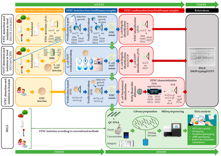
Gradually, conventional methods for foodborne pathogen typing are replaced by whole genome sequencing (WGS). Despite studies describing the overall benefits, National Reference Laboratories of smaller countries often show slower uptake of WGS, mainly because of significant investments required to generate and analyze data of a limited amount of samples. To facilitate this process and incite policy makers to support its implementation, a Shiga toxin-producing Escherichia coli (STEC) O157:H7 (stx1+, stx2+, eae+) outbreak (2012) and a STEC O157:H7 (stx2+, eae+) outbreak (2013) were retrospectively analyzed using WGS and compared with their conventional investigations. The corresponding results were obtained, with WGS delivering even more information, e.g., on virulence and antimicrobial resistance genotypes. Besides a universal, all-in-one workflow with less hands-on-time (five versus seven actual working days for WGS versus conventional), WGS-based cgMLST-typing demonstrated increased resolution. This enabled an accurate cluster definition, which remained unsolved for the 2013 outbreak, partly due to scarce epidemiological linking with the suspect source. Moreover, it allowed detecting two and one earlier circulating STEC O157:H7 (stx1+, stx2+, eae+) and STEC O157:H7 (stx2+, eae+) strains as closely related to the 2012 and 2013 outbreaks, respectively, which might have further directed epidemiological investigation initially. Although some bottlenecks concerning centralized data-sharing, sampling strategies, and perceived costs should be considered, we delivered a proof-of-concept that even in smaller countries, WGS offers benefits for outbreak investigation, if a sufficient budget is available to ensure its implementation in surveillance. Indeed, applying a database with background isolates is critical in interpreting isolate relationships to outbreaks, and leveraging the true benefit of WGS in outbreak investigation and/or prevention.

Keywords: STEC; Shiga toxin-producing Escherichia coli; WGS; food safety; foodborne outbreak investigation; surveillance; whole genome sequencing.


Conflict of interest statement

The authors declare no conflict of interest.

Figures

Figure 1
Figure 1
A schematic overview comparing the conventional methods and whole genome sequencing (WGS) workflow as applied in outbreak investigation. The routinely applied conventional methods for food samples (according to ISO/TS 13136:2012 (for detection and isolation of Shiga toxin-producing Escherichia coli (STEC)) and ISO 16654:2001 (for detection and isolation of Escherichia coli O157)) and for human samples are depicted. Shortly, according to ISO/TS 13136:2012, screening for STEC is performed through qPCR detection of stx1, stx2, and eae in enriched food or swab samples. In case of stx and eae detection, serogroup (i.e., O-type) determination is also performed with qPCR. STEC isolation is attempted through selective growth on CHROMagar STEC. Both qPCR assays are repeated on a sweep of colonies (pool) for STEC detection, and subsequently on separate colonies to enable STEC isolation. Identification of E. coli/Shigella is performed with MALDI-TOF mass spectrometry (MS), and the qPCRs are repeated for STEC identification and confirmation. When a STEC O157 is detected with the ISO/TS 13136:2012 workflow (through qPCR; depicted with a blue arrow), isolation can also be attempted through the serogroup specific workflow (ISO 16654:2001), as was performed for the food isolates of the outbreaks analyzed in this study. Shortly, according to ISO 16654, E. coli O157 in enriched food or swab samples are captured using immunomagnetic beads (i.e., immunomagnetic separation (IMS)), and selectively grown on a CT-SMAC plate for E. coli O157 detection. Through the Oxoid-Latex test, E. coli O157 identification is performed on single colonies, to enable their isolation. The isolated E. coli O157 is then confirmed for E. coli through MALDI-TOF MS, and for STEC O157 through PCR detection of stx1, stx2, eae, and O157 (e.g., rfbE). Human samples are selectively grown on (CT)-SMAC biplates for STEC detection. A sweep of colonies (pool) is tested for stx presence (including subtyping). STEC is verified through PCR on single colonies grown ((CT)-SMAC) from the PCR positive pool. Isolated STEC is then confirmed as E. coli through biochemical identification tests (citrate, mobility indole urease, indole, urease, β-glucuronidase, and sorbitol). Moreover, all human isolates are phenotypically tested for antimicrobial resistance (AMR) through disc diffusion. STEC characterization is further performed through PCR detection of multiple virulence genes, and serogroup determination through slide agglutination. IS629-typing and pulsed-field gel electrophoresis (PFGE) are performed to investigate the relationship between human and food isolates. Completion of these conventional workflows takes approximately 7 working days (indicated by the green time line). With WGS, STEC isolation (according to the conventional methods) is followed by DNA extraction, DNA quality control, and subsequent Nextera XT library preparation for MiSeq sequencing. From the obtained data, all isolate characteristics, and the relatedness between the isolates are retrieved with one approach, with a higher resolution (approximately 7 days (i.e., five working days as two days are used for sequencing), indicated by the green time line). More details concerning the applied workflows are available in the Materials and Methods section and the Supplementary Methods.
Figure 2
Figure 2
A cgMLST tree of all suspected outbreak isolates (Limburg 2012 and Flanders 2013) and the 41 background isolates. A minimum spanning tree was made based on the cgMLST allele matrices of all isolates using the MSTreeV2 method with GrapeTree. The outbreak clusters of Limburg 2012 and Flanders 2013 are outlined in green boxes. The cow carcass isolate that was discriminated from the Limburg 2012 outbreak cluster is visualized with a red box. Isolates that were identified with WGS to be closely related to the outbreak cluster are outlined in a yellow box. The two isolates of the 2013 outbreak in Flanders (EH2297 and TIAC1903) for which laboratory confirmation of the link was supported by epidemiological information are outlined in a blue box. Moreover, the isolates for which conventional PFGE and IS629 profiling was non-corresponding, are indicated with a red arrow. The scale bar represents the absolute number of cgMLST allele differences between isolates.
Figure 3
Figure 3
The IS629 fingerprints (a) and PFGE patterns (b) of all suspected isolates involved in the outbreak in Flanders (2013) are depicted. The scale bars represent the percentages of similarity between the isolates. All outbreak isolates clustering together (i.e., identical patterns) are outlined in a green box (identified as the outbreak cluster). The two isolates outlined in a blue box concern the human case for which the epidemiological link with the consumption of the contaminated food source (raw beef meat) was laboratory confirmed. Background isolates included in the initial outbreak investigation are outlined in a yellow box. Red arrows indicate the differences in the outbreak clusters obtained with both conventional methods.

References

    1. World Health Organization (WHO) Foodborne Disease Outbreaks: Guidelines for Investigation and Control. World Health Organization; Geneva, Switzerland: 2008.
    1. Baker C.A., Rubinelli P.M., Park S.H., Carbonero F., Ricke S.C. Shiga Toxin-Producing Escherichia coli in Food: Incidence, Ecology, and Detection Strategies. Food Control. 2016;59:407–419. doi: 10.1016/j.foodcont.2015.06.011. - DOI
    1. Amézquita-López B.A., Soto-Beltrán M., Lee B.G., Yambao J.C., Quiñones B. Isolation, Genotyping and Antimicrobial Resistance of Shiga Toxin-Producing Escherichia coli. J. Microbiol. Immunol. Infect. 2018;51:425–434. doi: 10.1016/j.jmii.2017.07.004. - DOI - PubMed
    1. Holmes A., Allison L., Ward M., Dallman T.J., Clark R., Fawkes A., Murphy L., Hanson M. Utility of Whole-Genome Sequencing of E. coli O157 for Outbreak Detection and Epidemiological Surveillance. J. Clin. Microbiol. 2015;53:3565–3573. doi: 10.1128/JCM.01066-15. - DOI - PMC - PubMed
    1. Ooka T., Ogura Y., Asadulghani M., Ohnishi M., Nakayama K., Terajima J., Watanabe H., Hayashi T. Inference of the Impact of Insertion Sequence (IS) Elements on Bacterial Genome Diversification through Analysis of Small-Size Structural Polymorphisms in Escherichia coli O157 Genomes. Genome Res. 2009;19:1809–1816. doi: 10.1101/gr.089615.108. - DOI - PMC - PubMed

LinkOut - more resources