. 2020 Aug 1;9(8):1033.
doi: 10.3390/foods9081033.

In Vivo Anti-inflammatory Potential of Viscozyme®-Treated Jujube Fruit

Affiliations

In Vivo Anti-inflammatory Potential of Viscozyme®-Treated Jujube Fruit

Yoonsu Kim et al. Foods. .

Abstract

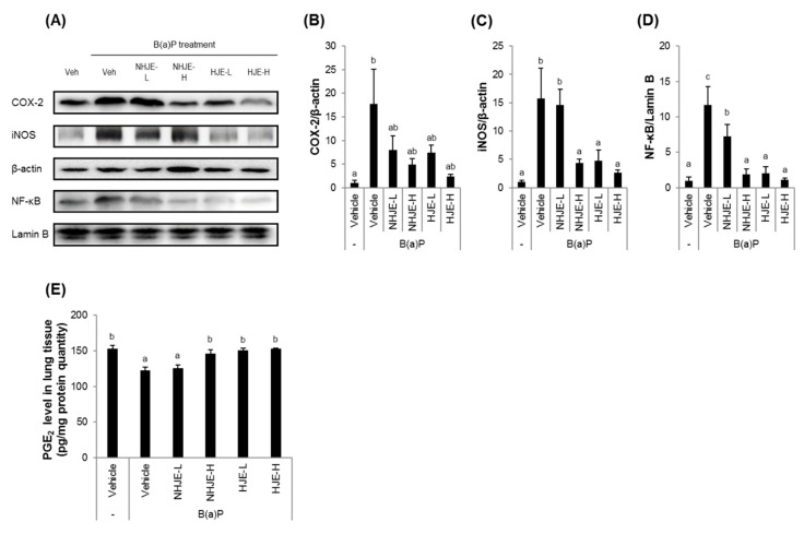
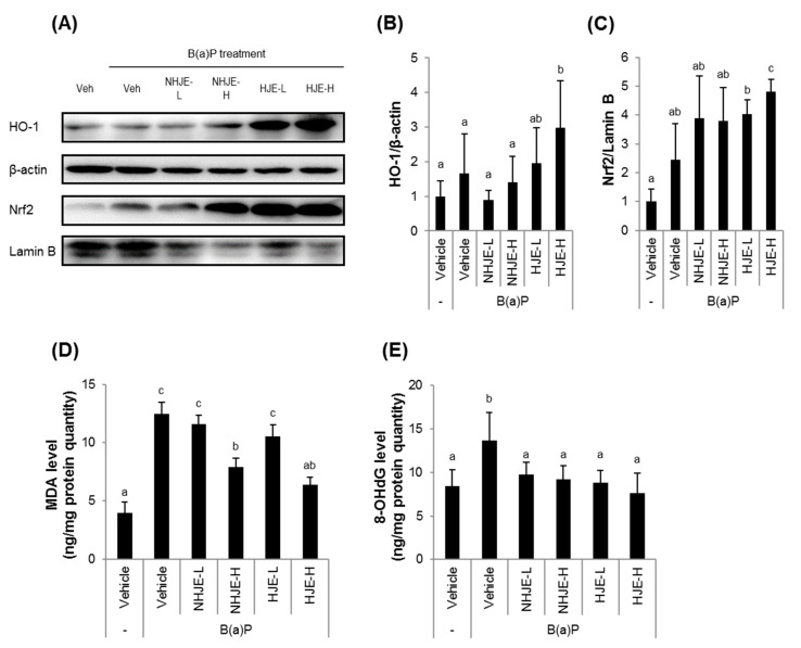
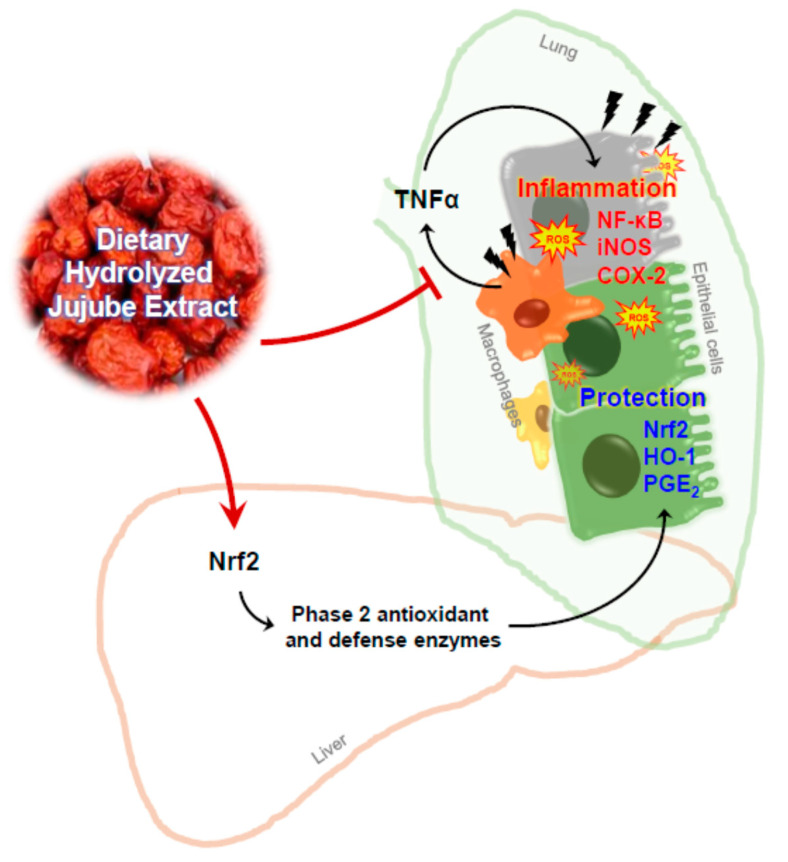
The fruit of Ziziphus jujuba, commonly called jujube, has long been consumed for its health benefits. The aim of this study was to examine the protective effect of dietary supplementation of enzymatically hydrolyzed jujube against lung inflammation in mice. The macerated flesh of jujube was extracted with aqueous ethanol before and after Viscozyme treatment. The extract of enzyme-treated jujube, called herein hydrolyzed jujube extract (HJE), contained higher levels of quercetin, total phenolics, and flavonoids, and exhibited more effective radical-scavenging abilities in comparison to non-hydrolyzed jujube extract (NHJE). HJE treatment decreased production of inflammation-associated molecules, including nitric oxide and pro-inflammatory cytokines from activated Raw 264.7 or differentiated THP-1 cells. HJE treatment also reduced expression of nuclear factor-κB and its downstream proteins in A549 human lung epithelial cells. Moreover, oral supplementation of 1.5 g of HJE per kg of body weight (BW) attenuated histological lung damage, decreased plasma cytokines, and inhibited expression of inflammatory proteins and oxidative stress mediators in the lungs of mice exposed to benzo(a)pyrene at 50 mg/kg BW. Expression levels of antioxidant and cytoprotective factors, such as nuclear factor erythroid-derived 2-related factor 2 and heme oxygenase-1, were increased in lung and liver tissues from mice treated with HJE, compared to mice fed NHJE. These findings indicate that dietary HJE can reduce benzo(a)pyrene-induced lung inflammation by inhibiting cytokine release from macrophages and promoting antioxidant defenses in vivo.

Keywords: HO-1; Jujube; NF-κB; Nrf2; anti-inflammation; hydrolysis; lung.


Conflict of interest statement

The authors have declared no conflict of interest.

Figures

Figure 1
Figure 1
HJE decreased nitric oxide (NO) production and increased antioxidant response element (ARE) transcription activity in cultured cells. (A) Raw 264.7 cells were stimulated by LPS and treated with NHJE and HJE for 24 h. The culture media were collected and measured for the levels of NO produced from the cells. (B) HepG2-ARE cells carrying the luciferase reporter gene linked to the ARE sequence were treated with NHJE and HJE for 24 h. ARE transcription activity was assessed by the activity of luciferase. SFN, sulforaphane. N = 3; error bars, mean ± SEM. Different alphabetical letters on the bars (a–c) indicate statistically significant difference from each other (p < 0.05).
Figure 2
Figure 2
HJE decreased secretions of pro-inflammatory cytokines from differentiated THP-1 cells and expression of inflammation-related proteins in A549 cells. (AC) THP-1 human monocytes were differentiated with tetradecanoylphorbol-13-acetate (TPA) (200 nM) and treated with either NHJE or HJE at a concentration of 500 µg/mL for 24 h. The pro-inflammatory cytokines, including TNFα (A), IL-1β (B), and IL-6 (C), in the culture media were quantified by enzyme-linked immunosorbent assay (ELISA). (DG) A549 human lung epithelial cells were cultured in conditioned media collected from TPA-differentiated THP-1 cells treated with either NHJE or HJE. After 24 h, A549 cells were collected and subjected to western blot analysis (D). The relative expression levels of inflammation-related proteins, including COX-2 (E), iNOS (F), and NF-κB (G), were densitometrically determined. N = 3; error bars, mean ± SEM. Different alphabetical letters (a–c) presented on the bars indicate statistically significant difference from each other (p < 0.05).
Figure 3
Figure 3
Oral administration of HJE alleviated B(a)P-induced lung injury in mice. C57BL/6J mice were fed either NHJE or HJE at two different doses, 0.75 g/kg BW or 1.5 g/kg BW, every day for 14 days. One day prior to sacrifice, B(a)P was given by intraperitoneal injection. (A) Experimental scheme. (B) Changes in average body weight during the experimental period. Values are means ± standard deviation (SD) (n = 8 mice per group). (C) Representative photographs of the dissected lungs. (D) Representative tissue sections stained with H&E (magnification, 40×). A, alveolar sac; AD, alveolar duct; B, respiratory bronchiole; TB, terminal bronchiole; bv, blood vessel. NHJE, non-hydrolyzed jujube extract; NHJE-L, NHJE at a low dose (0.75 g/kg BW); NHJE-H, NHJE at a high dose (1.5 g/kg BW); HJE, hydrolyzed jujube extract; HJE-L, HJE at a low dose (0.75 g/kg BW); HJE-H, HJE at a high dose (1.5 g/kg BW).
Figure 4
Figure 4
Oral administration of HJE lowered plasma levels of pro-inflammatory cytokines in B(a)P-injected mice. (AC) Levels of pro-inflammatory cytokines, TNFα (A), IL-1β (B), and IL-6 (C), in the mouse blood samples were measured by ELISA. Values are means ± SD (n = 8). Different alphabetical letters presented on the bars (a–d) indicate statistically significant difference from each other (p < 0.05). NS, not significant.
Figure 5
Figure 5
Oral administration of HJE suppressed expression of inflammation-related proteins in lung tissue. Expression levels of inflammation marker proteins in the lung tissues were examined. (A) Representative western blot images. (BD) Quantitative data for cytosolic COX-2 (B), cytosolic iNOS (C), and nuclear NF-κB (D). (E) PGE2 protein levels in dissected lung tissues were quantified using ELISA. Values are mean ± SD (n = 5). Different alphabetical letters on the bars (a–c) indicate statistically significant difference from each other (p < 0.05).
Figure 6
Figure 6
Oral administration of HJE increased Nrf2 and HO-1 expressions in lungs. Expression levels of a key antioxidant/defense transcription factor Nrf2 and its downstream protein HO-1 in the lung tissues were examined. (A) Representative western blot images. (BC) Quantitative data for cytosolic HO-1 (B) and nuclear Nrf2 (C). Values are mean ± SD (n = 5). Different alphabetical letters on the bars (a–c) indicate statistically significant difference from each other (p < 0.05).
Figure 7
Figure 7
Oral administration of HJE increased Nrf2 and HO-1 expression and reduced oxidative stress in hepatic tissue. Liver tissue homogenates were used for analyses of expression levels of antioxidant proteins and oxidative stress markers. (A) Representative western blot images. (B,C) Quantitative data for cytosolic HO-1 (B) and nuclear Nrf2 (C). (D,E) Oxidative stress markers including MDA (D) and 8-OHdG (E) were quantified by ELISA. MDA, malondialdehyde. 8-OHdG, 8-hydoxydeoxyguanosine. Values are mean ± SD (n = 8). Different alphabetical letters presented on the bars (a–c) indicate statistically significant difference from each other (p < 0.05).
Figure 8
Figure 8
Schematic illustration of a potential mechanism for the anti-inflammatory activity of HJE.

References

    1. Inoue K.-I., Takano H., Yanagisawa R., Hirano S., Sakurai M., Shimada A., Yoshikawa T. Effects of airway exposure to nanoparticles on lung inflammation induced by bacterial endotoxin in mice. Environ. Health Perspect. 2006;114:1325–1330. doi: 10.1289/ehp.8903. - DOI - PMC - PubMed
    1. Qamar W., Khan A.Q., Khan R., Lateef A., Tahir M., Rehman M.U., Ali F., Sultana S. Benzo(a)pyrene-induced pulmonary inflammation, edema, surfactant dysfunction, and injuries in rats: Alleviation by farnesol. Exp. Lung Res. 2012;38:19–27. doi: 10.3109/01902148.2011.632064. - DOI - PubMed
    1. Vlahos R., Bozinovski S., Jones J., Powell J., Gras J., Lilja A., Hansen M.J., Gualano R.C., Irving L., Anderson G. Differential protease, innate immunity, and NF-κB induction profiles during lung inflammation induced by subchronic cigarette smoke exposure in mice. Am. J. Physiol. Lung Cell Mol. Physiol. 2006;290:L931–L945. doi: 10.1152/ajplung.00201.2005. - DOI - PubMed
    1. Liu J., Cao X.T. Cellular and molecular regulation of innate inflammatory responses. Cell. Mol. Immunol. 2016;13:711–721. doi: 10.1038/cmi.2016.58. - DOI - PMC - PubMed
    1. Ware L.B., Matthay M.A. The acute respiratory distress syndrome. N. Engl. J. Med. 2000;342:1334–1349. doi: 10.1056/NEJM200005043421806. - DOI - PubMed

LinkOut - more resources