. 2020 Oct 9;295(41):14111-14124.
doi: 10.1074/jbc.RA120.015074. Epub 2020 Aug 4.

A non-GPCR-binding partner interacts with a novel surface on β-arrestin1 to mediate GPCR signaling

Affiliations

A non-GPCR-binding partner interacts with a novel surface on β-arrestin1 to mediate GPCR signaling

Ya Zhuo et al. J Biol Chem. .

Abstract

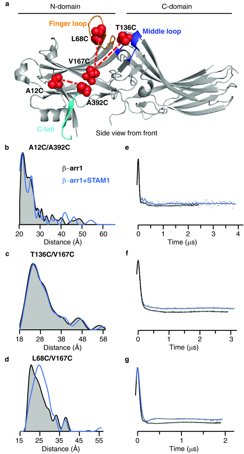
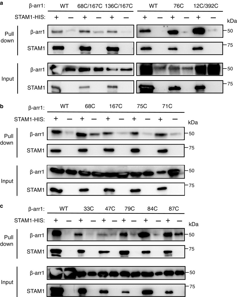
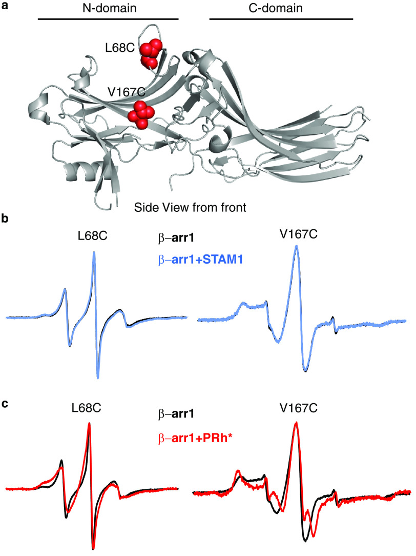
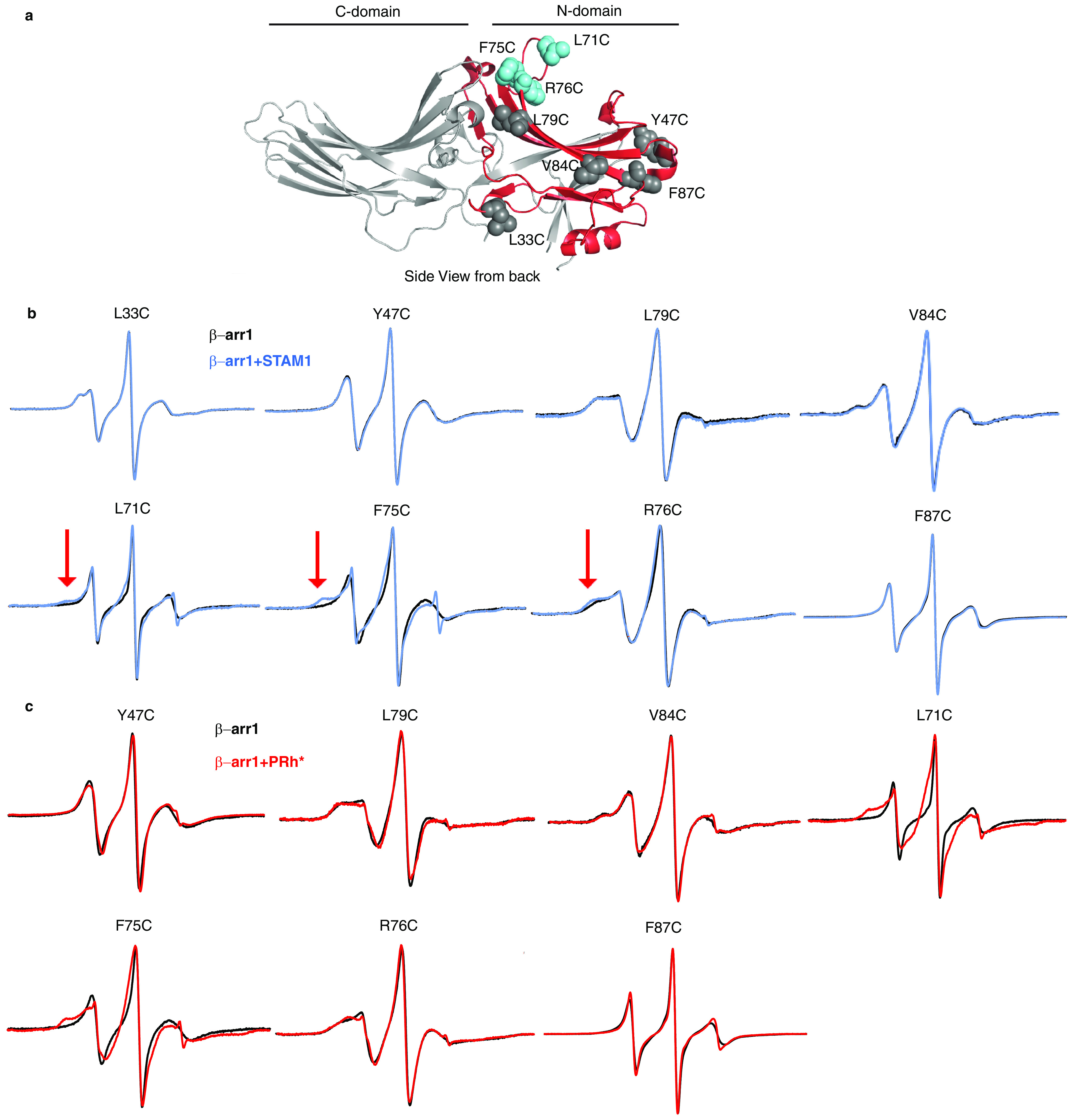
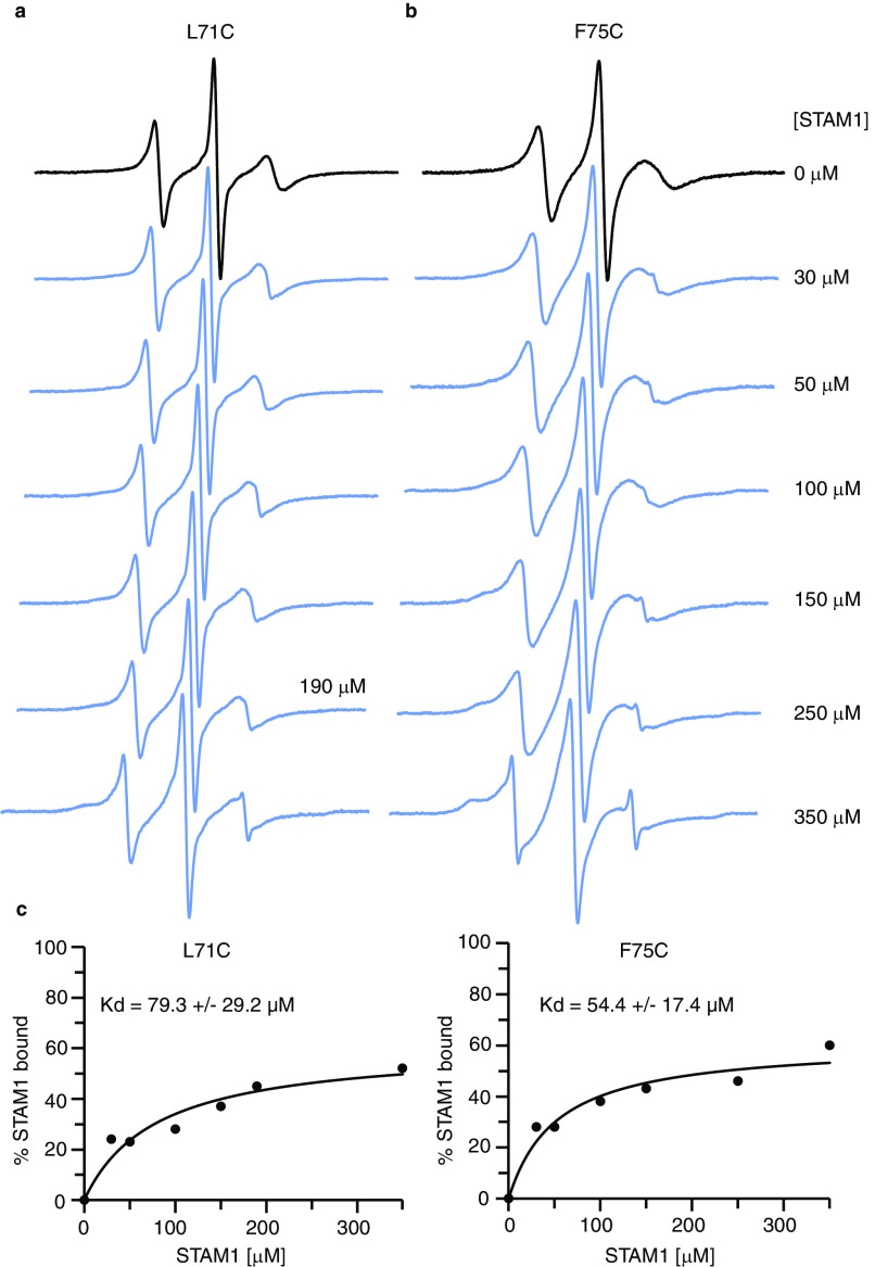
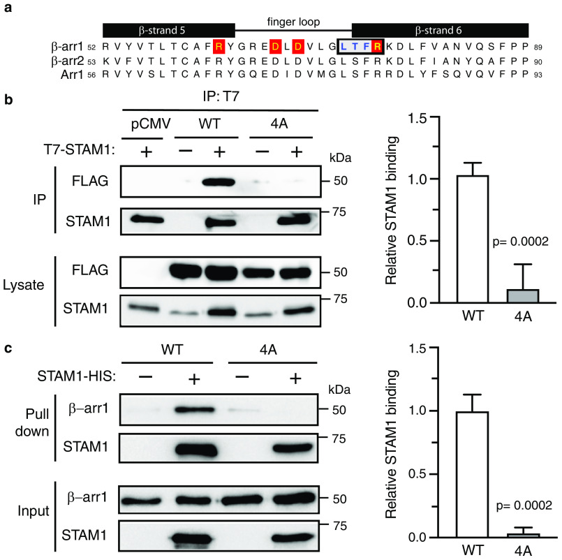
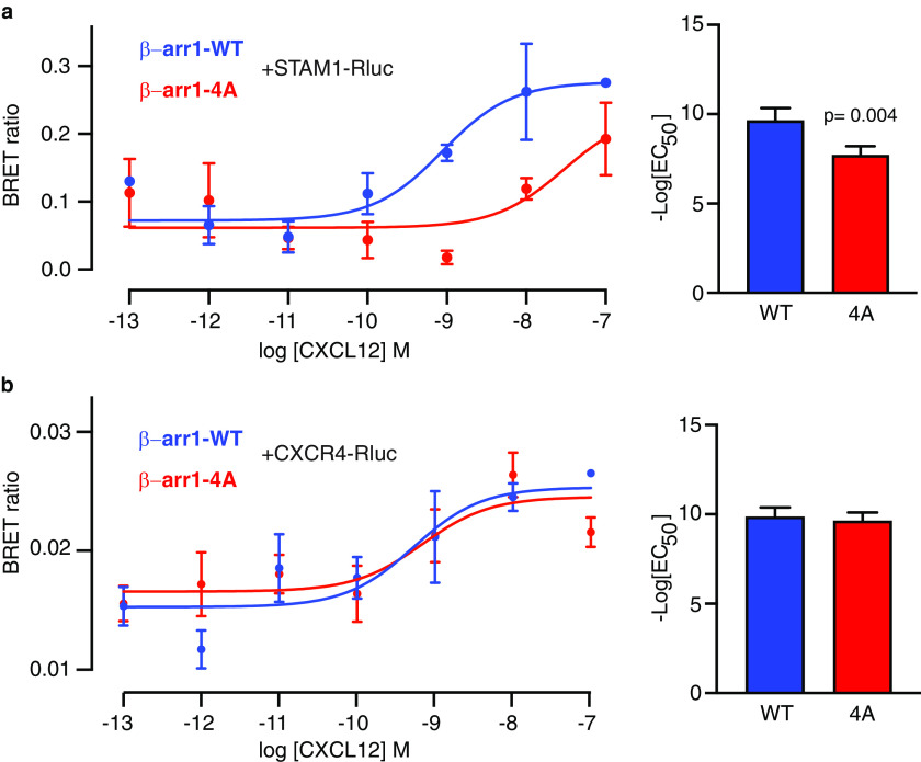
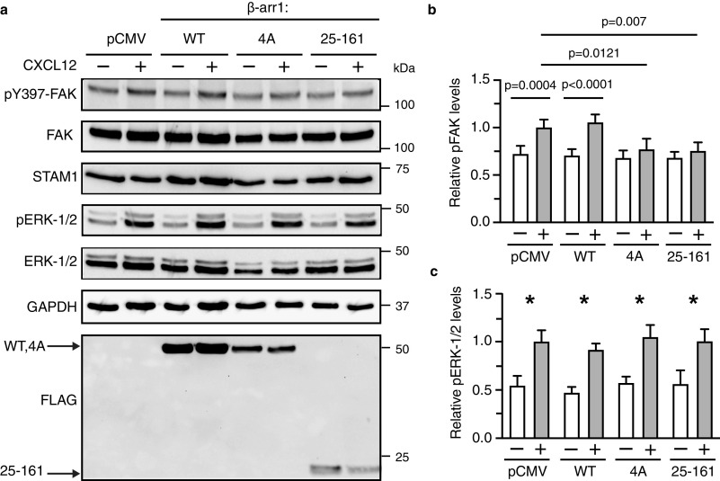
The multifaceted adaptor protein β-arr1 (β-arrestin1) promotes activation of focal adhesion kinase (FAK) by the chemokine receptor CXCR4, facilitating chemotaxis. This function of β-arr1 requires the assistance of the adaptor protein STAM1 (signal-transducing adaptor molecule 1) because disruption of the interaction between STAM1 and β-arr1 reduces CXCR4-mediated activation of FAK and chemotaxis. To begin to understand the mechanism by which β-arr1 together with STAM1 activates FAK, we used site-directed spin-labeling EPR spectroscopy-based studies coupled with bioluminescence resonance energy transfer-based cellular studies to show that STAM1 is recruited to activated β-arr1 by binding to a novel surface on β-arr1 at the base of the finger loop, at a site that is distinct from the receptor-binding site. Expression of a STAM1-deficient binding β-arr1 mutant that is still able to bind to CXCR4 significantly reduced CXCL12-induced activation of FAK but had no impact on ERK-1/2 activation. We provide evidence of a novel surface at the base of the finger loop that dictates non-GPCR interactions specifying β-arrestin-dependent signaling by a GPCR. This surface might represent a previously unidentified switch region that engages with effector molecules to drive β-arrestin signaling.

Keywords: CXC-chemokine receptor type 4 (CXCR4); G protein–coupled receptor (GPCR); PTK2 protein-tyrosine kinase 2 (PTK2); arrestin; arrestin signaling; bioluminescence resonance energy transfer (BRET); electron paramagnetic resonance (EPR); focal adhesion kinase (FAK).


Conflict of interest statement

Conflict of interest—The authors declare that they have no conflicts of interest with the contents of this article.

Figures

Figure 1.
Figure 1.
DEER measurements reveal conformational changes in β-arr1 induced by STAM1. a, structure of inactive β-arr1 (PDB code 1G4M (6)) highlighting the finger loop (orange), C-tail (cyan), and middle loop (blue). Residue pairs selected for DEER are connected by dotted lines. Each spin-labeled β-arr1 double cysteine mutant was analyzed by DEER with and without STAM1. Sample concentrations were as follows: 50 μm spin-labeled β-arr1 and 150 μm STAM1. b–d, DEER-derived distance probability distributions are shown as overlays for the free (gray area with black outline) and STAM1-bound (blue line) states. e–g, background-corrected dipolar evolution curves are plotted as overlays for the free (black line) and STAM1 bound states (blue line). Gray dots represent the raw data.
Figure 2.
Figure 2.
β-arr1 spin-labeled cysteine mutants maintained their ability to bind to STAM1, as assessed by pulldown experiments. Equimolar amounts of purified β-arr1-WT or indicated β-arr1 cysteine mutants (0.4 μm) were incubated with or without purified STAM1– (3 μm) for 20 min at 37˚C. Complexes were immobilized by incubation with Talon cobalt resin, and after washing, proteins were eluted in buffer with 200 mM imidazole and analyzed by immunoblotting. Representative blots are shown for β-arr1 double cysteine mutants (a) and single cysteine mutants (b and c).
Figure 3.
Figure 3.
Front surface N domain spin label mobility analysis of β-arr1 binding to STAM1 or GPCR. a, structure of inactive β-arr1 (PDB code 1G4M (6)) with spin label reporter sites 68 and 167 shown as red spheres. b, spectra for each spin-labeled β-arr1 in the absence (black) or presence of STAM1 (blue) are shown. Spectra obtained using 20 μm spin-labeled β-arr1 in the absence or presence of 400 μm STAM1 are overlaid. c, spectra in the absence or presence of 60 μm activated and phosphorylated rhodopsin (PRh*; red) in native disk membranes are overlaid. Spectra are normalized to the same center line height.
Figure 4.
Figure 4.
Back surface N domain spin label mobility analysis of β-arr1 binding to STAM1 or GPCR. a, Selected spin-labeled reporter sites are indicated in the β-arr1 crystal structure (PDB code 1G4M (6)). L33C, Y47C, L79C, V84C, and F87C are shown as gray spheres, and L71C, F75C, and R76C are shown as blue spheres. The previously determined primary STAM1-binding region on the N domain is shown in red. b, spectra for each spin-labeled β-arr1 in the absence (black) or presence of STAM1 (blue) are shown. Spectra obtained using 20 μm spin-labeled β-arr1 in the absence or presence of 400 μm STAM1 are overlaid. The red arrows indicate spectral line shape changes at the low field region caused by STAM1 binding. c, spectra in the absence or presence of 60 μm PRh* (red) in native disk membranes are overlaid. Spectra are normalized to the same center line height.
Figure 5.
Figure 5.
The affinity of STAM1 for β-arr1 determined by CW EPR spectroscopy. a and b, spectra obtained using 10 μm spin-labeled β-arr1 L71C (a) or F75C (b) in the presence of increasing concentrations of STAM1 are shown. c, graphs of STAM1 binding affinity curves for L71C and F75C. To quantify the unbound and bound population of spin-labeled β-arr1, EPR spectra were deconvoluted, and the resultant data points were fit to a one-site binding model.
Figure 6.
Figure 6.
Biochemical analysis of β-arr1 WT and finger-loop mutant (4A) binding to STAM1. a, sequence alignment of β-strand 5, finger loop, and β-strand 6 of bovine β-arr1 (residues 52–89), β-arr2 (residues 53–90), and visual arrestin (Arr1, residues 56–93). Boxed residues denote the four residues that were substituted to alanine residues in β-arr1. Letters highlighted in red denote residues that are identified to form two ionic locks to maintain the finger loop in an inactive bent conformation. b, cleared lysates from HEK293 cells co-transfected with T7–STAM1 and β-arr1–WT–FLAG, β-arr1–4A-FLAG, or empty vector (pCMV) were incubated with an anti-T7 antibody. Immunoprecipitates (IP) were analyzed by immunoblotting to detect bound β-arr1. Shown are representative immunoblots. Bound STAM1 was quantified by densitometric analysis. The graph represents the fraction of STAM1 bound to β-arr1–WT–FLAG from three independent experiments. The error bars represent the S.D. The data were analyzed by an unpaired t test. The p values are indicated. c, equimolar amounts of purified β-arr1–WT or β-arr1–4A (0.4 μm) were incubated with or without purified STAM1–HIS (3 μm) for 20 min at 37 °C. Complexes were captured by incubation with Talon cobalt resin, and after washing, the proteins were eluted in binding buffer supplemented with 200 mM imidazole and analyzed by immunoblotting. Representative blots are shown. Bound STAM1 was quantified by densitometric analysis. The graph represents the fraction of STAM1 bound to β-arr1 from three independent experiments. The error bars represent the S.D. The data were analyzed by an unpaired t test. The p values are indicated.
Figure 7.
Figure 7.
BRET analysis of β-arr1 binding to STAM1 or CXCR4 in cells. a and b, cells co-transfected with β-arr1–WT–GFP10 or β-arr1–4A–GFP10 with HA–CXCR4 and STAM1–Rluc (a) or CXCR4–Rluc (b) were stimulated with increasing concentrations of CXCL12 for 2 min before BRET measurements. DeepBlueC was added in the continuous presence of CXCL12, and BRET measurements were taken 30 min after the addition of the luciferase substrate. The data shown are from a representative experiment performed in triplicate ± S.D. The curves were fitted by nonlinear regression, assuming a single binding site (GraphPad Prism). The bar graphs represent the average EC50 values from four independent experiments. The error bars represent the S.D. The data were analyzed by unpaired t test. The p values are indicated.
Figure 8.
Figure 8.
The β-arr1–4A mutant attenuates CXCR4-promoted autophosphorylation of FAK in cells. a, HeLa cells transfected with empty vector (pCMV), FLAG-tagged β-arr1 WT, mutant (4A), or N domain fragment (residues 25–161) were stimulated with 30 nm CXCL12 for 15 min. Whole cell lysates were analyzed by immunoblotting for the indicated proteins. Representative immunoblots are shown. b and c, bars represent the average levels of pTyr397–FAK (pFAK) (b) or pERK-1/2 (c) relative to pCMV-transfected cells stimulated with CXCL12 from four independent experiments. The error bars represent the S.D. The data were analyzed by two-way analysis of variance followed by Bonferroni's multiple comparison test. The p values between indicated groups are shown (b), and the asterisks indicate p < 0.05 in each transfection condition.

References

    1. Carman C. V., and Benovic J. L. (1998) G-protein–coupled receptors: turn-ons and turn-offs. Curr. Opin. Neurobiol. 8, 335–344 10.1016/S0959-4388(98)80058-5 - DOI - PubMed
    1. Gurevich V. V., and Gurevich E. V. (2004) The molecular acrobatics of arrestin activation. Trends Pharmacol. Sci. 25, 105–111 10.1016/j.tips.2003.12.008 - DOI - PubMed
    1. Shenoy S. K., and Lefkowitz R. J. (2003) Multifaceted roles of β-arrestins in the regulation of seven-membrane-spanning receptor trafficking and signalling. Biochem. J. 375, 503–515 10.1042/BJ20031076 - DOI - PMC - PubMed
    1. DeWire S. M., Ahn S., Lefkowitz R. J., and Shenoy S. K. (2007) β-Arrestins and cell signaling. Annu. Rev. Physiol. 69, 483–510 10.1146/annurev.physiol.69.022405.154749 - DOI - PubMed
    1. Hirsch J. A., Schubert C., Gurevich V. V., and Sigler P. B. (1999) A Model for arrestin's regulation: the 2.8 Å crystal structure of visual arrestin. Cell 97, 257–269 10.1016/S0092-8674(00)80735-7 - DOI - PubMed

Publication types

MeSH terms

Associated data

LinkOut - more resources