. 2020 Sep 25;295(39):13651-13663.
doi: 10.1074/jbc.RA120.013275. Epub 2020 Aug 4.

Development of Noonan syndrome by deregulation of allosteric SOS autoactivation

Affiliations

Development of Noonan syndrome by deregulation of allosteric SOS autoactivation

Hope Gloria Umutesi et al. J Biol Chem. .

Abstract

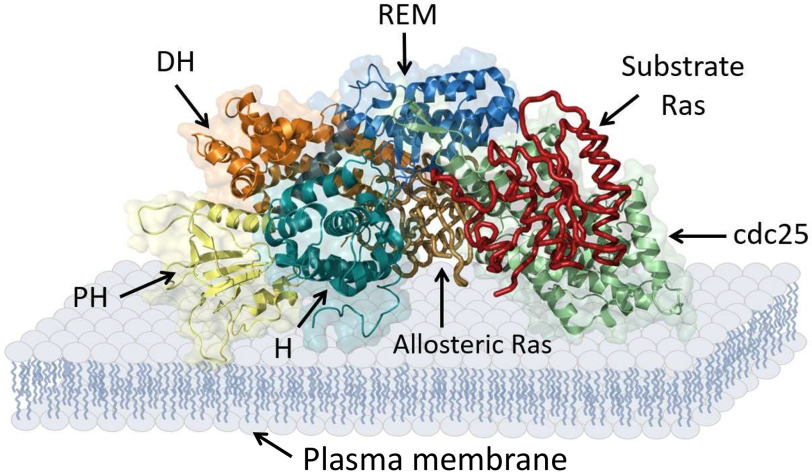
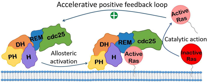
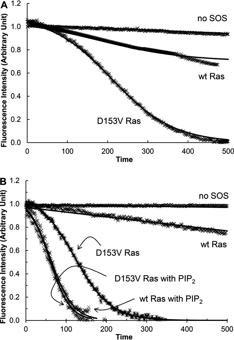
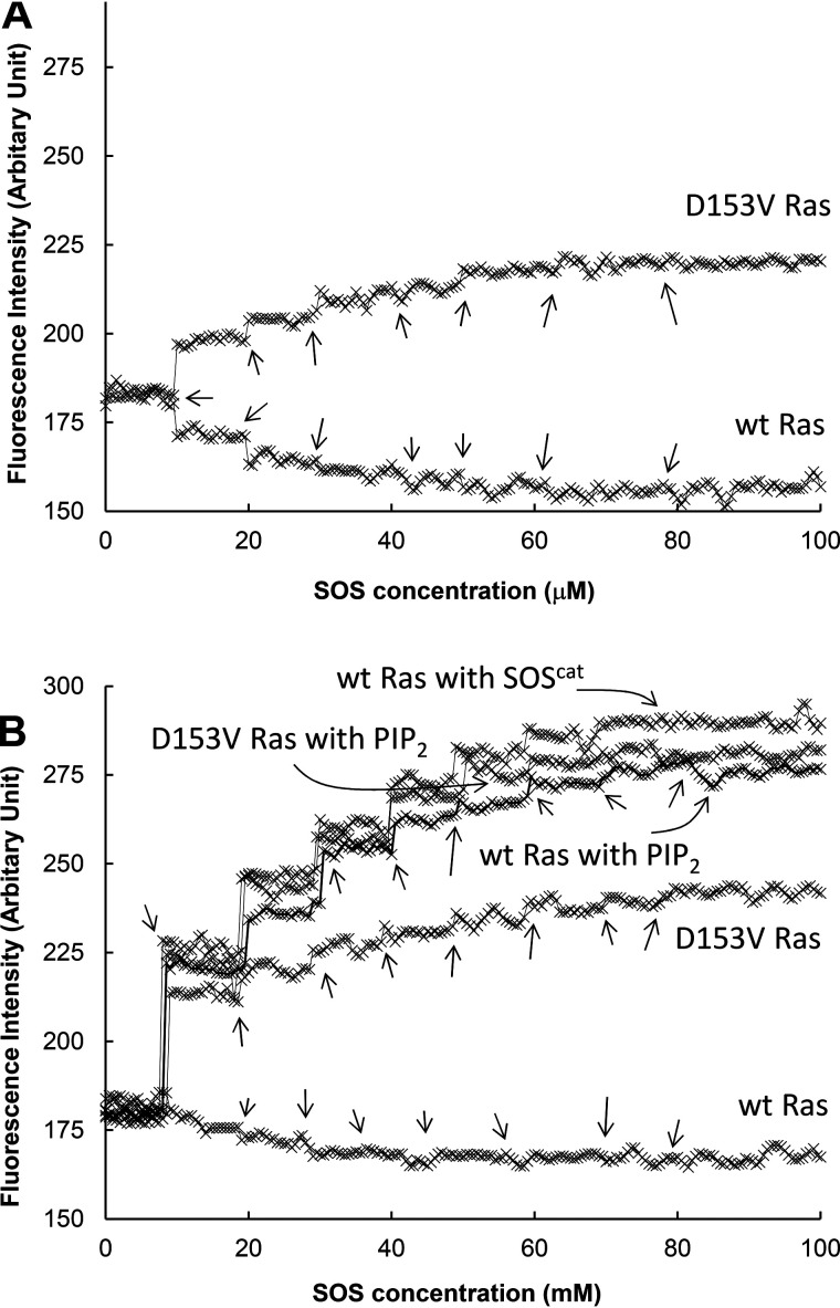
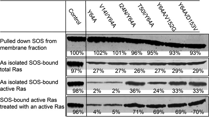
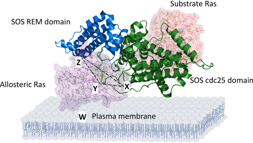
Ras family proteins play an essential role in several cellular functions, including growth, differentiation, and survival. The mechanism of action of Ras mutants in Costello syndrome and cancers has been identified, but the contribution of Ras mutants to Noonan syndrome, a genetic disorder that prevents normal development in various parts of the body, is unknown. Son of Sevenless (SOS) is a Ras guanine nucleotide exchange factor. In response to Ras-activating cell signaling, SOS autoinhibition is released and is followed by accelerative allosteric feedback autoactivation. Here, using mutagenesis-based kinetic and pulldown analyses, we show that Noonan syndrome Ras mutants I24N, T50I, V152G, and D153V deregulate the autoactivation of SOS to populate their active form. This previously unknown process has been linked so far only to the development of Noonan syndrome. In contrast, other Noonan syndrome Ras mutants-V14I, T58I, and G60E-populate their active form by deregulation of the previously documented Ras GTPase activities. We propose a novel mechanism responsible for the deregulation of SOS autoactivation, where I24N, T50I, V152G, and D153V Ras mutants evade SOS autoinhibition. Consequently, they are capable of forming a complex with the SOS allosteric site, thus aberrantly promoting SOS autoactivation, resulting in the population of active Ras mutants in cells. The results of this study elucidate the molecular mechanism of the Ras mutant-mediated development of Noonan syndrome.

Keywords: Noonan syndrome; Ras; Ras protein; SOS; Son of Sevenless; allosteric regulation; allostery; autoactivation; autoinhibition; catalysis; enzyme kinetics; guanine nucleotide exchange factor (GEF).


Conflict of interest statement

Conflict of interest—The authors declare that they have no conflicts of interest with the contents of this article.

Figures

Figure 1.
Figure 1.
SOS domains. Multimeric SOS domains with allosteric and substrate Ras-binding sites are shown. The scheme was generated using PyMOL with PDB entry 3KSY.
Figure 2.
Figure 2.
Model mechanism of SOS autoactivation. The allosteric SOS activation followed by the SOS catalytic action is illustrated. The action of the accelerative positive feedback loop between the SOS allostery and catalysis results in SOS autoactivation.
Figure 3.
Figure 3.
Autoactivation of SOS with WT and D153V mutant Ras. Fluorescence mant-based SOS autoactivation assays were performed with WT and D153V Ras in solution (A) and tethered to the lipid vesicle (B). Given that a reaction initiator is necessary to start SOS autoactivation, in all assays unless otherwise unnecessary, a GTP analog GppNHp-bound active Ras in solution or tethered to the lipid vesicle (30% of total Ras mole fraction) was added before the assay was initiated. Initiation was by the addition of SOSmemb-cat (1 μm) to an assay cuvette that contained Ras·mdGDP (1 μm) in solution or tethered to the lipid vesicle in the presence of excess GppNHp (1 mm) in an assay buffer. Once the assay was initiated, we monitored the change in the intensity of mant fluorescence over time as described under “Experimental procedures.” When necessary, Ras tethered to the PIP2-containing lipid vesicle, instead of Ras tethered to the lipid vesicle, was also used for the assay (B). The SOS autoactivation data fit was performed as described in the previous study (26) that gave the kauto values of SOSmemb-cat with WT and D153V Ras in solution as >1500 and 219 s, respectively. The kauto values of SOSmemb-cat with WT Ras tethered to the lipid vesicle (PIP2 lacking) and the PIP2-containing lipid vesicle were determined to be >1500 and 129 s, respectively. The kauto values of SOSmemb-cat with D153V Ras tethered to the lipid vesicle (that lacks PIP2) and the PIP2-containing lipid vesicle were determined to be 484 and 133 s, respectively. For all analyses, triplicate experiments were performed, and the graphic figures close to the average values are shown.
Figure 4.
Figure 4.
Fraction occupancies of SOS with WT Ras and various Ras mutants under the SOS autoinhibition conditions. SOSmemb-cat was titrated with Y64A and Y64A Ras (i.e. Y64A/D153V Ras) as described under “Experimental procedures.” For this titration, SOSmemb-cat was the titrant, whereas Ras·mdGDP in solution (A) as well as Ras·mdGDP tethered to the lipid vesicle and tethered to the PIP2-containing lipid vesicle (B) were receptors. The titration was performed by adding the titrant, as indicated with arrows, to the receptor repeatedly until the increase in mant fluorescence ended. As a control, SOScat was also used instead of SOSmemb-cat for the titration with Y64A Ras tethered to the lipid vesicle. The fluorescence-based titration results of these Ras proteins with SOSmemb-cat were normalized against the fluorescence-based titration results of Y64A Ras tethered to the lipid vesicle with SOScat. The normalized fluorescent intensities were plotted against the titrant concentrations of SOS and then fitted to saturation kinetics that gave the maximal fraction occupancy (Bmax) of the SOSmemb-cat allosteric site with Y64A and Y64A/D153V Ras in solution as 28 and 29%, respectively. The Bmax values of the SOSmemb-cat allosteric site with Y64A and Y64A/D153V Ras tethered to the lipid vesicle were determined to be 22 and 17%, respectively. The Bmax values of the SOSmemb-cat allosteric site with Y64A and Y64A/D153V Ras tethered to the lipid vesicle were determined to be 93 and 88%, respectively. All these reported values were averages of the three independent measurements, and the S.D. values are less than 15% of the value reported.
Figure 5.
Figure 5.
The SOS allosteric site–binding interactions with Y64A Ras mutants under the autoinhibition conditions in cells. Y64A and V14I/Y64A Ras as well as I24N/Y64A, T50I/Y64A, Y64A/V152G, and Y64A/D153V are Ras proteins that are, respectively, versions of SOS allosteric site-specific WT and GTPase-affecting V14I Ras as well as GEF-affecting I24N, T50I, V152G, and D153V Ras proteins. These Ras proteins were expressed in HEK293T cells as described under “Experimental procedures.” The SOS in the membrane fraction was isolated. Then the Ras fractions bound to SOS and their activity were determined as described under “Experimental procedures.” A portion of isolated SOS was pretreated with the lipid vesicle-tethered active Y64A Ras proteins before analysis of the Ras fractions bound to SOS and their activity. For a control, SOSmemb-cat fully loaded with the lipid vesicle–tethered active Y64A Ras was used. All these cell-based analyses were performed three times, and the graphic figures close to the average densitometric analysis values are shown. The figure's embedded triplicate mean values of the results of the densitometric analysis are reported as relative densitometry values compared with the control SOSmemb-cat that was set at 100% as indicated. The S.D. values of these measurements are less than 15% of the values noted.
Figure 6.
Figure 6.
Critical Ras SOS allosteric binding interfaces. Ras in the allosteric site and the catalytic site of SOS are shown with space fill structures, whereas SOS is shown with a strand structure. The Ras SOS interfaces with X, Y, and Z residues that also are indicated with arrows. The triangle configuration of the X, Y, and Z interfaces also is shown with a dotted line. An additional W interface is noted below the Ras SOS complex. PyMOL with PDB entry 1NVV was used to produce this scheme.

References

    1. Bos J. L. (1989) ras oncogenes in human cancer: a review. Cancer Res. 49, 4682–4689 - PubMed
    1. Geyer M., and Wittinghofer A. (1997) GEFs, GAPs, GDIs and effectors: taking a closer (3D) look at the regulation of Ras-related GTP-binding proteins. Curr. Opin. Struct. Biol. 7, 786–792 10.1016/S0959-440X(97)80147-9 - DOI - PubMed
    1. Barbacid M. (1987) ras genes. Annu. Rev. Biochem. 56, 779–827 10.1146/annurev.bi.56.070187.004023 - DOI - PubMed
    1. Kinbara K., Goldfinger L. E., Hansen M., Chou F. L., and Ginsberg M. H. (2003) Ras GTPases: integrins' friends or foes? Nat. Rev. Mol. Cell Biol. 4, 767–776 10.1038/nrm1229 - DOI - PubMed
    1. Colicelli J. (2004) Human RAS superfamily proteins and related GTPases. Sci. STKE 2004, RE13 10.1126/stke.2502004re13 - DOI - PMC - PubMed

Publication types

Substances

LinkOut - more resources