. 2020 Oct 2;295(40):13838-13849.
doi: 10.1074/jbc.RA120.012801. Epub 2020 Aug 4.

AI26 inhibits the ADP-ribosylhydrolase ARH3 and suppresses DNA damage repair

Affiliations

AI26 inhibits the ADP-ribosylhydrolase ARH3 and suppresses DNA damage repair

Xiuhua Liu et al. J Biol Chem. .

Abstract

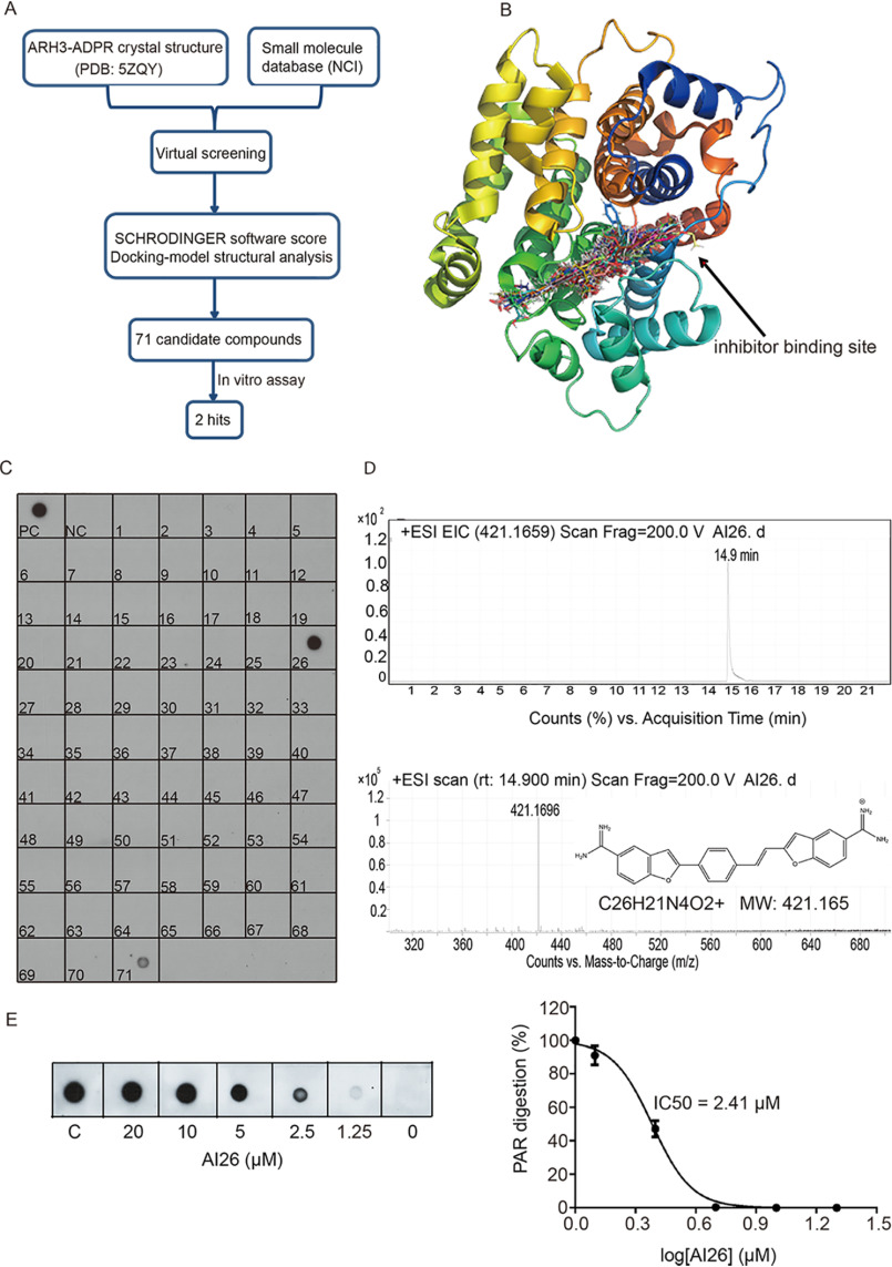
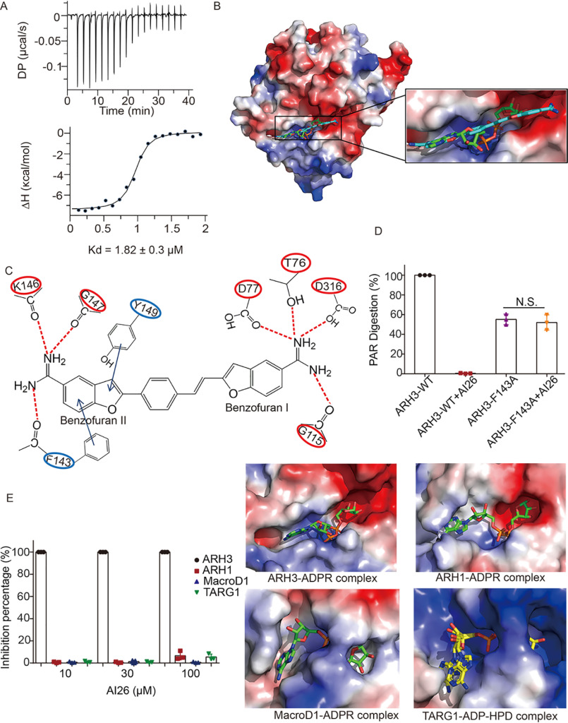
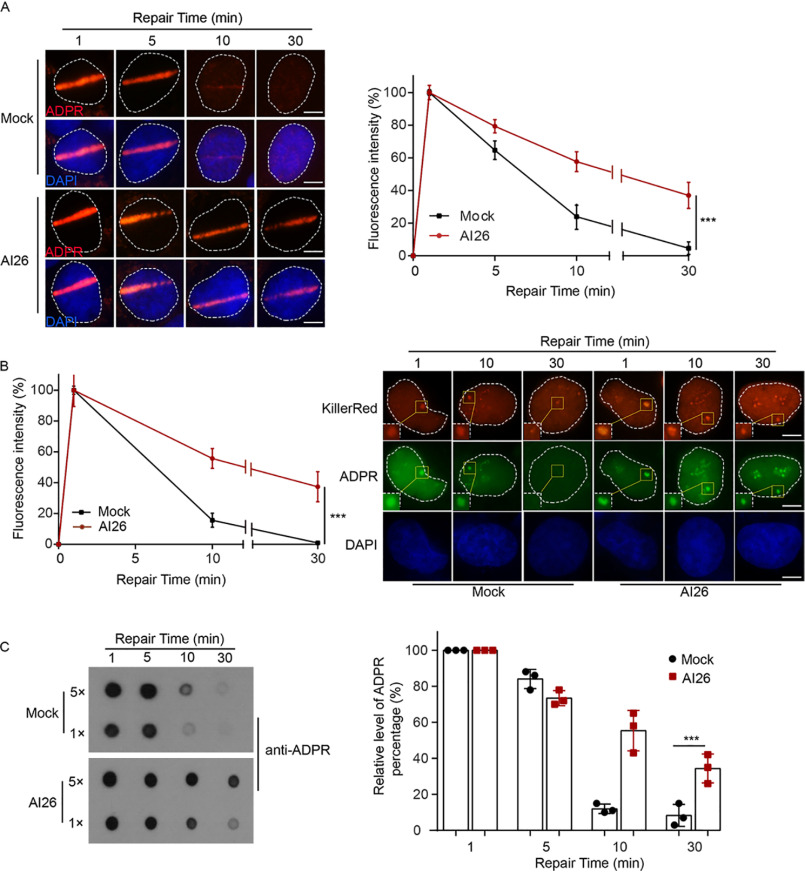
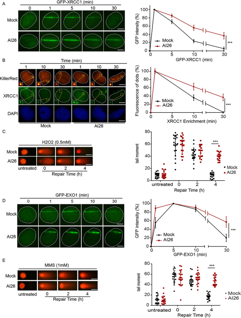
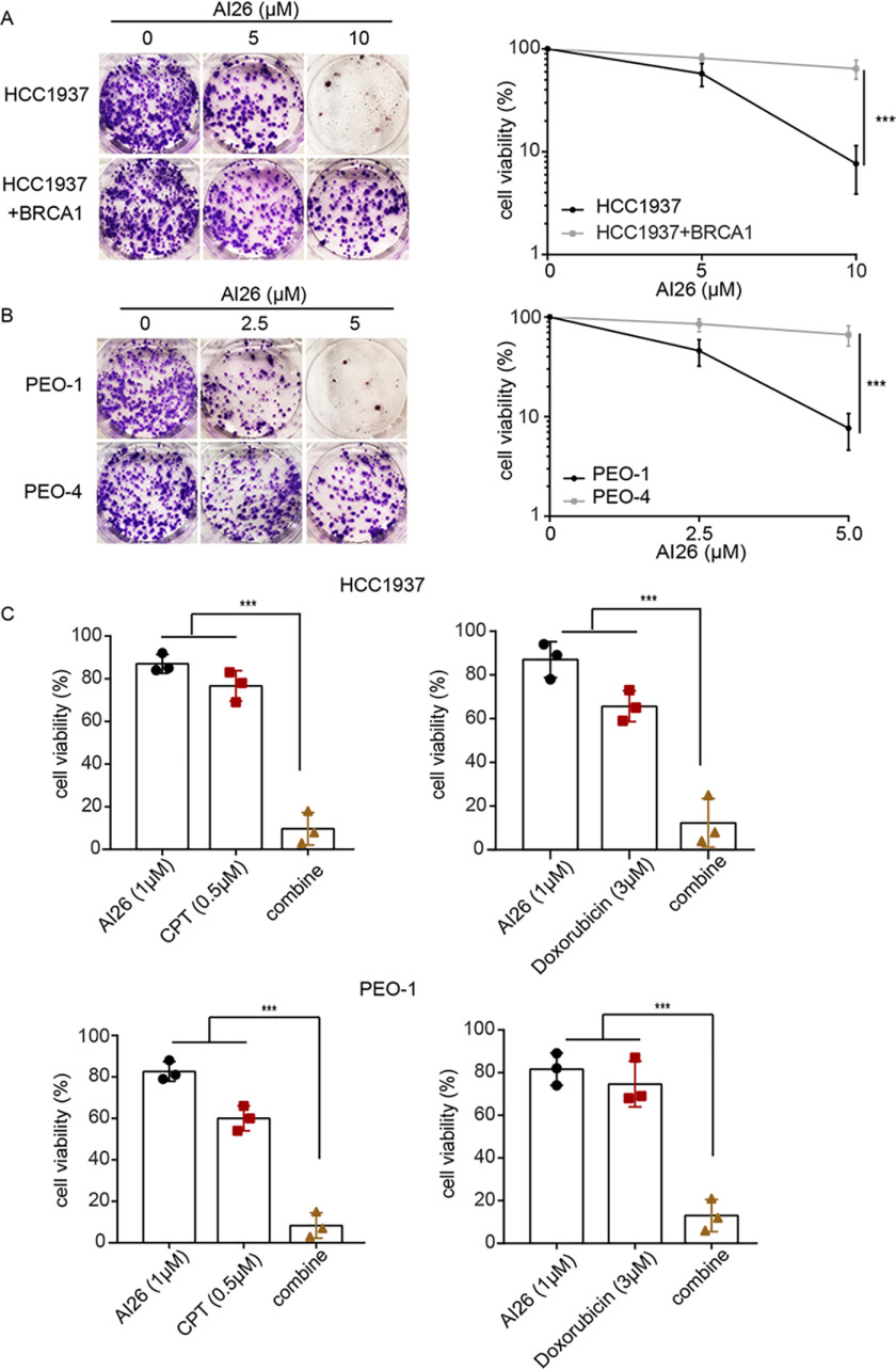
The ADP-ribosylhydrolase ARH3 plays a key role in DNA damage repair, digesting poly(ADP-ribose) and removing ADP-ribose from serine residues of the substrates. Specific inhibitors that selectively target ARH3 would be a useful tool to examine DNA damage repair, as well as a possible strategy for tumor suppression. However, efforts to date have not identified any suitable compounds. Here, we used in silico and biochemistry screening to search for ARH3 inhibitors. We discovered a small molecule compound named ARH3 inhibitor 26 (AI26) as, to our knowledge, the first ARH3 inhibitor. AI26 binds to the catalytic pocket of ARH3 and inhibits the enzymatic activity of ARH3 with an estimated IC50 of ∼2.41 μm in vitro Moreover, hydrolysis of DNA damage-induced ADP-ribosylation was clearly inhibited when cells were pretreated with AI26, leading to defects in DNA damage repair. In addition, tumor cells with DNA damage repair defects were hypersensitive to AI26 treatment, as well as combinations of AI26 and other DNA-damaging agents such as camptothecin and doxorubicin. Collectively, these results reveal not only a chemical probe to study ARH3-mediated DNA damage repair but also a chemotherapeutic strategy for tumor suppression.

Keywords: ADP-ribosylation; ARH3; DNA damage response; DNA repair; cancer therapy; dePARylation; inhibitor.


Conflict of interest statement

Conflict of interest—The authors declare that they have no conflicts of interest with the contents of this article.

Figures

Figure 1.
Figure 1.
Identification of ARH3 small-molecule inhibitors. A, a diagram of screening approach for the ARH3 inhibitors. B, the docking models of small molecules fitting into the catalytic pocket of ARH3. The structure of ARH3 is displayed in rainbow cartoon, and the small molecule compounds are shown in stick. C, biochemical screening of the ARH3 inhibitor. 71 candidates from NCI library were examined. ARH3 (1 μm) was incubated with PAR (10 μm) for 30 min at room temperature in the presence of each compound (100 μm). PAR digestion results were measured by dot-blotting assays with anti-PAR antibody. PC and NC indicate the positive control and negative control, respectively. D, AI26 was examined by LC–MS. From top to bottom, HPLC chromatogram, extracted ion chromatogram, and chemical structure of AI26. EIC, extracted ion chromatogram, ESI, electron spray ionization, MW, molecular weight. E, the estimated IC50 of AI26 was calculated from the in vitro PAR digestion assay (n = 3 independent experiments). AI26 with the indicated concentration was incubated with ARH3 (0.5 μm) and PAR (10 μm). The dot-blotting assays were performed with anti-PAR antibody to examine the in vitro PAR digestion. The IC50 value was determined using GraphPad Prism 7 software and the equation: log (inhibitor) versus normalized response – variable slope.
Figure 2.
Figure 2.
AI26 occupies the catalytic pocket of ARH3. A, the binding affinity between AI26 and recombinant ARH3 was measured using ITC. The Kd value was the average of three independent experiments shown in Fig. S1. B, a computer modeling of AI26 in the catalytic pocket of ARH3. The ARH3 is shown in electrostatic potential map, the AI26 is in cyan stick, and the ADPR is shown in green stick. C, schematic representation of the interaction between AI26 and ARH3. The dashed lines represent the predicted hydrogen bonds. The arrows represent the predicted π–π stacking interaction. D, AI26 does not suppress the enzymatic activity of the F143A mutant. AI26 (100 μm) was incubated with ARH3 (1 μm) or the F143A mutant (1 μm) and PAR (10 μm). The PAR digestion was measured by dot-blotting assays with anti-PAR antibody (n = 3 independent experiments). N.S., nonsignificant. E, AI26 specifically suppresses ARH3 but not other ADPR hydrolases. The relative ADPR digestion inhibition by AI26 treatment on each ADPR hydrolases was examined. The detailed approaches are included under “Experimental procedures.” Three independent experiments were performed on each inhibition assay, and the results are shown in a histogram (left panel). The catalytic pockets of ADPR hydrolases are shown in an electrostatic potential map. The ARH3–ADPR complex (PDB code 5ZQY), ARH1–ADPR complex (PDB code 6IUX), MacroD1–ADPR complex (PDB code 6LH4), and TARG1–ADP–HPD complex (PDB code 4J5R) are included in the right panels. ADPR is shown in green stick. The ADPR analog ADP-HPD is in yellow stick, and its adenine base exhibits two alternate conformations in the binding pocket.
Figure 3.
Figure 3.
AI26 suppresses hydrolysis of ADP-ribosylation in cells. A, AI26 treatment suppresses the degradation of ADPR at laser strips. U2OS cells were with or without 10 μm AI26 followed by laser microirradition. ADP-ribosylation at DNA lesions was examined by IF with anti-ADPR antibody. The accumulation kinetics of ADP-ribosylation at DNA lesions was examined in 50 cells (n = 3 independent experiments). The results were summarized as means ± S.D. ***, P < 0.001. The scale bar represents 5 μm. B, AI26 treatment suppresses the digestion of ADPR at oxidative lesions. U2OS cells expressing KillerRed were treated with or without 10 μm AI26 for 1 h, following white light treatment at 25 °C for 10 min. The location of DNA lesions were indicated by IF with anti-KillerRed antibody. The kinetics of ADP-ribosylation at DNA lesions was examined by IF with anti-ADPR antibody. The results are summarized from 50 cells (n = 3 independent experiments) and shown as means ± S.D. ***, P < 0.001. The scale bar represents 5 μm. C, 293T cells were pretreated with or without 10 μm AI26 for 1 h, followed by 0.5 mm H2O2 treatment for 5 min at 37 °C. The cellular levels of ADP-ribosylation were examined by dot-blotting assays with anti-ADPR antibody (left panel). The histograms represent the time course results from three independent experiments (right panel). The results were summarized as means ± S.D. ***, P < 0.001. DAPI, 4[prime],6[prime]-diamino-2-phenylindole.
Figure 4.
Figure 4.
AI26 treatment impairs DNA damage repair. A and B, AI26 treatment traps XRCC1 at DNA lesions. U2OS cells expressing GFP-XRCC1 were pretreated with or without 10 μm AI26 for 1 h, and the retention of XRCC1 at laser strip was examined with live cell imaging (A). U2OS cells expressing KillerRed were pretreated with or without 10 μm AI26 for 1 h, and the endogenous XRCC1 at the oxidative damage sites was examined by IF with anti-XRCC1 antibodies. The scale bar represents 5 μm. B, results are displayed as means ± S.D. from 50 cells (n = 3 independent experiments). ***, P < 0.001. The scale bar represents 5 μm. C, AI26 treatment suppresses SSBR. U2OS cells were pretreated with or without 10 μm AI26 for 1 h, followed by 0.5 mm H2O2 for 5 min. Alkaline comet assays were performed to examine the rate of SSBR in time course experiments. The tail moments were determined from at least 50 cells at each time point in each experiment, and three independent experiments were carried out. ***, P < 0.001. The scale bar represents 30 μm. D, AI26 treatment traps EXO1 at DNA lesions. U2OS cells expressing GFP-EXO1 were pretreated with or without 10 μm AI26 for 1 h, and the retention of EXO1 at laser strip was examined with live cell imaging. Three independent experiments were carried out. ***, P < 0.001. The scale bar represents 5 μm. E, AI26 treatment suppresses DSBR. U2OS cells were pretreated with or without 10 μm AI26 for 1 h, followed by 1 mm MMS for 30 min. Neutral comet assays were performed to examine the rate of DSBR in time course experiments. The scale bar represents 30 μm.
Figure 5.
Figure 5.
AI26 selectively kills tumor cells with DNA damage repair defects. A and B, AI26 suppresses the growth of tumor cells with BRCA1 or BRCA2 mutations. HCC1937, a BRCA1-null triple-negative breast cancer cell line (A), and PEO-1, a BRCA2-deificent ovarian cancer cell line (B), were treated with the indicated doses of AI26. BRCA1-reconstituted HCC1937 (HCC1937 + BRCA1) and PEO-4 (BRCA2-proficient ovarian cancer cell line) were used as controls, respectively. The cells were stained using crystal violet after 14 days of culture. Cell viability is displayed as means ± S.D. from three independent experiments. ***, P < 0.001. C, AI26 sensitizes tumor cells to camptothecin or doxorubicin. HCC1937 or PEO-1 cells were treated with the indicated dose of compounds. Cell viabilities from three independent experiments are displayed in the histograms. ***, P < 0.001. CPT, camptothecin.

References

    1. Rouleau M., Patel A., Hendzel M. J., Kaufmann S. H., and Poirier G. G. (2010) PARP inhibition: PARP1 and beyond. Nat. Rev. Cancer 10, 293–301 10.1038/nrc2812 - DOI - PMC - PubMed
    1. Gupte R., Liu Z., and Kraus W. L. (2017) PARPs and ADP-ribosylation: recent advances linking molecular functions to biological outcomes. Genes Dev. 31, 101–126 10.1101/gad.291518.116 - DOI - PMC - PubMed
    1. Hassa P. O., and Hottiger M. O. (2008) The diverse biological roles of mammalian PARPS, a small but powerful family of poly-ADP-ribose polymerases. Front. Biosci. 13, 3046–3082 10.2741/2909 - DOI - PubMed
    1. Aguiar R. C. T., Takeyama K., He C. Y., Kreinbrink K., and Shipp M. A. (2005) B-aggressive lymphoma family proteins have unique domains that modulate transcription and exhibit poly(ADP-ribose) polymerase activity. J. Biol. Chem. 280, 33756–33765 10.1074/jbc.M505408200 - DOI - PubMed
    1. Vyas S., Chesarone-Cataldo M., Todorova T., Huang Y. H., and Chang P. (2013) A systematic analysis of the PARP protein family identifies new functions critical for cell physiology. Nat. Commun. 4, 2240 10.1038/ncomms3240 - DOI - PMC - PubMed

Publication types

MeSH terms

LinkOut - more resources