. 2020 Aug 4;11(4):e01728-20.
doi: 10.1128/mBio.01728-20.

PPP6C Negatively Regulates STING-Dependent Innate Immune Responses

Affiliations

PPP6C Negatively Regulates STING-Dependent Innate Immune Responses

Guoxin Ni et al. mBio. .

Abstract

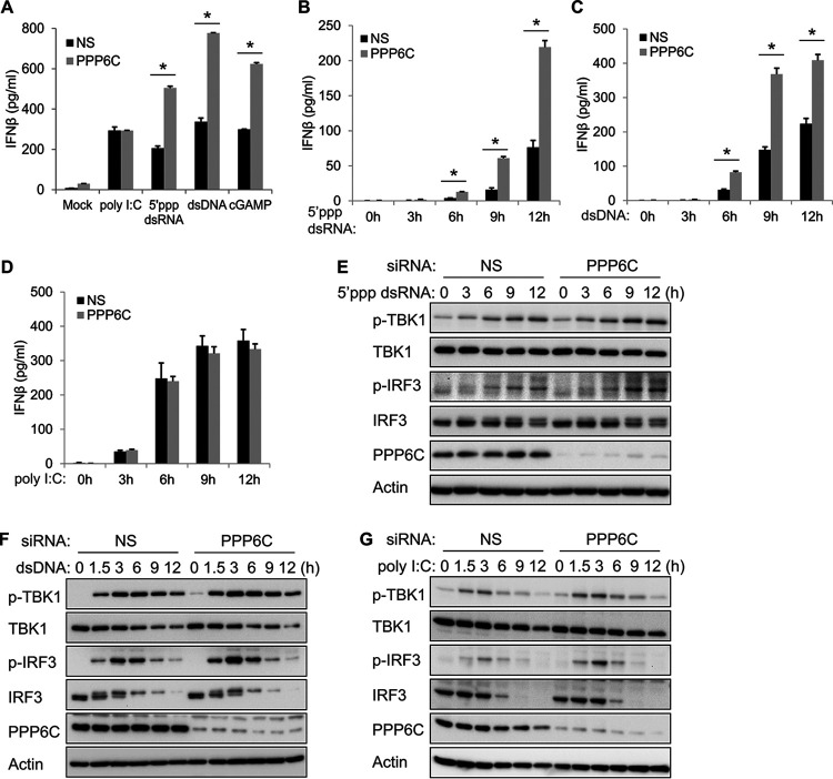
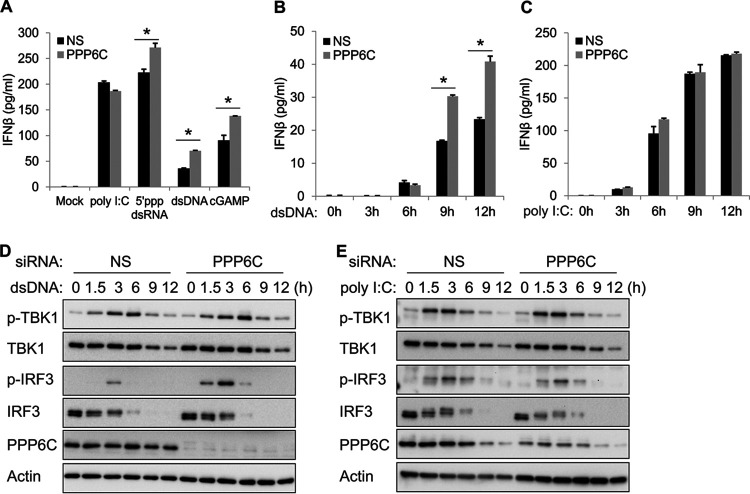
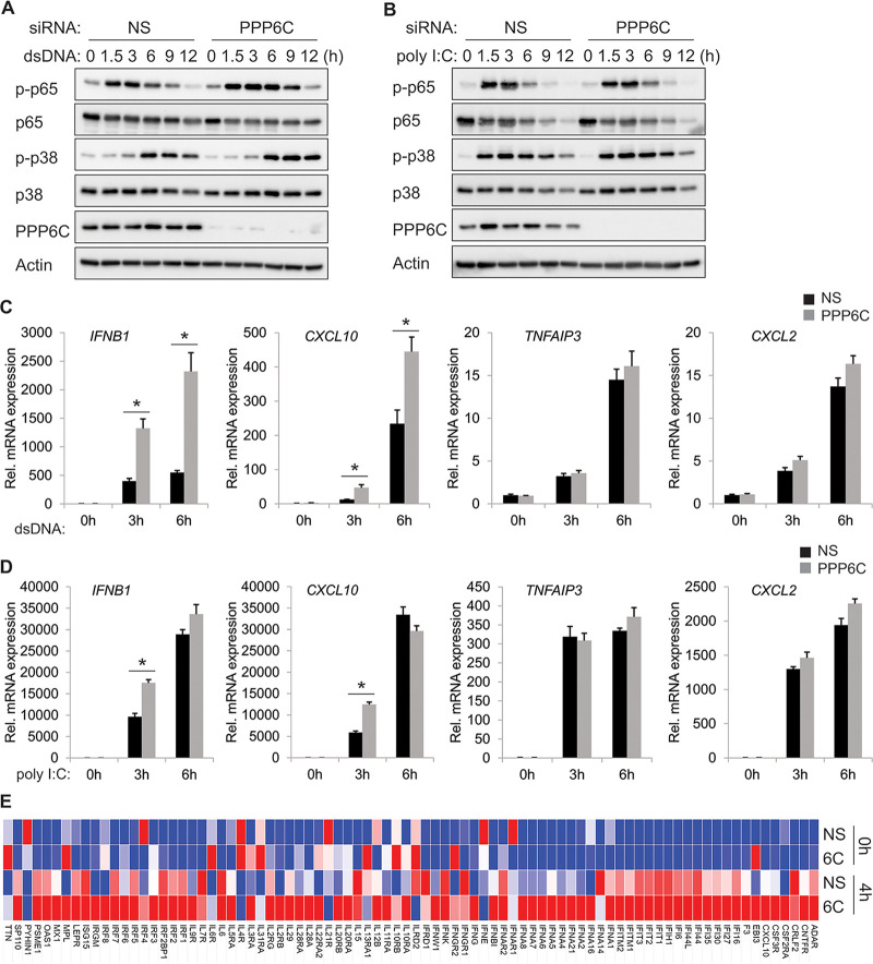
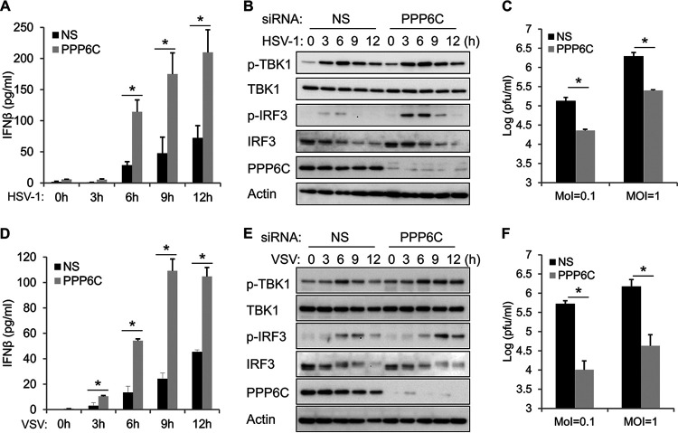
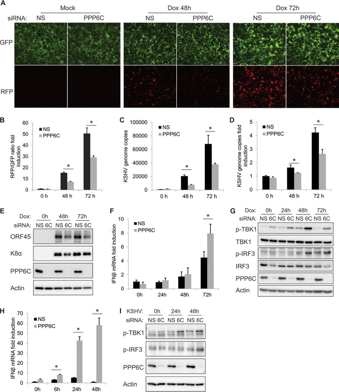
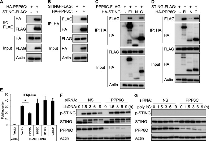
Stimulator of interferon genes (STING) is an essential adaptor protein of the innate DNA-sensing signaling pathway, which recognizes genomic DNA from invading pathogens to establish antiviral responses in host cells. STING activity is tightly regulated by several posttranslational modifications, including phosphorylation. However, specifically how the phosphorylation status of STING is modulated by kinases and phosphatases remains to be fully elucidated. In this study, we identified protein phosphatase 6 catalytic subunit (PPP6C) as a binding partner of Kaposi's sarcoma-associated herpesvirus (KSHV) open reading frame 48 (ORF48), which is a negative regulator of the cyclic GMP-AMP synthase (cGAS)-STING pathway. PPP6C depletion enhances double-stranded DNA (dsDNA)-induced and 5'ppp double-stranded RNA (dsRNA)-induced but not poly(I:C)-induced innate immune responses. PPP6C negatively regulates dsDNA-induced IRF3 activation but not NF-κB activation. Deficiency of PPP6C greatly inhibits the replication of herpes simplex virus 1 (HSV-1) and vesicular stomatitis virus (VSV) as well as the reactivation of KSHV, due to increased type I interferon production. We further demonstrated that PPP6C interacts with STING and that loss of PPP6C enhances STING phosphorylation. These data demonstrate the important role of PPP6C in regulating STING phosphorylation and activation, which provides an additional mechanism by which the host responds to viral infection.IMPORTANCE Cytosolic DNA, which usually comes from invading microbes, is a dangerous signal to the host. The cGAS-STING pathway is the major player that detects cytosolic DNA and then evokes the innate immune response. As an adaptor protein, STING plays a central role in controlling activation of the cGAS-STING pathway. Although transient activation of STING is essential to trigger the host defense during pathogen invasion, chronic STING activation has been shown to be associated with several autoinflammatory diseases. Here, we report that PPP6C negatively regulates the cGAS-STING pathway by removing STING phosphorylation, which is required for its activation. Dephosphorylation of STING by PPP6C helps prevent the sustained production of STING-dependent cytokines, which would otherwise lead to severe autoimmune disorders. This work provides additional mechanisms on the regulation of STING activity and might facilitate the development of novel therapeutics designed to prevent a variety of autoinflammatory disorders.

Keywords: HSV-1; KSHV; Kaposi's sarcoma-associated herpesvirus; PP6C; PPP6C; STING; VSV; herpes simplex virus; interferon; interferons; phosphatase; phosphorylation; protein phosphorylation; vesicular stomatitis virus.


Figures

FIG 1
FIG 1
PPP6C depletion enhances 5′ppp dsRNA- and dsDNA-induced but not poly(I:C)-induced innate immune responses. (A) EA.hy926 cells were transfected with control nonspecific (NS) or PPP6C siRNA for 72 h and then transfected with poly(I:C) (0.5 μg/ml), 5′ppp dsRNA (4 μg/ml), dsDNA (4 μg/ml), or cGAMP (4 μg/ml) for 16 h. IFN-β production in the supernatant was measured by ELISA. (B to D) EA.hy926 cells were transfected with NS or PPP6C siRNA for 72 h and then transfected with 5′ppp dsRNA (B), dsDNA (C), or poly(I:C) (D) for the indicated time periods. IFN-β production in the supernatant was measured by ELISA. (E to G) EA.hy926 cells were transfected with NS or PPP6C siRNA for 72 h and then transfected with 5′ppp dsRNA (E), dsDNA (F), or poly(I:C) (G) for the indicated times. Cell lysates were immunoblotted with the indicated antibodies. The data shown are representative of three independent experiments. Data in panels A to D are presented as mean + SD. *, P < 0.05 by Student's t test.
FIG 2
FIG 2
Deficiency of PPP6C enhances dsDNA- but not poly(I:C)-induced innate immune responses in primary HUVEC. (A) Primary HUVEC were transfected with NS or PPP6C siRNA for 72 h and then transfected with poly(I:C) (0.5 μg/ml), 5′ppp dsRNA (4 μg/ml), dsDNA (4 μg/ml), or cGAMP (4 μg/ml) for 16 h. IFN-β production in the supernatant was measured by ELISA. (B and C) Primary HUVEC were transfected with NS or PPP6C siRNA for 72 h and then transfected with dsDNA (B) or poly(I:C) (C) for the indicated time periods. IFN-β production in the supernatant was measured by ELISA. (D and E) Primary HUVEC were transfected with NS or PPP6C siRNA for 72 h and then transfected with dsDNA (D) or poly(I:C) (E) for the indicated time periods. Cell lysates were immunoblotted with the indicated antibodies. The data shown are representative of three independent experiments. Data in panels A to C are presented as mean + SD. *, P < 0.05 by Student's t test.
FIG 3
FIG 3
PPP6C regulates dsDNA-induced IRF3 activity but not NF-κB activity. (A and B) EA.hy926 cells were transfected with NS or PPP6C siRNA for 72 h and then transfected with dsDNA (A) or poly(I:C) (B) for the indicated times. Cell lysates were immunoblotted with the indicated antibodies. (C and D) EA.hy926 cells were transfected with NS or PPP6C siRNA for 72 h and then transfected with dsDNA (C) or poly(I:C) (D) for the indicated time periods. mRNA expression of the indicated genes was measured by real-time PCR, and fold changes were normalized to β-actin mRNA. (E) Heat map of the human interferon and receptor PCR array. EA.hy926 cells were transfected with NS or PPP6C siRNA for 72 h and then transfected with dsDNA for 0 or 4 h. RNA was extracted from the indicated samples, and mRNA levels of indicated genes were analyzed using the human interferon and receptor RT2 Profiler PCR array. mRNA levels of genes were normalized to β-actin. The heat map was generated using the web-based tool Morpheus (https://software.broadinstitute.org/morpheus/). The data shown are representative of three independent experiments, except in panel E, which is representative of two independent experiments. Data in panels C and D are presented as mean + SD. *, P < 0.05 by Student's t test. 6C, PPP6C.
FIG 4
FIG 4
Knockdown of PPP6C enhances HSV-1- and VSV-induced innate immune responses and inhibits virus replication. (A and B) EA.hy926 cells were transfected with NS or PPP6C siRNA for 72 h and then infected with HSV-1 (MOI = 10) for the indicated times. IFN-β production in the supernatant was measured by ELISA (A), and cell lysates were immunoblotted with the indicated antibodies (B). (C) EA.hy926 cells were transfected with NS or PPP6C siRNA for 72 h and then infected with HSV-1 (MOI = 0.1 or 1) for 24 h. The viral titer in the supernatant was measured by plaque assay. (D and E) EA.hy926 cells were transfected with NS or PPP6C siRNA for 72 h and then infected with VSV (MOI = 10) for the indicated time periods. IFN-β production in the supernatant was measured by ELISA (D), and cell lysates were immunoblotted with the indicated antibodies (E). (F) EA.hy926 cells were transfected with NS or PPP6C siRNA for 72 h and then infected with VSV (MOI = 0.1 or 1) for 24 h. The viral titer in the supernatant was measured by plaque assay. The data shown are representative of three independent experiments. Data in panels A, C, D, and F are presented as mean + SD. *, P < 0.05 by Student's t test.
FIG 5
FIG 5
Suppression of PPP6C inhibits KSHV lytic reactivation due to an increased innate immune response. iSLK.219 cells were transfected with NS siRNA or PPP6C siRNA for 48 h and then treated with doxycycline (Dox; 1 μg/ml). (A) GFP- and RFP-positive cells were imaged at 0, 48, and 72 h after Dox treatment, and a representative image of each sample is shown. (B) GFP and RFP fluorescence intensities were measured by a Clariostar plate reader, and the RFP/GFP ratio was calculated at the indicated time points. (C) KSHV genome copy numbers in the supernatants were measured by real-time PCR. (D) Relative KSHV genome copy numbers in the cells were measured by real-time PCR and normalized to β-actin. (E) Cell lysates were collected at the indicated time points, and immunoblots were performed with the indicated antibodies. (F) IFN-β mRNA fold induction was measured by real-time PCR and normalized to β-actin mRNA. (G) Cell lysates were collected at the indicated time points, and immunoblots were performed with the indicated antibodies. (H and I) EA.hy926 cells were transfected with NS or PPP6C siRNA for 72 h and then infected with KSHV for the indicated times. IFN-β mRNA fold induction was measured by real-time PCR (H), and cell lysates were immunoblotted with the indicated antibodies (I). The data shown are representative of three independent experiments. Data in panels B, C, D, F, and H are presented as mean + SD. *, P < 0.05 by Student's t test. 6C, PPP6C.
FIG 6
FIG 6
PPP6C interacts with STING and dephosphorylates STING. (A and B) HA-tagged STING and FLAG-tagged PPP6C were transfected individually or together into HEK293T cells for 30 h. Cell lysates were immunoprecipitated with anti-FLAG (A) or anti-HA (B) antibodies and then immunoblotted with the indicated antibodies. (C) FLAG-tagged PPP6C was cotransfected with full-length (FL), N-terminal (N; aa 1 to 195), or C-terminal (C; aa 181 to 379) HA-tagged STING into HEK293T cells for 30 h. Cell lysates were immunoprecipitated with anti-HA antibody and then immunoblotted with the indicated antibodies. (D) FLAG-tagged STING was cotransfected with full-length (FL), N-terminal (N; aa 1 to 160), or C-terminal (C; aa 151 to 305) HA-tagged PPP6C into HEK293T cells for 30 h. Cell lysates were immunoprecipitated with anti-HA antibody and then immunoblotted with the indicated antibodies. (E) HEK293T cells were cotransfected as indicated with IFN-β-luc, pRL-TK, cGAS and STING plasmids along with plasmids expressing wild-type PPP6C or the indicated PPP6C mutants. Luciferase activity was measured after 30 h. (F and G) EA.hy926 cells were transfected with NS or PPP6C siRNA for 72 h and then transfected with dsDNA (F) or poly(I:C) (G) for the indicated times. Cell lysates were immunoblotted with the indicated antibodies. The data shown are representative of three independent experiments. Data in panel E are presented as mean + SD. *, P < 0.05 by Student's t test.

References

    1. Kawasaki T, Kawai T, Akira S. 2011. Recognition of nucleic acids by pattern‐recognition receptors and its relevance in autoimmunity. Immunol Rev 243:61–73. doi: 10.1111/j.1600-065X.2011.01048.x. - DOI - PMC - PubMed
    1. Wu J, Chen ZJ. 2014. Innate immune sensing and signaling of cytosolic nucleic acids. Annu Rev Immunol 32:461–488. doi: 10.1146/annurev-immunol-032713-120156. - DOI - PubMed
    1. Sun L, Wu J, Du F, Chen X, Chen ZJ. 2013. Cyclic GMP-AMP synthase is a cytosolic DNA sensor that activates the type I interferon pathway. Science 339:786–791. doi: 10.1126/science.1232458. - DOI - PMC - PubMed
    1. Wu J, Sun L, Chen X, Du F, Shi H, Chen C, Chen ZJ. 2013. Cyclic GMP-AMP is an endogenous second messenger in innate immune signaling by cytosolic DNA. Science 339:826–830. doi: 10.1126/science.1229963. - DOI - PMC - PubMed
    1. Rongvaux A, Jackson R, Harman CC, Li T, West AP, de Zoete MR, Wu Y, Yordy B, Lakhani SA, Kuan CY, Taniguchi T, Shadel GS, Chen ZJ, Iwasaki A, Flavell RA. 2014. Apoptotic caspases prevent the induction of type I interferons by mitochondrial DNA. Cell 159:1563–1577. doi: 10.1016/j.cell.2014.11.037. - DOI - PMC - PubMed

Publication types

MeSH terms

LinkOut - more resources