Coevolutionary Analysis Reveals a Conserved Dual Binding Interface between Extracytoplasmic Function σ Factors and Class I Anti-σ Factors
- PMID: 32753504
- PMCID: PMC7406223
- DOI: 10.1128/mSystems.00310-20
Coevolutionary Analysis Reveals a Conserved Dual Binding Interface between Extracytoplasmic Function σ Factors and Class I Anti-σ Factors
Abstract
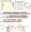
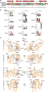
Extracytoplasmic function σ factors (ECFs) belong to the most abundant signal transduction mechanisms in bacteria. Among the diverse regulators of ECF activity, class I anti-σ factors are the most important signal transducers in response to internal and external stress conditions. Despite the conserved secondary structure of the class I anti-σ factor domain (ASDI) that binds and inhibits the ECF under noninducing conditions, the binding interface between ECFs and ASDIs is surprisingly variable between the published cocrystal structures. In this work, we provide a comprehensive computational analysis of the ASDI protein family and study the different contact themes between ECFs and ASDIs. To this end, we harness the coevolution of these diverse protein families and predict covarying amino acid residues as likely candidates of an interaction interface. As a result, we find two common binding interfaces linking the first alpha-helix of the ASDI to the DNA-binding region in the σ4 domain of the ECF, and the fourth alpha-helix of the ASDI to the RNA polymerase (RNAP)-binding region of the σ2 domain. The conservation of these two binding interfaces contrasts with the apparent quaternary structure diversity of the ECF/ASDI complexes, partially explaining the high specificity between cognate ECF and ASDI pairs. Furthermore, we suggest that the dual inhibition of RNAP- and DNA-binding interfaces is likely a universal feature of other ECF anti-σ factors, preventing the formation of nonfunctional trimeric complexes between σ/anti-σ factors and RNAP or DNA.IMPORTANCE In the bacterial world, extracytoplasmic function σ factors (ECFs) are the most widespread family of alternative σ factors, mediating many cellular responses to environmental cues, such as stress. This work uses a computational approach to investigate how these σ factors interact with class I anti-σ factors-the most abundant regulators of ECF activity. By comprehensively classifying the anti-σs into phylogenetic groups and by comparing this phylogeny to the one of the cognate ECFs, the study shows how these protein families have coevolved to maintain their interaction over evolutionary time. These results shed light on the common contact residues that link ECFs and anti-σs in different phylogenetic families and set the basis for the rational design of anti-σs to specifically target certain ECFs. This will help to prevent the cross talk between heterologous ECF/anti-σ pairs, allowing their use as orthogonal regulators for the construction of genetic circuits in synthetic biology.
Keywords: RNA polymerase; coevolutionary analysis; comparative genomics; computational biology; direct coupling analysis; gene regulation; transcription factors.
Copyright © 2020 Casas-Pastor et al.
Figures


Similar articles
-
Extracytoplasmic Function σ Factors as Tools for Coordinating Stress Responses.Int J Mol Sci. 2021 Apr 9;22(8):3900. doi: 10.3390/ijms22083900. Int J Mol Sci. 2021. PMID: 33918849 Free PMC article. Review.
-
Characterization of the Widely Distributed Novel ECF42 Group of Extracytoplasmic Function σ Factors in Streptomyces venezuelae.J Bacteriol. 2018 Oct 10;200(21):e00437-18. doi: 10.1128/JB.00437-18. Print 2018 Nov 1. J Bacteriol. 2018. PMID: 30126941 Free PMC article.
-
ECF σ factors with regulatory extensions: the one-component systems of the σ universe.Mol Microbiol. 2019 Aug;112(2):399-409. doi: 10.1111/mmi.14323. Epub 2019 Jun 26. Mol Microbiol. 2019. PMID: 31175685 Review.
-
The role of C-terminal extensions in controlling ECF σ factor activity in the widely conserved groups ECF41 and ECF42.Mol Microbiol. 2019 Aug;112(2):498-514. doi: 10.1111/mmi.14261. Epub 2019 Jun 11. Mol Microbiol. 2019. PMID: 30990934
-
Past, Present, and Future of Extracytoplasmic Function σ Factors: Distribution and Regulatory Diversity of the Third Pillar of Bacterial Signal Transduction.Annu Rev Microbiol. 2023 Sep 15;77:625-644. doi: 10.1146/annurev-micro-032221-024032. Epub 2023 Jul 12. Annu Rev Microbiol. 2023. PMID: 37437215 Review.
Cited by
-
PG1659 functions as anti-sigma factor to extracytoplasmic function sigma factor RpoE in Porphyromonas gingivalis W83.Mol Oral Microbiol. 2021 Feb;36(1):80-91. doi: 10.1111/omi.12329. Epub 2021 Jan 13. Mol Oral Microbiol. 2021. PMID: 33377315 Free PMC article.
-
Extracytoplasmic Function σ Factors as Tools for Coordinating Stress Responses.Int J Mol Sci. 2021 Apr 9;22(8):3900. doi: 10.3390/ijms22083900. Int J Mol Sci. 2021. PMID: 33918849 Free PMC article. Review.
References
-
- Lonetto MA, Brown KL, Rudd KE, Buttner MJ. 1994. Analysis of the Streptomyces coelicolor sigE gene reveals the existence of a subfamily of eubacterial RNA polymerase sigma factors involved in the regulation of extracytoplasmic functions. Proc Natl Acad Sci U S A 91:7573–7577. doi:10.1073/pnas.91.16.7573. - DOI - PMC - PubMed
LinkOut - more resources
Full Text Sources
Research Materials
