. 2020 Aug 4;10(1):13077.
doi: 10.1038/s41598-020-69805-2.

IL-17F induces inflammation, dysfunction and cell death in mouse islets

Affiliations

IL-17F induces inflammation, dysfunction and cell death in mouse islets

Tara Catterall et al. Sci Rep. .

Abstract

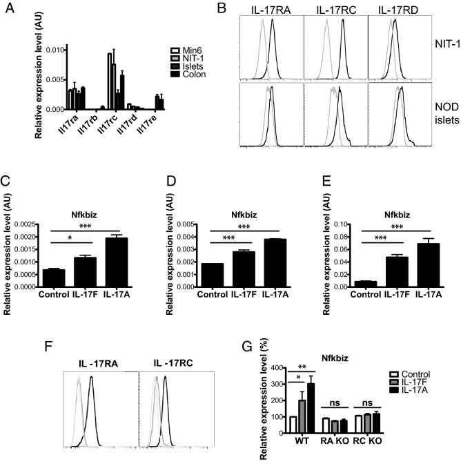
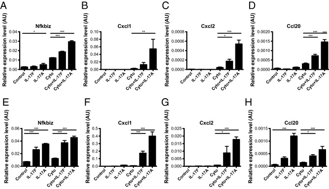
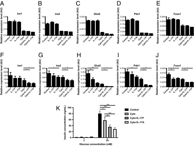
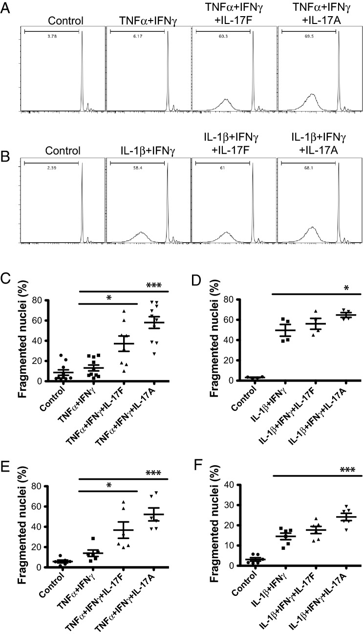
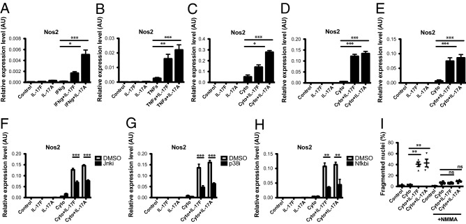
Type 17 immune responses, typified by the production of the cytokines IL-17A and IL-17F, have been implicated in the development of type 1 diabetes in animal models and human patients, however the underlying pathogenic mechanisms have not been clearly elucidated. While previous studies show that IL-17A enhances inflammatory gene expression and cell death in mouse β-cells and human islets, the function of IL-17F in pancreatic β-cells is completely untested to date. Here we show that IL-17F exhibits potent pathogenic effects in mouse β-cell lines and islets. IL-17F signals via the IL-17RA and -RC subunits in β-cells and in combination with other inflammatory cytokines induces expression of chemokine transcripts, suppresses the expression of β-cell identity genes and impairs glucose stimulated insulin secretion. Further IL-17F induces cell death in primary mouse islets. This occurs via Jnk, p38 and NF-κB dependent induction of Nos2 and is completely ablated in the presence of an inducible nitric oxide synthase (iNOS) inhibitor. Together these data indicate that IL-17F possesses similar pathogenic activities to IL-17A in mouse β-cell lines and islets and is likely to be a type 17 associated pathogenic factor in type 1 diabetes.


Conflict of interest statement

The authors declare no competing interests.

Figures

Figure 1
Figure 1
Pancreatic β-cells respond to IL-17F signals via IL-17RA and -RC. (A) Total RNA was isolated from Min6, NIT-1, pancreatic islets and colon tissue and the expression of Il17ra-e measured by Taqman quantitative RT-PCR. (B) NIT-1 cells and primary NOD islets were stained with antibodies against IL-17RA, RC and RD and cell surface expression quantified using flow cytometry (black line = antibody stained, grey line = unstained control). (C) NIT-1 cells were stimulated with IL-17F or IL-17A for 2 h and Nfkbiz expression measured by Taqman quantitative RT-PCR (n = 4 for all groups), p < 0.05 (*), p < 0.001 (***) One-Way ANOVA with Bonferonni’s Multiple Comparison test. (D) Min6 cells were stimulated with IL-17F or IL-17A for 4 h and Nfkbiz expression measured by Taqman quantitative RT-PCR (n = 4 for all groups), p < 0.001 (***) One-Way ANOVA with Bonferonni’s Multiple Comparison test. (E) NOD islets were stimulated with IL-17F or IL-17A for 4 h and Nfkbiz expression measured by Taqman quantitative RT-PCR (n = 3 for all groups), p < 0.001 (***) One-Way ANOVA with Bonferonni’s Multiple Comparison test. (F) IL-17RA knockout (left panel) and RC knockout (right panel) NIT-1 cells were generated with CRISPR/Cas9 and stained with antibodies against IL-17RA (left panel) and RC (right panel) (black = parental control cell line, grey = receptor knockout cell line, light grey = unstained control). (E) Wildtype, IL-17RA KO and IL-17RC KO NIT-1 cells were stimulated with IL-17F or IL-17A for 2 h and Nfkbiz expression measured by Taqman quantitative RT-PCR (n = 2 for all groups), p < 0.05 (*), p < 0.01 (**) Two-Way ANOVA with Bonferonni’s Multiple Comparison test.
Figure 2
Figure 2
IL-17F induces chemokine expression in mouse β-cell lines and islets. Min6 cells were stimulated with IL-17F or IL-17A in the presence of TNFα + IFNγ (Cyto) for 24 h and (A) Nfkbiz, (B) Cxcl1, (C) Cxcl2, (D) Ccl20 expression measured by Taqman quantitative RT-PCR (n = 4 for all groups), p < 0.05 (*), p < 0.01 (**), p < 0.001 (***) One-Way ANOVA with Bonferonni’s Multiple Comparison test. NOD islets were stimulated with IL-17F or IL-17A in the presence of TNFα + IFNγ (Cyto) for 24 h and (E) Nfkbiz, (F) Cxcl1, (G) Cxcl2, (H) Ccl20 expression measured by Taqman quantitative RT-PCR (n = 6 for all groups), p < 0.05 (*), p < 0.001 (***) One-Way ANOVA with Bonferonni’s Multiple Comparison test.
Figure 3
Figure 3
IL-17F suppresses expression of β-cell identity genes and glucose stimulated insulin secretion in mouse islets. Min6 cells were stimulated with IL-17F or IL-17A in the presence of TNFα + IFNγ (Cyto) for 24 h and (A) Ins1, (B) Ins2, (C) Glut2, (D) Pdx1 and (E) Foxo1 expression measured by Taqman quantitative RT-PCR (n = 4 for all groups). NOD islets were stimulated with IL-17F or IL-17A in the presence of TNFα + IFNγ (Cyto) for 24 h and (F) Ins1, (G) Ins2, (H) Glut2, (I) Pdx1 and (J) Foxo1 expression measured by Taqman quantitative RT-PCR (n = 6 for all groups), p < 0.05 (*), p < 0.01 (**), p < 0.001 (***) One-Way ANOVA with Bonferonni’s Multiple Comparison test. (K) NOD islets were stimulated with IL-17F or IL-17A in the presence of TNFα + IFNγ (Cyto) for 48 h and glucose stimulated insulin secretion assay performed (3 independent experiments, n = 4 for all groups), p < 0.05 (*), p < 0.01 (**), p < 0.001 (***) Two-Way ANOVA with Bonferonni’s Multiple Comparison test.
Figure 4
Figure 4
IL-17F induces cell death in mouse islets. NOD islets were stimulated with IL-17F or IL-17A in the presence of (A,C) TNFα + IFNγ or (B,D) IL-1β + IFNγ for 4 days and cell death quantified by DNA fragmentation (n = 5 for TNFα + IFNγ and n = 2 for IL-1β + IFNγ), p < 0.05 (*), p < 0.001 (***) One-Way ANOVA with Bonferonni’s Multiple Comparison test. (C) C57Bl/6 islets were stimulated with IL-17F or IL-17A in the presence of (C) TNFα + IFNγ or (D) IL-1β + IFNγ for 4 days and cell death quantified by DNA fragmentation (n = 3 for all groups), p < 0.05 (*), p < 0.001 (***) One-Way ANOVA with Bonferonni’s Multiple Comparison test.
Figure 5
Figure 5
IL-17F induced cell death is iNOS dependent in mouse islets. NOD islets were stimulated with IL-17F or IL-17A in the presence of (A) IFNγ or (B) TNFα for 4 h and Nos2 expression measured by Taqman quantitative RT-PCR (n = 5 for all groups), p < 0.05 (*), p < 0.01 (**) One-Way ANOVA with Bonferonni’s Multiple Comparison test. (C) Min6 cells, (D) NOD islets and (E) C57Bl/6 islets were stimulated with IL-17F or IL-17A in the presence of TNFα + IFNγ (Cyto) for 24 h and Nos2 expression measured by Taqman quantitative RT-PCR (n = 4 for Min6 cells and C57Bl/6 islets, n = 6 for NOD islets), p < 0.05 (*), p < 0.001 (***) One-Way ANOVA with Bonferonni’s Multiple Comparison test. (F) NOD islets were stimulated with IL-17F or IL-17A and TNFα + IFNγ (Cyto) +/− Jnk inhibitor (SP600125) at 50 μM for 24 h and Nos2 expression measured by Taqman quantitative RT-PCR (n = 2 for all groups), p < 0.001 (***) Two-Way ANOVA with Bonferonni’s Multiple Comparison test. (G) NOD islets were stimulated with IL-17F or IL-17A and TNFα + IFNγ (Cyto) ± p38 inhibitor (SB203580) at 20 μM for 24 h and Nos2 expression measured by Taqman quantitative RT-PCR (n = 2 for all groups), p < 0.001 (***) Two-Way ANOVA with Bonferonni’s Multiple Comparison test. (H) NOD islets were stimulated with IL-17F or IL-17A and TNFα + IFNγ (Cyto) ± NF-κB inhibitor (BAY11-7082) at 10 μM for 24 h and Nos2 expression measured by Taqman quantitative RT-PCR (n = 2 for all groups), p < 0.01 (**) Two-Way ANOVA with Bonferonni’s Multiple Comparison test. (I) NOD islets were stimulated with IL-17F or IL-17A in the presence of TNFα + IFNγ (Cyto) for 4 days ± NMMA (1 mM) and cell death quantified by DNA fragmentation (n = 2 for all groups), p < 0.01 (**) One-Way ANOVA with Bonferonni’s Multiple Comparison test.

References

    1. Graham KL, et al. Pathogenic mechanisms in type 1 diabetes: the Islet is both target and driver of disease. Rev. Diabet. Stud. 2012;9:148–168. doi: 10.1900/RDS.2012.9.148. - DOI - PMC - PubMed
    1. Mannering SI, Pathiraja V, Kay TW. The case for an autoimmune aetiology of type 1 diabetes. Clin. Exp. Immunol. 2016;183:8–15. doi: 10.1111/cei.12699. - DOI - PMC - PubMed
    1. Nordmann TM, et al. The role of inflammation in beta-cell dedifferentiation. Sci. Rep. 2017;7:6285. doi: 10.1038/s41598-017-06731-w. - DOI - PMC - PubMed
    1. Rui J, et al. Beta cells that resist immunological attack develop during progression of autoimmune diabetes in NOD mice. Cell Metab. 2017;25:727–738. doi: 10.1016/j.cmet.2017.01.005. - DOI - PMC - PubMed
    1. Thomas HE, McKenzie MD, Angstetra E, Campbell PD, Kay TW. Beta cell apoptosis in diabetes. Apoptosis. 2009;14:1389–1404. doi: 10.1007/s10495-009-0339-5. - DOI - PubMed

Publication types

MeSH terms

Substances