. 2020 Jul 1:15:4739-4752.
doi: 10.2147/IJN.S253990. eCollection 2020.

Engineering Thermo-pH Dual Responsive Hydrogel for Enhanced Tumor Accumulation, Penetration, and Chemo-Protein Combination Therapy

Affiliations

Engineering Thermo-pH Dual Responsive Hydrogel for Enhanced Tumor Accumulation, Penetration, and Chemo-Protein Combination Therapy

Xiuping Pang et al. Int J Nanomedicine. .

Abstract

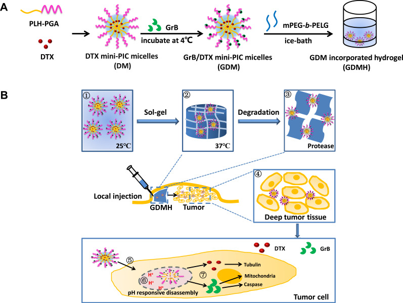
Purpose: Combined chemotherapeutic drug and protein drug has been a widely employed strategy for tumor treatment. To realize both tumor accumulation and deep tumor penetration for drugs with different pharmacokinetics, we propose a structure-transformable, thermo-pH dual responsive co-delivery system to co-load granzyme B/docetaxel (GrB/DTX).

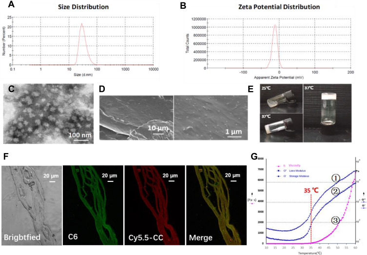
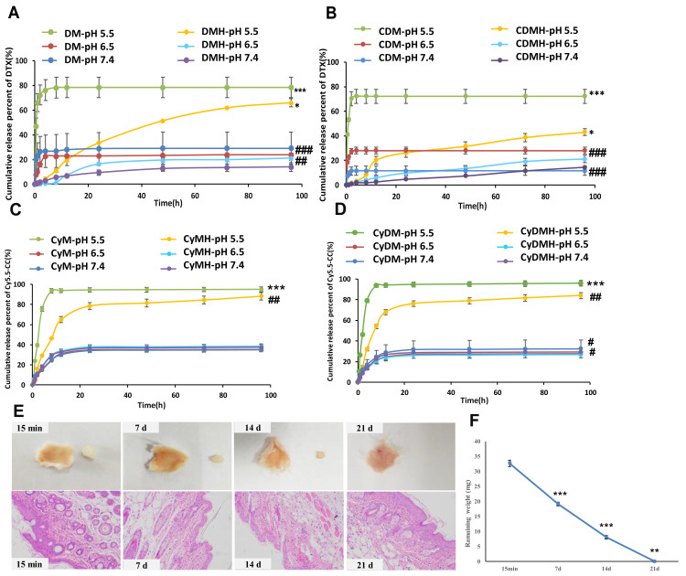
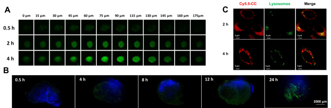
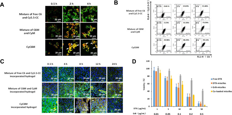
Methods: Thermo-sensitive hydrogels based on diblock copolymers (mPEG-b-PELG) were synthesized through ring opening polymerization. GrB/DTX mini micelles (GDM) was developed by co-loading these two drugs in pH-sensitive mini micelles, and the GDM-incorporated thermo-sensitive hydrogel (GDMH) was constructed. The thermo-induced gelation behavior of diblock copolymers and the physiochemical properties of GDMH were characterized. GDMH degradation and deep tumor penetration of released mini micelles were confirmed. The pH-sensitive disassembly and lysosomal escape abilities of released mini micelles were evaluated. In vitro cytotoxicity was studied using MTT assays and the in vivo antitumor efficacy study was evaluated in B16-bearing C57BL/6 mice.

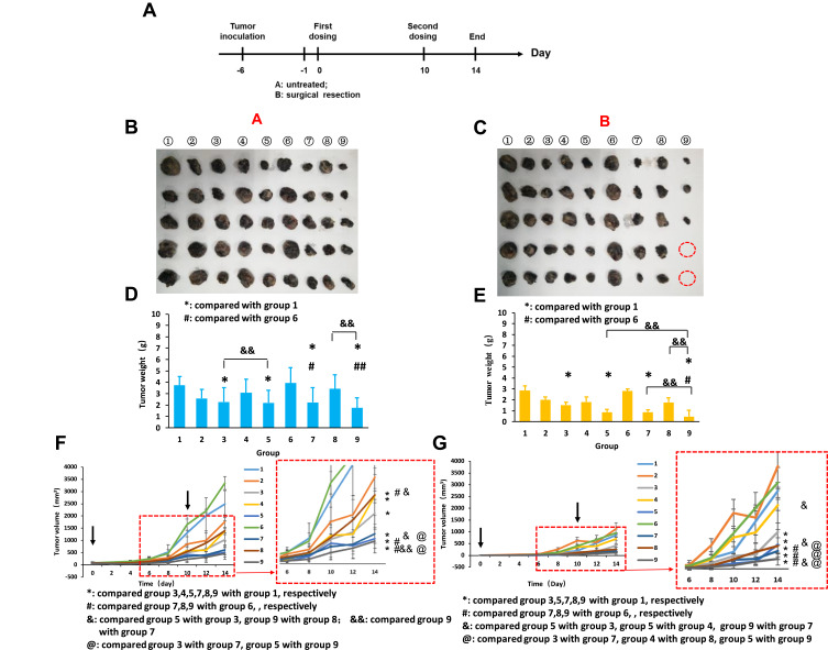
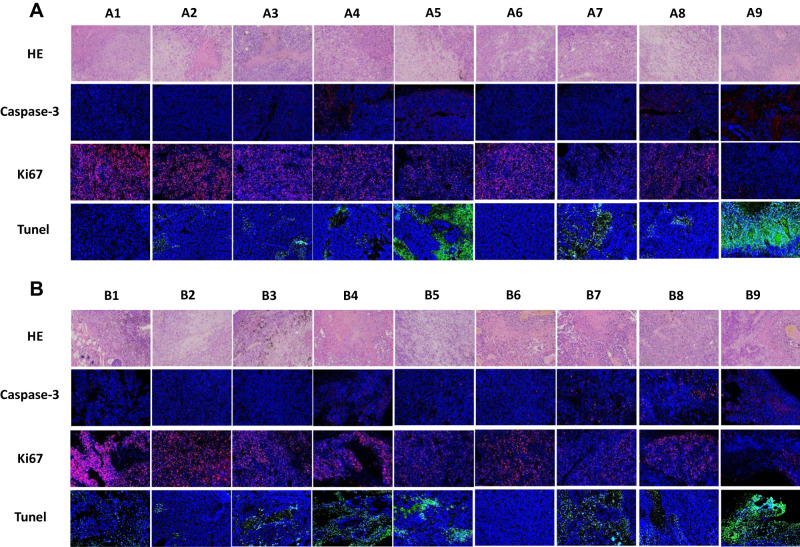
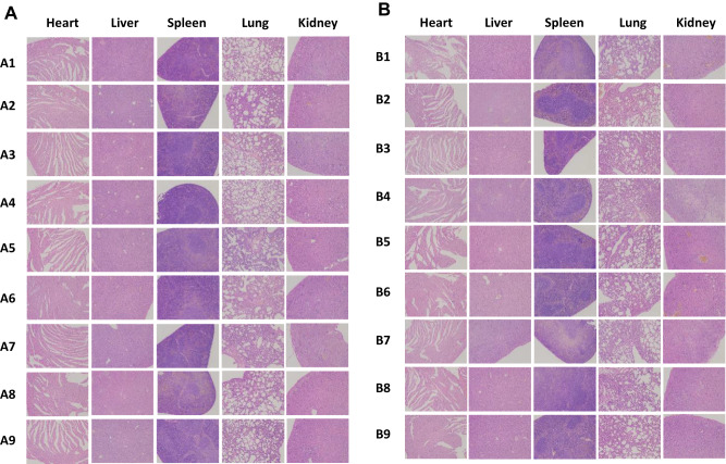
Results: GDMH was gelatinized at body temperature and can be degraded by proteinase to release mini micelles. The mini micelles incorporated in GDMH can achieve deep tumor penetration and escape from lysosomes to release GrB and DTX. MTT results showed that maximum synergistic antitumor efficacy of GrB and DTX was observed at mass ratio of 1:100. Our in vivo antitumor efficacy study showed that GDMH inhibited tumor growth in the subcutaneous tumor model and in the post-surgical recurrence model.

Conclusion: The smart-designed transformable GDMH can facilitate tumor accumulation, deep tumor penetration, and rapid drug release to achieve synergistic chemo-protein therapy.

Keywords: chemo-protein combination therapy; hydrogel; structure-transformable; thermo-pH dual responsive.


Conflict of interest statement

The authors declare that there are no conflicts of interest.

Figures

Figure 1
Figure 1
Schematic diagram of transformable injectable hydrogel as the local GrB/DTX co-delivery system for combination therapy. (A) The construction of structure-transformable hydrogel (GDMH). (B) The structure-transformable hydrogel could achieve sequentially responsive delivery: ①–③ The GrB/DTX pH-sensitive mini micelles (GDM) were incorporated into thermo-sensitive hydrogel (GDMH); ④ GDMH was degraded by proteinase and released mini micelles to achieve deep tumor penetration after be injected peritumorally; ⑤–⑦ Mini micelles disassembled and escaped from lysosomes via proton sponge effect to release GrB and DTX, then GrB and DTX played synergistic chemo-protein antitumor efficacy.
Figure 2
Figure 2
Characterizations of physical and chemical properties of CDM and CDMH. (A) Size distribution of CDM, for which the average size was 26.9±0.99 nm. (B) Zeta potential distribution of CDM, for which the average zeta potential was −14.53±0.58 mV. (C) Representative transmission electron microscope (TEM) images of CDM. (D) SEM images of lyophilized CDMH. (E) Photographs of the CDMH sol-to-gel transition with the increasing of temperature. (F) CLSM images of CyC6M-incorporated hydrogel, for which red, green and yellow color represent the fluorescence of Cy5.5-CC, C6 and the merged images, respectively. Based on the SEM images and CLSM, micelles could be homogeneously distributed in the hydrogel and could maintain the original structure for co-delivery. (G) Rheological properties of CDMH, for which G’ (①), G” (②) and η (③) represent the loss modulus, storage modulus and viscosity, for which 35°C could be the turning point of the CDMH system.
Figure 3
Figure 3
Characterizations of hydrogel for in vitro cumulative release profiles and in vivo degradation. (A) Cumulative release (%) behavior of DTX from DM and DMH at different pH values. (B) Cumulative release (%) behavior of DTX from CDM and CDMH at different pH values. (C) Cumulative release (%) behavior of Cy5.5-CC from CyM and CyMH at different pH values. (D) Cumulative release (%) behavior of Cy5.5-CC from CyDM and CyDMH at different pH values. (E) In situ hydrogel formation and in vivo hydrogel degradation. Histological images of the subcutaneous tissues surrounding the hydrogels (HE staining) (200×). (F) In vivo remaining weight for the in situ-forming mPEG-b-PELG (6.0 wt%) hydrogel. Notes: For (A and B), ***p<0.001, DM pH=5.5 versus pH=6.5 and 7.4; CDM pH=5.5 versus pH=6.5 and 7.4; ###p<0.001, DM pH= 7.4 versus DMH pH=7.4; CDM pH= 7.4 versus CDMH pH= 7.4; CDM pH= 6.5 versus CDMH pH=6.5; ##p<0.01, DM pH= 6.5 versus DMH pH= 6.5; *p< 0.05, DMH pH=5.5 versus pH=6.5 and 7.4; CDMH pH=5.5 versus pH=6.5 and 7.4. For (C and D), ***p<0.001, CyM pH=5.5 versus pH=6.5 and 7.4; CyDM pH=5.5 versus pH=6.5 and 7.4; ##p< 0.01, CyMH pH=5.5 versus pH=6.5 and 7.4; CyDMH pH=5.5 versus pH=6.5 and 7.4; #p< 0.05, CyDMH pH=6.5 versus CyDMH 7.4, and CyDM pH=6.5 versus CyDM 7.4; For (F), the significant difference was calculated by comparing the result of current weight with that of last weight, ***p<0.001, current weight (7 d) versus last weight (15 min), current weight (14 d) versus last weight (7 d); **p<0.01, current weight (21 d) versus last weight (14 d).
Figure 4
Figure 4
Characterizations of deep tumor penetration and lysosomal escape ability of mini micelles. (A) In vitro penetration of C6M into the B16-tumor sphere at different depths after incubation for 0.5,2 and 4 h, for which green color was from C6. (B) The sliced tumor tissues at different time points were recorded. The nuclei were stained with DAPI (blue), green color was from C6 which loaded by micelles-incorporated in hydrogel. Scale bar: 2000 μm. (C) Lysosomal escape of CyM on B16 cells at 2 and 4 h, in which red, green and blue colors represent CyM, lysosomes and the nucleus, respectively (magnification 40×, bar represents 5 μm).
Figure 5
Figure 5
Characterizations of in vitro and in vivo co-delivery ability and in vitro cytotoxicity. For co-delivery study, Cy5.5-CC was selected for taking the place of GrB and C6 was selected for taking the place of DTX, respectively. For (A and B), first group was mixture of free C6 and Cy5.5-CC, the second group was mixture of C6M and CyM, and the third group was CyC6M. For (C), first group was mixture of free C6 and Cy5.5-CC-incorporated hydrogel, the second group was mixture of C6M and CyM-incorporated hydrogel, and the third group was CyC6M-incorporated hydrogel. (A) CLSM images of cellular uptake at different time intervals on B16 cells, in which red, green and yellow colors represent Cy5.5-CC, C6 and the merged images, respectively (magnification 63×, bar represents 20 μm, white arrow marked the two fluorescent drugs were delivered to the same cell). (B) Co-delivery efficiency evaluation of the mini micelles by flow cytometric analysis in B16 cells. (C) CLSM images of tumor cyro-sections after the mice were administrated with mixture of free drug (Cy5.5-CC and C6)-incorporated hydrogel (Lane 1), mixture of Cy5.5-CC-micelles and C6-micelles-incorporated hydrogel (Lane 2), and CyC6MH (Lane 3), for which blue, red, green and yellow color represent the fluorescence of DAPI, Cy5.5-CC, C6 and the merged images, respectively (magnification 63×, bar represents 20 μm). (D) In vitro cytotoxicity study of different formulations against B16 cells. Data were given as mean ± SD (n = 3). The concentrations of DTX and GrB were shown in x-axis.
Figure 6
Figure 6
In vivo antitumor efficacy study. A: without surgery after tumor bearing; B: with surgery after tumor bearing. Nine groups in A or B including ① Normal Solution; ② Free DTX; ③ DM; ④ GM; ⑤ GDM; ⑥ Blank hydrogel; ⑦ DMH; ⑧ GMH; ⑨ GDMH. (A) Time points of in vivo anti-tumor activity study. (B) Photographs of tumors in A. (C) Photographs of tumors in B. (D) &&: p<0.01, group 5 versus group 3, group 9 versus group 8; ## :p<0.01, group 9 versus group 6; #: p<0.05, group 7 versus group 6 *: p<0.05, group 3 versus group 1, group 5 versus group 1, group 7 versus group 1, group 9 versus group 1. (E) &&: p<0.01, group 9 versus group 5, group 9 versus group 7, group 9 versus group 8; #: p<0.05, group 9 versus group 6; *: p<0.05, group 3 versus group 1, group 5 versus group 1, group 7 versus group 1, group 9 versus group 1. (F) Tumor volume of Group A. (G) Tumor volume of Group B. Data were given as mean ± SD (n=5).
Figure 7
Figure 7
Representative microscopy images of H&E (200×), Caspase-3 (200×), Ki67 (200×) and Tunel (200×) stained tumor histological sections after treatment with different formulations (A was for Group A, B was for Group B). ① Normal Solution; ② Free DTX; ③ DM; ④ GM; ⑤GDM; ⑥ Blank hydrogel; ⑦ DMH; ⑧ GMH; ⑨ GDMH.
Figure 8
Figure 8
In vivo security evaluation. Representative microscopy images of H&E stained histological sections on mice after treatment with different formulations (100×) (A was for Group A, B was for Group B). ① Normal Solution; ② Free DTX; ③ DM; ④ GM; ⑤ GDM; ⑥ Blank hydrogel; ⑦ DMH; ⑧ GMH; ⑨ GDMH.

References

    1. Enriquez-Navas PM, Wojtkowiak JW, Gatenby RA. Application of evolutionary principles to cancer therapy. Cancer Res. 2015;75(22):4675–4680. doi:10.1158/0008-5472.CAN-15-1337 - DOI - PMC - PubMed
    1. He CL, Tang ZH, Tian HY, Chen XS. Co-delivery of chemotherapeutics and proteins for synergistic therapy. Adv Drug Deliver Rev. 2016;98:64–76. doi:10.1016/j.addr.2015.10.021 - DOI - PubMed
    1. Hiller JG, Perry NJ, Poulogiannis G, Riedel B, Sloan EK. Perioperative events influence cancer recurrence risk after surgery. Nat Rev Clin Oncol. 2018;15(4):205–218. doi:10.1038/nrclinonc.2017.194 - DOI - PubMed
    1. Gao WP, Sun MM. Drug nanorods are potential new nanocarriers for intracellular protein delivery. Theranostics. 2018;8(14):3872–3873. doi:10.7150/thno.27815 - DOI - PMC - PubMed
    1. Schoonen L, van Hest JCM. Functionalization of protein-based nanocages for drug delivery applications. Nanoscale. 2014;6(13):7124–7141. doi:10.1039/C4NR00915K - DOI - PubMed