Review
. 2020 Jul 10:14:691.
doi: 10.3389/fnins.2020.00691. eCollection 2020.

Evolution of Orexin Neuropeptide System: Structure and Function

Affiliations
Review

Evolution of Orexin Neuropeptide System: Structure and Function

Shingo Soya et al. Front Neurosci. .

Abstract

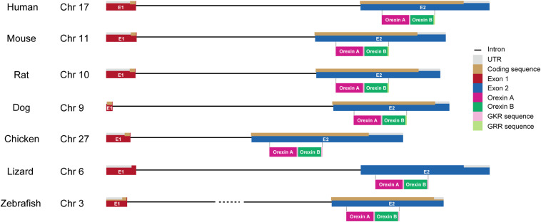
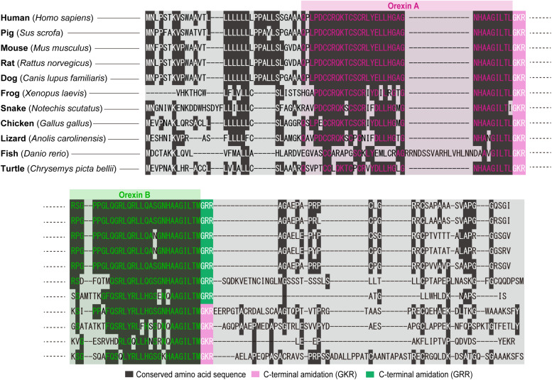
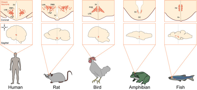
Orexins are hypothalamic neuropeptides that were initially identified in the rat brain as endogenous ligands for an (previously) orphan G-protein-coupled receptor (GPCR). They are multitasking peptides involved in many physiological functions, including regulation of feeding behavior, wakefulness and autonomic/neuroendocrine functions, and sleep/wakefulness states in mammals. There are two isopeptides of orexin, orexin A and orexin B, which are produced from a common precursor peptide, prepro-orexin. Structures of orexins, as well as orexin genes, are highly conserved throughout mammalian species, suggesting strong evolutionary pressure that maintains the structures. Their lengths and structure suggested that orexin B is the ancestral form of the orexin neuropeptide. In mammals, orexins bind to two subtypes of GPCRs, i.e., orexin 1 receptor (OX1R) and orexin 2 receptor (OX2R). Phylogenetically, the orexin system is present exclusively in vertebrates. In genomes of species outside mammals, there is only one orexin receptor, which is similar to OX2R, suggesting that OX2R is the prototype receptor for orexins. OX1R is likely to have evolved during early mammalian evolution. Orexin-producing neurons (orexin neurons) are mainly located in the lateral hypothalamic area (LHA) in mammals and are also found in hypothalamic regions in many other vertebrates. Orexins are likely to be closely related to the regulation of active, motivated behavior in many species. The orexin system seems to have evolved as a system that supports active and purposeful behavior which is closely related with wakefulness.

Keywords: OX1R; OX2R; hypothalamus; neuropeptide; orexin; vertebrate.


Figures

FIGURE 1
FIGURE 1
Gene structure of orexin in vertebrates. Chromosome numbers (Chr) in which the orexin gene is located are shown in each vertebrate. The drawing shows the length of gene structures of orexin including intron (black line), UTR (gray), coding sequences (brown), exon 1 (E1; red), and exon 2 (E2; blue) in different species (human, mouse, rat, dog, chicken, lizard, and zebrafish). Amino acid sequences of orexin A (magenta) and orexin B (dark green) with GKR (pink) and/or GRR (light green) amidation are also shown under their coding sequences.
FIGURE 2
FIGURE 2
Overview of amino acid sequences of orexins in vertebrates. The cartoon shows the sequences of amino acids coding orexins A and B in different species (human: Homo sapiens; pig: Sus scrofa; mouse: Mus musculus; rat: Rattus norvegicus; dog: Canis lupus familiaris; frog: Xenopus laevis; snake: Notechis scutatus; chicken: Gallus gallus; lizard: Anolis carolinensis; fish: Danio rerio; and turtle: Chrysemys picta bellii). Conserved amino acid sequence (dark gray), C-terminal amidation (GKR, pink), and C-terminal amidation (GRR, green) are highlighted. Amino acid sequences are aligned by ClustalW algorithm using MEGA software (Stecher et al., 2020).
FIGURE 3
FIGURE 3
Neuronal system of orexin is highly conserved in vertebrates. Upper squares show a coronal view of the brain focusing on orexin neurons (red dots) in different species [human (Peyron et al., 2000), rat (Nambu et al., 1999), bird (Ohkubo et al., 2002), amphibian (López et al., 2009), and fish (Appelbaum et al., 2009)]. Lower squares show a sagittal view of the brain focusing on the populations of orexin neurons in different species. Arrows show the anatomical orientation (A, anterior; P, posterior; D, dorsal; and V, ventral). DMH, dorsomedial nuclei of hypothalamus; LHA, lateral hypothalamic area; PaF, parafornical nucleus; 3V, third ventricle; PeF, perifornical hypothalamus; PHN, periventricular hypothalamic nucleus; VM, ventromedial thalamic nucleus; SC, suprachiasmatic nucleus; OC, optic chiasm; Hd, dorsal zone of periventricular hypothalamus; Hv, ventral zone of periventricular hypothalamus.

References

    1. Abbas M. G., Shoji H., Soya S., Hondo M., Miyakawa T., Sakurai T. (2015). Comprehensive behavioral analysis of Ox1r-/- mice showed implication of orexin receptor-1 in mood, anxiety and social behavior. Front. Behav. Neurosci. 9:324. 10.3389/fnbeh.2015.00324 - DOI - PMC - PubMed
    1. Amiya N., Amano M., Oka Y., Iigo M., Takahashi A., Yamamori K. (2007). Immunohistochemical localization of orexin/hypocretin-like immunoreactive peptides and melanin-concentrating hormone in the brain and pituitary of medaka. Neurosci. Lett. 427 16–21. 10.1016/j.neulet.2007.07.043 - DOI - PubMed
    1. Appelbaum L., Wang G. X., Maro G. S., Mori R., Tovin A., Marin W., et al. (2009). Sleep – wake regulation and hypocretin – melatonin interaction in zebrafish. Proc. Natl. Acad. Sci. U.S.A. 10 1–6. 10.1073/pnas.906637106 - DOI - PMC - PubMed
    1. Bentzley B. S., Aston-jones G. (2015). Orexin-1 receptor signaling increases motivation for cocaine-associated cues. Eur. J. Neurosci. 41 1149–1156. 10.1111/ejn.12866 - DOI - PMC - PubMed
    1. Burdakov D., Gerasimenko O., Verkhratsky A. (2005). Physiological changes in glucose differentially modulate the excitability of hypothalamic melanin-concentrating hormone and orexin neurons in situ. J. Neurosci. 25 2429–2433. 10.1523/JNEUROSCI.4925-04.2005 - DOI - PMC - PubMed

LinkOut - more resources