. 2020 Jul 11;10(19):8834-8850.
doi: 10.7150/thno.45158. eCollection 2020.

LXR activation potentiates sorafenib sensitivity in HCC by activating microRNA-378a transcription

Affiliations

LXR activation potentiates sorafenib sensitivity in HCC by activating microRNA-378a transcription

Zhongjie Lin et al. Theranostics. .

Abstract

Sorafenib resistance is a major obstacle to the treatment of advanced hepatocellular carcinoma (HCC). MicroRNAs (miRNAs) are multifunctional regulators of gene expression with profound impact for human disease. Therefore, better understanding of the biological mechanisms of abnormally expressed miRNAs is critical to discovering novel, promising therapeutic targets for HCC treatment. This study aimed to investigate the role of miR-378a-3p in the sorafenib resistance of HCC and elucidate the underlying molecular mechanisms. Methods: A novel hub miR-378a-3p was identified based on miRNA microarray and bioinformatics analysis. The abnormal expression of miR-378-3p was validated in different HCC patient cohorts and sorafenib-resistant (SR) HCC cell lines. The functional role of miR-378a-3p and its downstream and upstream regulatory machinery were investigated by gain-of-function and loss-of-function assays in vitro and in vivo. Interactions among miR-378a-3p, LXRα, and IGF1R were examined by a series of molecular biology experiments. Then, the clinical relevance of miR-378a-3p and its targets were evaluated in HCC samples. HCC patient-derived xenograft (PDX) model was used to assess the therapeutic value of LXRα and its downstream miR-378a-3p. Results: miR-378a-3p expression was frequently reduced in established sorafenib-resistant HCC cell lines. The decreased miR-378a-3p levels correlated with poor overall survival of HCC patients following sorafenib treatment. miR-378a-3p overexpression induced apoptosis in SR HCC cells, whereas miR-378a-3p silencing exerted the opposite effects. IGF1R was identified as a novel target of miR-378a-3p. Furthermore, the primary miR-378 level was not consistent with its precursor miRNA level in SR HCC cells, which was attributed to the downregulation of exportin5 (XPO5) and subsequently reduced nuclear export of precursor miR-378 and restrained maturation of miR-378-3p. In this context, we combined an agonist GW3965 of liver X receptor alpha (LXRα), which functioned as a transcription activator of miRNA-378a, and its activation re-sensitized sorafenib-resistant cells to sorafenib treatment in vitro and in vivo. Conclusions: Our finding suggested decreased expression of XPO5 prevents maturation of miR-378a-3p, which leaded to the overexpression of IGF-1R and counteracted the effects of sorafenib-induced apoptosis. LXRα was able to activate miRNA-378a-3p transcription in HCC cells and could be a potential combinable treatment strategy with sorafenib to suppress HCC progression.

Keywords: IGF1R; LXRα; XPO5; hepatocellular carcinoma; microRNA; sorafenib resistance.


Conflict of interest statement

Competing Interests: The authors have declared that no competing interest exists.

Figures

Figure 1
Figure 1
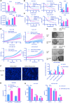
miR-378-3p expression is reduced in sorafenib-resistant cells and may participate in resistance development. (A) The process of the establishment of SR cells; (B) Cell viability was measured by CCK-8 assay at different concentration of sorafenib after 48 hours of dosing. The proliferation of resistant cells was all greater than that of their parental cells at different concentration of sorafenib; (C) Flow cytometry showed the apoptosis in sorafenib-resistant cells was less than parental cells; (D) Three sorafenib-resistant cells and parental cells were treated by sorafenib (10 μM). Colony-formation efficiency was carried out. Drug treatments were repeated every 3 days until colonies were visible; the emergency of sorafenib-resistant coloines was more than that of their parental cells; (E) miRNA sequencing of Huh7-SR and Huh7-WT showed a total of 39 miRNAs exhibited significantly different expression between the two groups; (F)Five miRNAs (miR-23b-3p, miR-342-3p, miR-708-3p, miR-797-3p, and miR-378a-3p, fold change >1.5 and p<0.01) were significantly reduced in Huh7-SR cells; (G) RT-qPCR evaluated the expression of the five candidates in all three resistant cell lines. The results confirmed that these five miRNAs were all downregulated in sorafenib-resistant cells. (H) HCC prognosis data obtained from KM-plotter showed patients with higher miR-378a-3p levels in cancer tissue had significantly better overall (HR=0.42; 95%CI: 0.25 to 0.71; P=2.0e-0.4) and disease-free survival (HR=0.53; 95%CI: 0.32 to 0.88; P=0.0069).
Figure 2
Figure 2
miR-378a-3p could sensitize cellular response to sorafenib. (A) The expression of miR-378a-3p in Huh7 cells transfected with miR-378a-3p mimic or miR-378a-3p inhibitor; (B,C) The proliferation of Huh-SR cells and HCCLM3-SR cells was all greater than that transfected with miR-378a-3p mimic at different concentration of sorafenib; Meanwhile, the proliferation of Huh7-WT cells and HCCLM3-WT cells which transfected with miR-378a-3p inhibitor was all greater than that Huh7-WT cells at different concentration of sorafenib; (D) Real-time cellular analysis showed Huh7-SR cells and HCCLM3-SR cells which transfected with miRNA-378a-3p mimics were more sensitive to sorafenib than control cells; (E) Colony-formation efficiency showed the emergency of sorafenib-resistant colonies which transfected with miR-378a-3p mimics was less than that of their parental cells; (F) Fluorescence microscopy image post-miR-378a-3p transfection and incubation with sorafenib for 48 h. (G) Flow cytometry showed miR-378a- 3p transfection could induce apoptosis in Huh-SR cells and HCCLM3-SR cells; (H) Flow cytometry showed miR-378a- 3p transfection did not significantly affect cell-cycle distribution in Huh-SR cells and HCCLM3-SR cells.
Figure 3
Figure 3
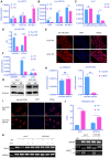
miR-378a-3p participated in sorafenib resistance most likely by targeting IGF1R. (A) The process of research for target genes; (B) RT-qPCR revealed mRNA levels of IGF1R were significantly higher in Huh7-SR, HCCLM3-SR and SK-Hep-1-SR cells compared with their parental lines; (C)WB showed IGF1R were significantly upregulated in resistant cell lines along with their common downstream target pERK/p-AKT; (D) The conserved miR-378a-3p cognate site in 3′-UTR of IGF-1R;Verification of the conserved miR-122 cognate site in 3′-UTR of IGF-1R. Luciferase activity driven by 3′-UTR of IGF-1R was inhibited by ectopic expression of miR-122. Huh7 cells were co-transfected with firefly luciferase-3′-UTR-IGF-1Ror mutant 3′-UTR of IGF-1R with the miR-378a-3p complementary site deleted and miR-122 mimics or control RNA (100 nM) along with a control plasmid; Reduction of luciferase activity driven by IGF-1R-3′UTR was observed (left panel). The IGF-1R-3′UTR-Mutant construct failed to suppress the luciferase activity (right panel) in Huh7 and HCCLM3; (E) The process of RNA pulldown; RNA pulldown assay revealed miR-378a-3p could bind to both sites in IGF1R 3'UTR; (F) IGF1R knockdown significantly blocked the miR-378a-3p inhibitor-mediated sorafenib resistance of HCC cells; (G) HCC prognosis data in TCGA obtained from KM-plotter showed patients with lower IGF1R levels in cancer tissue had significantly better overall (HR=1.58; 95%CI: 1.11 to 2.25; p=0.01); there was negative correlation between miR-378a and IGF1R in the TCGA LIHC dataset (R=-0.330, p=7.53e-11); (H) HCC prognosis data obtained from KM-plotter showed patients with lower IGF1R levels in cancer tissue had significantly better overall (HR=4.96; 95%CI: 2.69 to 9.14; p<0.0001) and disease-free survival (HR=2.63; 95%CI: 1.49 to 4.61; p<0.0001).
Figure 4
Figure 4
attenuated XPO5-mediated export of pre-miR-378a. (A) RT-qPCR revealed the levels of the pri-miR-378a were no different in Huh7-SR, HCCLM3-SR and SK-Hep-1-SR cells compared with their parental lines. (B) RT-qPCR revealed the levels of pre-miR-378a were lower in Huh7-SR, HCCLM3-SR and SK-Hep-1-SR cells compared with their parental lines. (C)There was a obvious uncouple of pri-miR-378 and pre-miR-378a in Huh7-SR cells compared with Huh7-WT cells; (D) RT-qPCR revealed the content of precursors in Huh7-SR cytoplasm decreased significantly; (E) FISH showed the pre-miR-378a in Huh7-SR was trapped in the nucleus; (F) RT-qPCR revealed the levels of XPO5 was lower in Huh7-SR compared with Huh7-WT. (G) WB showed XPO5 were significantly down regulated in Huh7-SR and HCCLM3-SR. (H) After knockdown XPO5 in Huh7-WT, RT-qPCR revealed the levels of pre-miR-378a was reduced. (I) After knockdown XPO5 in Huh7-WT, the pre-miR-378a was also trapped in the nucleus. (J) DNA methylation was detected in Huh7-SR cells, Huh7-WT cells HCCLM3-SR cells and HCCLM3-WT cells by MSP; (K) RT-PCR showed that XPO5 mRNA expression was restored after treatment with demethylation agent 5-aza-2′-deoxycytidine (5-Aza) in Huh7-SR cells, Huh7-WT cells HCCLM3-SR cells and HCCLM3-WT cells.
Figure 5
Figure 5
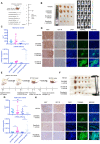
LXRα agonist could increase miR-378a-3p and potentiated sorafenib sensitivity. (A) miR-378 possesses its own transcription machinery independent of its host gene; (B) LXRα agonist could increase the level of miR-378a-3p in Huh7-SR. (C) GW3965 could increase the effect of sorafenib in Huh7-SR and HCCLM3-SR; (D) Flow cytometry showed the combination of sorafenib and GW3965 could induce apoptosis in Huh7-SR and HCCLM3-SR; (E) Colony-formation efficiency showed the emergency of Huh7-SR coloines which treated with sorafenib and GW3965 was less than that treated with sorafenib alone; (F) Cells were seeded in 24-well plates (5X10³cells/well) and incubated overnight for attachment, and were then treated with indicated doses of GW3965 and sorafenib for 48 hr. The impact of Sorafenib, GW3965 and the combination therapy on cell viability in Huh7-SR and HCCLM3-SR cells were determined by CCK8 assay. The CI values of each dose were calculated by the CompuSyn software and CI < 1 indicated synergism; (G) WB showed IGF1R were upregulated in combination along with their common downstream target pERK/p-AKT and combination treatment also could induce apoptosis; (H) Transfected with miR-378a-3p inhibitor could partially rescue the effect of combination therapy in Huh7-SR and HCCLM3-SR.
Figure 6
Figure 6
in vivo experiment showed the combination sorafenib and GW3965 could suppress HCC proliferation. (A) Schematic representation of the in vivo model timeline; The IVIS monitor showed combination therapy could suppress HCCLM3 proliferation (B) gross view of tumors from four groups; (C) The total volume of tumors from four groups; miRNA levels of four groups were verified using RT-qPCR; (D) Immunohistochemistry showed combination therapy could suppress HCC proliferation and IGF1R level in vivo; Meanwhile, TUNEL assay showed combination therapy induced more apoptosis; (E) Schematic representation of the SR-PDX model timeline; (F) gross view of tumors from four groups; Liver view of tumors from four groups; (G) The total volume of tumors from four groups; miRNA levels of four groups were verified using RT-qPCR; (H) Immunohistochemistry showed combination therapy could suppress HCC proliferation, IGF1R level and TUNEL assay in SR-PDX model also showed combination therapy induced more apoptosis.

References

    1. Cheng AL, Kang YK, Chen Z, Tsao CJ, Qin S, Kim JS. et al. Efficacy and safety of sorafenib in patients in the Asia-Pacific region with advanced hepatocellular carcinoma: a phase III randomised, double-blind, placebo-controlled trial. Lancet Oncol. 2009;10:25–34. - PubMed
    1. Wilhelm SM, Adnane L, Newell P, Villanueva A, Llovet JM, Lynch M. Preclinical overview of sorafenib, a multikinase inhibitor that targets both Raf and VEGF and PDGF receptor tyrosine kinase signaling. Mol Cancer Ther. 2008;7:3129–40. - PubMed
    1. Xia S, Pan Y, Liang Y, Xu J, Cai X. The microenvironmental and metabolic aspects of sorafenib resistance in hepatocellular carcinoma. EBioMedicine. 2020;51:102610. - PMC - PubMed
    1. Bartel DP. MicroRNAs: target recognition and regulatory functions. Cell. 2009;136:215–33. - PMC - PubMed
    1. Wen J, Xiong K, Aili A, Wang H, Zhu Y, Yu Z. et al. PEX5, a novel target of microRNA-31-5p, increases radioresistance in hepatocellular carcinoma by activating Wnt/β-catenin signaling and homologous recombination. Theranostics. 2020;10:5322–40. - PMC - PubMed

Publication types

MeSH terms