. 2020 Sep;3(3):203-226.
doi: 10.1007/s42242-020-00084-6. Epub 2020 Jun 30.

Engineered Hydrogels for Brain Tumor Culture and Therapy

Affiliations

Engineered Hydrogels for Brain Tumor Culture and Therapy

Jai Thakor et al. Biodes Manuf. 2020 Sep.

Abstract

Brain tumors' severity ranges from benign to highly aggressive and invasive. Bioengineering tools can assist in understanding the pathophysiology of these tumors from outside the body and facilitate development of suitable antitumoral treatments. Here, we first describe the physiology and cellular composition of brain tumors. Then, we discuss the development of three-dimensional tissue models utilizing brain tumor cells. In particular, we highlight the role of hydrogels in providing a biomimetic support for the cells to grow into defined structures. Microscale technologies, such as electrospinning and bioprinting, and advanced cellular models aim to mimic the extracellular matrix and natural cellular localization in engineered tumor tissues. Lastly, we review current applications and prospects of hydrogels for therapeutic purposes, such as drug delivery and co-administration with other therapies. Through further development, hydrogels can serve as a reliable option for in vitro modeling and treatment of brain tumors for translational medicine.

Keywords: Brain tumor; bioengineering; cancer cells; drug delivery; hydrogel.


Figures
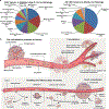
Fig. 1.
Fig. 1.
(a) Types of CNS tumors and incidence by age group among children (ages 0–14) and adults (Adapted from McNeill et al., 2016 [37]), (b) Multiple steps of metastatic colonization including pre-colonization phase, which occurs in minutes to hours, followed by colonization phase, which occurs in years. Pre-colonization steps include intravasation of cancer cells into the tumor vasculature, entry into the circulatory system, and extravasation into the parenchyma of target tissues or organs (Adapted from Massague et al., 2016 [40]).
Fig. 2.
Fig. 2.
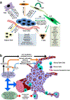
(a) Mechanism of interaction of tumor cells with stromal cells in tumor microenvironment promoting tumor growth, invasion, and metastasis (Adapted from Mao et al., 2013 [52]), (b) The glioma perivascular niche. Vascular endothelial cells provide chemotactic signals to migrating glioma cells in order to attract them to blood vessels. GSCs may also migrate to the site and differentiate to multiple cell types due to signals in the PVN (Adapted from Diksin et al., 2017 [56]).
Fig. 3.
Fig. 3.
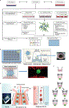
(a) In vitro brain tumor models. Models consist of 2D and 3D platforms, subdivided into: (i) anchorage independent, which do not utilize scaffolds; [79] anchorage-dependent, (iii) integrating scaffolds for culture; and (iv) specialized devices. Non-scaffold methods to generate 3D masses use hanging-drop method, low-attachment plates, or magnetic levitation, while scaffold-based include hydrogels. Specialized platforms include microfluidic devices and micropatterned plates (Adapted from Langhans et al., 2018 [58]), (b) Incorporation of brain glioblastoma spheroids within a microfluidic 3D culture platform of 96 well plates containing a tapered hole in the center of a rail for brain glioblastoma spheroids to generate angiogenic sprouting patterns (Adapted from Ko et al., 2019 [69]), (c) A 3D model generated by combination of transwell-96 well-plates and 96-well spheroid microplates to simulate penetration of anti-cancer drugs through the BBB (Adapted from Sherman et al., 2019 [71]), (d) Generation of brain organoids on a brain organoid-on-a chip device to investigate the effect of nicotine on early brain development (Adapted from Wang et al., 2018 [74]).
Fig. 4.
Fig. 4.
Microscale procedures for directing cellular growth. (a) Microchannels patterned with laminin cause glioma cell adhesion and alignment as cell density increases (Adapted from Monzo et al., 2016 [111]), (b) Aligned PCL nanofibers promote directional contact guidance to glioma cells seeded on their surface, while randomly oriented fibers produce nonspecific cell populations on their surface (Adapted from Agudelo-Garcia et al., 2016 [76]), (c) Microfluidic devices are compartmentalized and consist of wells which allow for GBM cell aggregation as the flow of media provides nutrients as cells proliferate (Adapted from Fan et al., 2016 [21]), (d) Bioprinting extrudes of alginate/gelatin/fibrin gel which encapsulates cells and causes them to grow in distinct arrays (Adapted from Wang et al., 2018 [112]).
Fig. 5.
Fig. 5.
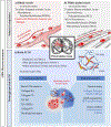
Physiological structures in GBM microenvironment promote cancer invasion: (a) White matter tracts facilitating communication between brain regions serve as invasion routes for GBM cells. Tracts recapitulated on nanofiber-coated and pattered surfaces allow for cell adhesion and migration. (b) Blood vessels are invasion routes for migrating GBM cells. PDMS devices micropatterned with ECM proteins mimic elongated structures of blood vessels. (C) Brain ECM composed of high HA and other glycosaminoglycans in tumor stroma and invasive fronts. Polymers are able to mimic components and properties of brain ECM (Adapted from Cha et al., 2017 [17]).
Fig. 6.
Fig. 6.
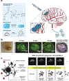
(a) Hydrogel composition and routes of administration of hydrogels for glioblastoma treatment. Macroscopic hydrogels containing therapeutic agents with various chemical compositions were locally implanted or injected in the tumor site, while hydrogel nanoparticles were intravenously administered (Adapted from Li et al., 2016 [132], and Basso et al., 2018 [8]), (b) Insertion of a PCL/polyurethane carrier conduit containing aligned PCL nanofiber films, and cyclopamine (anti-cancer)-conjugated collagen hydrogel serving as an apoptotic tumor sink in brain. Migration of tumor cells along the aligned films throughout the cross-section of conduit was observed at different distances from the interface of the tumor in the brain (Adapted from Jain et al., 2014 [141], (c) Stereotactical injection of drug-loaded magnetic hydrogel, and simultaneous magnetic resonance images of treated region over time by degradation of hydrogel and sustained release of drug (Adapted from Kim et al., 2012 [144]).

References

    1. Lu QR, Qian L, Zhou X (2019) Developmental origins and oncogenic pathways in malignant brain tumors. Wiley Interdiscip Rev Dev Biol 8 (4):e342. doi:10.1002/wdev.342 - DOI - PMC - PubMed
    1. Madhusoodanan S, Opler MG, Moise D, Gordon J, Danan DM, Sinha A, Babu RP (2010) Brain tumor location and psychiatric symptoms: is there any association? A meta-analysis of published case studies. Expert Rev Neurother 10 (10):1529–1536. doi:10.1586/ern.10.94 - DOI - PubMed
    1. Perkins A, Liu G (2016) Primary brain tumors in adults: diagnosis and treatment. Am Fam Physician 93 (3):211–217 - PubMed
    1. Lefranc F, Sadeghi N, Camby I, Metens T, Dewitte O, Kiss R (2006) Present and potential future issues in glioblastoma treatment. Expert Rev Anticancer Ther 6 (5):719–732. doi:10.1586/14737140.6.5.719 - DOI - PubMed
    1. Puente P, Fettig N, Luderer MJ, Jin A, Shah S, Muz B, Kapoor V, Goddu SM, Salama NN, Tsien C, Thotala D, Shoghi K, Rogers B, Azab AK (2018) Injectable Hydrogels for Localized Chemotherapy and Radiotherapy in Brain Tumors. J Pharm Sci 107 (3):922–933. doi:10.1016/j.xphs.2017.10.042 - DOI - PMC - PubMed

LinkOut - more resources