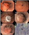
Esophageal Adenocarcinoma in the Proximal Esophageal Segment: A Unique Presentation in a Male With Alcohol Abuse
- PMID: 32754402
- PMCID: PMC7386092
- DOI: 10.7759/cureus.8863
Esophageal Adenocarcinoma in the Proximal Esophageal Segment: A Unique Presentation in a Male With Alcohol Abuse
Abstract
Esophageal adenocarcinoma (EAC) is a malignancy classically seen in the distal esophagus. While many risk factors associated with the condition have been reported, the most common among them are gastroesophageal reflux disease (GERD) and obesity. Histological changes range from metaplasia within the esophagus from stratified squamous epithelium to non-ciliated columnar cells with goblet cells. In contrast, squamous cell carcinoma (SCC) is classically found in the proximal portion of the esophagus and its risk factors include tobacco and alcohol use. We present a unique case of a 59-year-old African American male who presented to the ED with dysphagia, weight loss, and multiple episodes of emesis. Notable medical history included tobacco abuse, alcohol abuse, and alcoholic cirrhosis. Currently, there are numerous case reports delineating unique presentations of esophageal cancers; however, there are few case reports that demonstrate EAC affecting the proximal segment of the esophagus.
Keywords: esophageal adenocarcinoma.
Copyright © 2020, Shiflett et al.
Conflict of interest statement
The authors have declared that no competing interests exist.
Figures
Similar articles
-
Gastroesophageal reflux disease, proton-pump inhibitor use and Barrett's esophagus in esophageal adenocarcinoma: Trends revisited.Surgery. 2013 Oct;154(4):856-64; discussion 864-6. doi: 10.1016/j.surg.2013.07.020. Surgery. 2013. PMID: 24074425
-
The histologic squamo-oxyntic gap: an accurate and reproducible diagnostic marker of gastroesophageal reflux disease.Am J Surg Pathol. 2010 Nov;34(11):1574-81. doi: 10.1097/PAS.0b013e3181f06990. Am J Surg Pathol. 2010. PMID: 20871393
-
A New Pathologic Assessment of Gastroesophageal Reflux Disease: The Squamo-Oxyntic Gap.Adv Exp Med Biol. 2016;908:41-78. doi: 10.1007/978-3-319-41388-4_4. Adv Exp Med Biol. 2016. PMID: 27573767 Review.
-
Short segment Barrett's esophagus and distal gastric intestinal metaplasia.Arq Gastroenterol. 2006 Apr-Jun;43(2):117-20. doi: 10.1590/s0004-28032006000200011. Arq Gastroenterol. 2006. PMID: 17119666
-
Epidemiology of Barrett's Esophagus and Esophageal Adenocarcinoma.Gastroenterol Clin North Am. 2015 Jun;44(2):203-31. doi: 10.1016/j.gtc.2015.02.001. Epub 2015 Apr 9. Gastroenterol Clin North Am. 2015. PMID: 26021191 Free PMC article. Review.
Cited by
-
Proximal esophageal adenocarcinoma: A rare case report.Int J Surg Case Rep. 2024 Jul;120:109868. doi: 10.1016/j.ijscr.2024.109868. Epub 2024 Jun 6. Int J Surg Case Rep. 2024. PMID: 38852572 Free PMC article.
-
A Combat Journey of Rehabilitation in Pre- and Post-chemotherapy for Esophagus Carcinoma.Cureus. 2024 Apr 13;16(4):e58202. doi: 10.7759/cureus.58202. eCollection 2024 Apr. Cureus. 2024. PMID: 38741852 Free PMC article.
References
Publication types
LinkOut - more resources
Full Text Sources
Research Materials

