. 2020 Jul 24;6(30):eaba6493.
doi: 10.1126/sciadv.aba6493. eCollection 2020 Jul.

Expression of inhibitory receptors by B cells in chronic human infectious diseases restricts responses to membrane-associated antigens

Affiliations

Expression of inhibitory receptors by B cells in chronic human infectious diseases restricts responses to membrane-associated antigens

Abhijit A Ambegaonkar et al. Sci Adv. .

Abstract

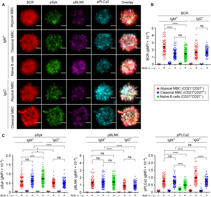
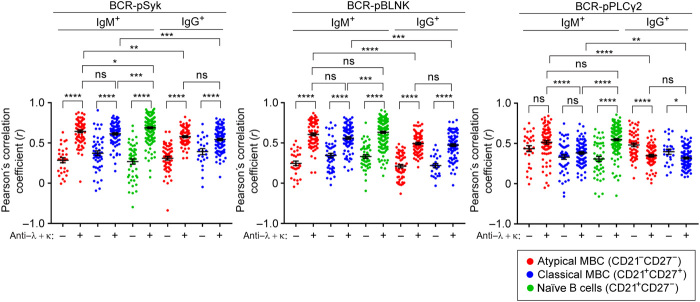
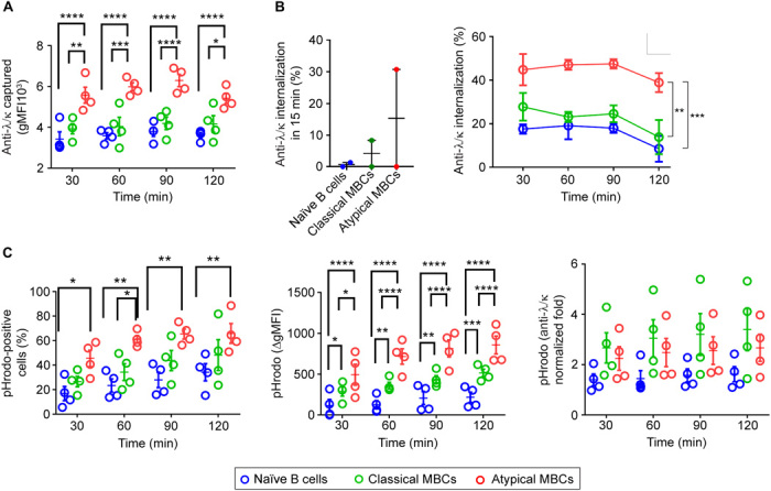
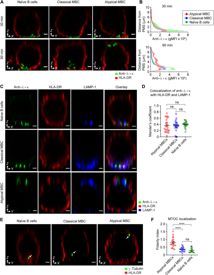
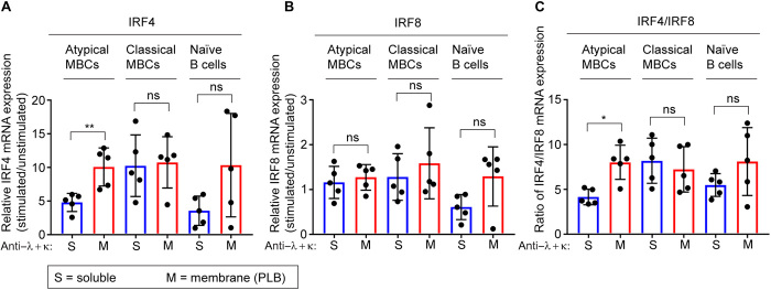
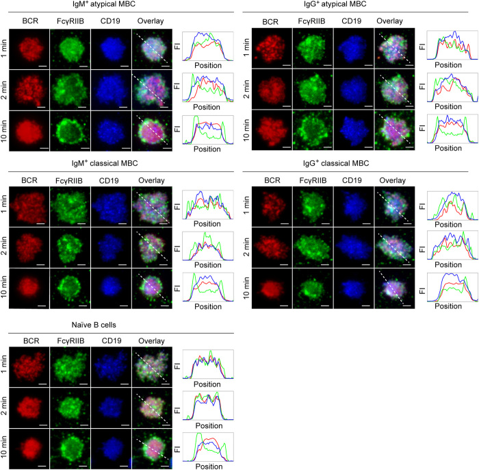
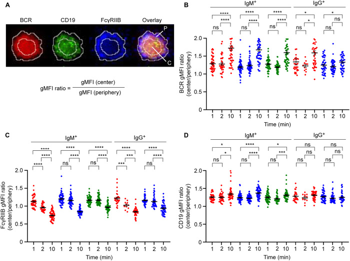
Chronic human infectious diseases, including malaria, are associated with a large expansion of a phenotypically and transcriptionally distinct subpopulation of B cells distinguished by their high expression of a variety of inhibitory receptors including FcγRIIB. Because these B cells, termed atypical memory B cells (MBCs), are unable to respond to soluble antigens, it was suggested that they contributed to the poor acquisition of immunity in chronic infections. Here, we show that the high expression of FcγRIIB restricts atypical MBC responses to membrane-associated antigens that function to actively exclude FcγRIIB from the B cell immune synapse and include the co-receptor CD19, allowing B cell antigen receptor signaling and differentiation toward plasma cells. Thus, chronic infectious diseases result in the expansion of B cells that robustly respond to antigens that associate with cell surfaces, such as antigens in immune complexes, but are unable to respond to fully soluble antigens, such as self-antigens.


Figures

Fig. 1
Fig. 1. Atypical MBCs signal robustly through their BCR in response to PLB-associated anti-λ/κ.
Atypical MBCs (CD19+ CD21 CD27), classical MBCs (CD19+ CD21+ CD27+), and naïve B cells (CD19+ CD21+ CD27) were fluorescence-activated cell sorting (FACS)–sorted from PBMCs, stained with DyLight 594–conjugated Fab fragments of either anti-IgM or anti-IgG, and placed on either PLBs alone or on PLBs containing anti-λ/κ for 10 min, fixed and stained with antibodies specific for the BCR and pSyk and for pBLNK and pPLC-γ2, and imaged by TIRF microscopy (see also fig. S1). (A) Representative TIRF microscopy images indicating accumulation of the BCR (IgM or IgG) (red), pSyk (green), pBLNK (magenta), and pPLC-γ2 (cyan) in the immune synapses formed by atypical MBCs, classical MBCs, and naïve B cells activated on PLBs containing anti-λ/κ (scale bar, 2 μm). (B and C) Quantification of mean fluorescence intensity (MFI) of BCR (B) and pSyk, pBLNK, and pPLC-γ2 (C) accumulated in the immune synapse of atypical MBCs (red dots), classical MBCs (blue dots), and naïve B cells (green dots) incubated on either PLBs alone or on PLBs containing anti-λ/κ. Data are representative of three experiments. The error bars indicate SEM data were analyzed using unpaired t test. *P < 0.05; ***P < 0.001; ****P < 0.0001; ns, not significant.
Fig. 2
Fig. 2. Synaptic colocalization of BCR and phosphorylated signaling molecules in atypical MBCs is enhanced in response to PLB-associated anti-λ/κ.
Atypical MBCs (CD19+ CD21 CD27), classical MBCs (CD19+ CD21+ CD27+), and naïve B cells (CD19+ CD21+ CD27) were FACS-sorted from PBMCs, stained with DyLight 594–conjugated Fab fragments of either anti-IgM or anti-IgG ,and placed on either PLBs alone or on PLBs containing anti-λ/κ for 10 min, fixed and stained with antibodies specific for the BCR and pSyk and for pBLNK and pPLC-γ2 and imaged by TIRF microscopy (as in Fig. 1). Colocalization of BCR with pSyk, pBLNK, or pPLC-γ2 within the immune synapse formed by atypical MBCs (red dots), classical MBCs (blue dots), and naïve B cells (green dots) following incubation on either PLBs alone or on PLBs containing anti-λ/κ. Data are representative of three experiments. The error bars indicate SEM. Data were analyzed using unpaired t test. *P < 0.05; **P < 0.01; ***P < 0.001; ****P < 0.0001; ns, not significant.
Fig. 3
Fig. 3. Atypical MBCs rapidly capture and internalize PMS-bound anti-λ/κ.
PBMCs were incubated for increasing lengths of time (30 to 120 min) at 37°C on PMS containing biotinylated anti-λ/κ conjugated to DyLight 650 and avidin-pHrodo. At each time point, cells were harvested and stained with streptavidin-Alexa488 and antibodies specific for CD19, CD21, and CD27 to identify naïve B cells (CD19+ CD21+ CD27), classical MBCs (CD19+ CD21+ CD27+), and atypical MBCs (CD19+ CD21 CD27). Antigen capture, internalization, and trafficking into the acidic compartments were analyzed by flow cytometry as described in the text and detailed in Materials and Methods (n = 4). In all cases: atypical MBCs (red circles), classical MBCs (green circles), and naïve B cells (blue circles) (see also fig. S2). (A) Comparison of the total amount of anti-λ/κ captured from PMS over time indicated by the MFI of DyLight 650. (B) Comparison of the amount of anti-λ/κ internalized in 15 min (left) and over time (right). (C) Comparison of anti-λ/κ trafficking to acidic compartment over time indicated by the percentage of cells positive for pHrodo or by the increases in pHrodo MFI from time 0. Data were analyzed using paired t test (A and C) or two-way analysis of variance (ANOVA) and Tukey’s multiple comparison test (B). *P < 0.05; **P < 0.01; ***P < 0.001; ****P < 0.0001.
Fig. 4
Fig. 4. Atypical MBCs show a distinct orientation when gathering, internalizing, and trafficking anti-λ/κ from PMS.
Atypical MBC, classical MBCs, and naïve B cells were FACS-sorted from PBMCs and activated for 30 or 90 min at 37°C on PMS containing anti-λ/κ conjugated to DyLight 550 and fixed and stained with various antibodies imaged by confocal microscope. (A) Representative images showing anti-λ/κ (green) and HLA-DR (red) in orthogonal confocal sections of cells incubated on PMS containing anti-λ/κ for 30 and 90 min (scale bar, 1 μm). (B) Quantification of the distance of the anti-λ/κ from the PMS after 30 and 90 min of incubation of anti-λ/κ–containing PMS. (C) Representative images showing anti-λ/κ (green), HLA-DR (red), and LAMP-1 (blue) in orthogonal confocal sections of cells incubated on PMS containing anti-λ/κ for 90 min (scale bar, 1 μm). (D) 3D colocalization of anti-λ/κ with HLA-DR and LAMP-1 (Mander’s coefficient) in each B cell subpopulation. (E) Representative images indicating γ-tubulin (green) and HLA-DR (red) in orthogonal confocal sections of cells incubated on PMS containing anti-λ/κ for 90 min. The arrows indicate the location of MTOCs in the cell (scale bar, 1 μm). (F) Polarity index for the localization of MTOCs in each B cell subpopulation. Data are representative of two experiments. The error bars indicate SEM. Data were analyzed using a one-way ANOVA and Tukey’s multiple comparison test. ****P < 0.0001; ns, not significant.
Fig. 5
Fig. 5. Atypical MBCs express IRF4 upon responding to PLB-associated anti-λ/κ.
Relative mRNA expression level of IRF4 (A), IRF8 (B), and ratio of IRF4/IRF8 (C) in FACS-sorted atypical MBC, classical MBCs, and naïve B cells after in vitro activation with PLB-bound anti-λ/κ or soluble anti-λ/κ, along with TLR9 agonist CpG, BAFF, and cytokines IL-2, IL-10, and IL-21 (n = 5). The error bars indicate SD. Data were analyzed using nonparametric Mann-Whitney test. *P < 0.05; **P < 0.01; ns, not significant.
Fig. 6
Fig. 6. Atypical MBCs rapidly segregate FcγRIIB to periphery of immune synapse formed on PLB-associated anti-λ/κ.
Atypical MBCs, classical MBCs, and naïve B cells were FACS-sorted from PBMCs, stained with DyLight 594–conjugated Fab fragments of either anti-IgM or anti-IgG and Alexa Fluor 488–conjugated Fab fragments of anti-CD19 antibodies, and activated on PLBs containing anti-λ/κ for 1 to 10 min, fixed and stained with antibodies recognizing FcγRIIB, and imaged by TIRF microscopy. Representative TIRF microscopy images indicating localization of the BCR (IgM or IgG) (red), FcγRIIB (green), and CD19 (blue) within immune synapses formed by atypical MBCs, classical MBCs, and naïve B cells activated on PLBs containing anti-λ/κ. Relative FI along the dashed white line is given (scale bar, 2 μm).
Fig. 7
Fig. 7. Segregation of FcγRIIB to the cell periphery is accompanied by accumulation of BCR and CD19 in the immune synapse of atypical MBCs responding to PLB-associated anti-λ/κ.
Atypical MBCs, classical MBCs, and naïve B cells were FACS-sorted from PBMCs, stained with DyLight 594–conjugated Fab fragments of either anti-IgM or anti-IgG and Alexa Fluor 488–conjugated Fab fragments of anti-CD19 antibodies, and activated on PLBs containing anti-λ/κ for 1 to 10 min, fixed and stained with antibodies recognizing FcγRIIB, and imaged by TIRF microscopy (as in Fig. 6) (see also fig. S3). (A) Representative TIRF microscopy image indicating localization of the BCR (red), CD19 (green), and FcγRIIB (blue) within immune synapses. White line indicates the ROI selected for calculating the MFI in the center (shown as C in overlay) and in the periphery (shown as P in overlay) of the immune synapse. (B to D) Quantification of ratio of MFI in center versus periphery of the immune synapse for BCR (B), FcγRIIB (C), and CD19 (D) in atypical MBCs (red dots), classical MBCs (blue dots), and naïve B cells (green dots) activated on PLBs containing anti-λ/κ. Data are representative of two experiments. The error bars indicate SEM. Data were analyzed using unpaired t test. *P < 0.05; ***P < 0.001; ****P < 0.0001; ns, not significant.

References

    1. World Health Organization, World Malaria Report 2018 (World Health Organization, Geneva, Switzerland, 2018), 165 pp.
    1. Langhorne J., Ndungu F. M., Sponaas A.-M., Marsh K., Immunity to malaria: More questions than answers. Nat. Immunol. 9, 725–732 (2008). - PubMed
    1. Crompton P. D., Moebius J., Portugal S., Waisberg M., Hart G., Garver L. S., Miller L. H., Barillas-Mury C., Pierce S. K., Malaria immunity in man and mosquito: Insights into unsolved mysteries of a deadly infectious disease. Annu. Rev. Immunol. 32, 157–187 (2014). - PMC - PubMed
    1. Weiss G. E., Traore B., Kayentao K., Ongoiba A., Doumbo S., Doumtabe D., Kone Y., Dia S., Guindo A., Traore A., Huang C.-Y., Miura K., Mircetic M., Li S., Baughman A., Narum D. L., Miller L. H., Doumbo O. K., Pierce S. K., Crompton P. D., The Plasmodium falciparum-specific human memory B cell compartment expands gradually with repeated malaria infections. PLOS Pathog. 6, e1000912 (2010). - PMC - PubMed
    1. Tran T. M., Li S., Doumbo S., Doumtabe D., Huang C. Y., Dia S., Bathily A., Sangala J., Kone Y., Traore A., Niangaly M., Dara C., Kayentao K., Ongoiba A., Doumbo O. K., Traore B., Crompton P. D., An intensive longitudinal cohort study of malian children and adults reveals no evidence of acquired immunity to Plasmodium falciparum infection. Clin. Infect. Dis. 57, 40–47 (2013). - PMC - PubMed

Publication types