. 2020 Jun 30;6(4):e468.
doi: 10.1212/NXG.0000000000000468. eCollection 2020 Aug.

Congenital myasthenic syndrome caused by a frameshift insertion mutation in GFPT1

Affiliations

Congenital myasthenic syndrome caused by a frameshift insertion mutation in GFPT1

Szabolcs Szelinger et al. Neurol Genet. .

Abstract

Objective: Description of a new variant of the glutamine-fructose-6-phosphate transaminase 1 (GFPT1) gene causing congenital myasthenic syndrome (CMS) in 3 children from 2 unrelated families.

Methods: Muscle biopsies, EMG, and whole-exome sequencing were performed.

Results: All 3 patients presented with congenital hypotonia, muscle weakness, respiratory insufficiency, head lag, areflexia, and gastrointestinal dysfunction. Genetic analysis identified a homozygous frameshift insertion in the GFPT1 gene (NM_001244710.1: c.686dupC; p.Arg230Ter) that was shared by all 3 patients. In one of the patients, inheritance of the variant was through uniparental disomy (UPD) with maternal origin. Repetitive nerve stimulation and single-fiber EMG was consistent with the clinical diagnosis of CMS with a postjunctional defect. Ultrastructural evaluation of the muscle biopsy from one of the patients showed extremely attenuated postsynaptic folds at neuromuscular junctions and extensive autophagic vacuolar pathology.

Conclusions: These results expand on the spectrum of known loss-of-function GFPT1 mutations in CMS12 and in one family demonstrate a novel mode of inheritance due to UPD.


Figures

Figure 1
Figure 1. Clinical neurophysiology
Decremental response on 3 Hz repetitive nerve stimulation of hand muscle of patient A showing a postjunctional neuromuscular deficit by postactivation facilitation and then exhaustion; decrement of 13% at rest (A), 7% immediately after 30 seconds exercise (B), and 15% at 360 seconds after the exercise (C). Stimulated (15 c/s) single-fiber EMG study on extensor digitorum communis muscle on patient B1 demonstrating a large jitter on the last unit (D).
Figure 2
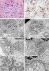
Figure 2. Muscle biopsy pathology of patient B2
The cryosection H&E image (A) shows increased variation in fiber size and a mild degree of regeneration (arrow), suggesting that the patient has a mild, necrotizing myopathy. AChE enzyme histochemistry (B) highlights numerous cytoplasmic autophagic vacuoles with sarcolemmal features (arrows). Extensive autophagy is confirmed by electron microscopy (C). av occupy most of the intracellular space of the muscle fiber in the upper portion of the micrograph. Only rare sarcomeres are present, as noted by 2 Z-lines (z) in this image. Extrusion of autophagic vacuoles (E) is evident at the surface of this muscle fiber. Intramuscular nerve twigs appear normal (D). Two of numerous NMJs observed in the biopsy are illustrated in panels (E–H). There are varying degrees of postsynaptic fold attenuation (arrows), milder in panels E and G and complete loss of NMJ endplate folds in panels F and H. Nerve terminals contain numerous sv. Portions of muscle fiber nuclei are present along the top of panels E, F, and H (n). AChE = acetylcholinesterase; av = autophagic vacuoles; H&E = hematoxylin & eosin; NMJ = neuromuscular junction; sv = synaptic vesicles.
Figure 3
Figure 3. GFPT1 gene and previously reported, ClinVar pathogenic and likely pathogenic variants in CMS12
BAF plot from WES of patient A1 (top) demonstrates long continuous regions of homozygosity (blue dots with BAF ∼1 or BAF ∼0) that overlaps with previous array CGH data (green horizontal bars). Red, vertical bar at 2p13.3 on the cytoband indicates the position of GFPT1 gene within the LCRH. Below the cytoband, domain structure of GFPT1 and ClinVar pathogenic and likely pathogenic mutations in relation to GFPT1 domains of NM_001244710 and to p.Arg230* (underlined) identified in this study. Variants are colored based on their functional impact (green = missense, black = frameshift, red = nonsense, and blue = splice). BAF = B allele frequency; CGH = comparative genomic hybridization; GFPT1 = glutamine-fructose-6-phosphate transaminase 1; LCRH = long contiguous regions of homozygosity; SIS = sugar isomerase domain; WES = whole-exome sequencing.
Figure 4
Figure 4. Sequence traces of p.Arg230*
(A) Sanger sequencing-confirmed exome sequencing findings for members of family A and for patient B1 with the red arrow indicating the frameshift insertion. Mother is heterozygous indicated by the overlapping peaks, whereas father was homozygous reference. (B) PacBio sequence traces showing patient B2 homozygosity. GFPT1 = glutamine-fructose-6-phosphate transaminase 1.

References

    1. de Souza PVS, Batistella GN de R, Lino VC, de Rezende Pinto WBV, Annes M, Oliveira ASB. Clinical and genetic basis of congenital myasthenic syndromes. Arq Neuro-Psiquiatr 2016;74:750–760. - PubMed
    1. Senderek J, Müller JS, Dusl M, et al. . Hexosamine biosynthetic pathway mutations cause neuromuscular transmission defect. Am J Hum Genet 2011;88:162–172. - PMC - PubMed
    1. Rodríguez Cruz P, Palace J, Beeson D. The neuromuscular junction and wide heterogeneity of congenital myasthenicsyndromes. Int J Mol Sci 2018;19:1677–1723. - PMC - PubMed
    1. Engel AG, Shen XM, Selcen D, Sine SM. Congenital myasthenic syndromes: pathogenesis, diagnosis, and treatment. Lancet Neurol 2015;14:420–434. - PMC - PubMed
    1. Guergueltcheva V, Müller JS, Dusl M, et al. . Congenital myasthenic syndrome with tubular aggregates caused by GFPT1 mutations. J Neurol 2011;259:838–850. - PubMed