. 2020 Dec;215(6):1421-1429.
doi: 10.2214/AJR.20.23313. Epub 2020 Oct 14.

Using Deep Learning to Accelerate Knee MRI at 3 T: Results of an Interchangeability Study

Affiliations

Using Deep Learning to Accelerate Knee MRI at 3 T: Results of an Interchangeability Study

Michael P Recht et al. AJR Am J Roentgenol. 2020 Dec.

Abstract

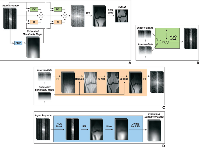
OBJECTIVE. Deep learning (DL) image reconstruction has the potential to disrupt the current state of MRI by significantly decreasing the time required for MRI examinations. Our goal was to use DL to accelerate MRI to allow a 5-minute comprehensive examination of the knee without compromising image quality or diagnostic accuracy. MATERIALS AND METHODS. A DL model for image reconstruction using a variational network was optimized. The model was trained using dedicated multisequence training, in which a single reconstruction model was trained with data from multiple sequences with different contrast and orientations. After training, data from 108 patients were retrospectively undersampled in a manner that would correspond with a net 3.49-fold acceleration of fully sampled data acquisition and a 1.88-fold acceleration compared with our standard twofold accelerated parallel acquisition. An interchangeability study was performed, in which the ability of six readers to detect internal derangement of the knee was compared for clinical and DL-accelerated images. RESULTS. We found a high degree of interchangeability between standard and DL-accelerated images. In particular, results showed that interchanging the sequences would produce discordant clinical opinions no more than 4% of the time for any feature evaluated. Moreover, the accelerated sequence was judged by all six readers to have better quality than the clinical sequence. CONCLUSION. An optimized DL model allowed acceleration of knee images that performed interchangeably with standard images for detection of internal derangement of the knee. Importantly, readers preferred the quality of accelerated images to that of standard clinical images.

Keywords: MRI; acceleration; deep learning; internal derangement; knee.


Figures

Fig. 1—
Fig. 1—
Structure of network used for deep learning reconstruction of 3-T knee MR images. A, Block diagram shows structure of our model, which takes undersampled k-space as input and applies several iterative refinements (R). Each refinement includes residual connection, R module, and data consistency (DC) module. Inverse Fourier transform (IFT) followed by root-sum-of-squares (RSS) transform is applied after final refinement to obtain reconstructed image. SME = sensitivity map estimation. B, Diagram shows DC module that computes correction map that brings intermediate k-space data closer to input k-space data. Correction is computed only at k-space locations where measurements have been performed. C, Diagram shows R module that converts multicoil k-space data into single image, applies U-Net, and then converts output back to multicoil k-space data. In first step, IFT is applied to obtain multicoil images, which are then multiplied by conjugate of sensitivity maps and added (Reduce). In final step, image is multiplied by sensitivity maps (Expand) followed by Fourier transform (FT). D, Diagram shows SME module that estimates sensitivity maps used in R modules. SME selects only autocalibration signal (ACS) lines from input k-space and applies IFT and then U-Net. Finally, output of U-Net is normalized by dividing each individual sensitivity map voxelwise by RSS of all maps.
Fig. 2—
Fig. 2—
34-year-old man with acute knee injury. A–C, Coronal fat-suppressed proton density–weighted images with no added noise (A), baseline noise value (σ) = 0.015 (B), and σ = 0.05 (C) show effect of dithering.
Fig. 3—
Fig. 3—
Graph shows structural similarity index (SSIM) score (or negative loss) as function of training time. SSIM score is computed both on training set (solid line) and on validation set (dashed line).
Fig. 4—
Fig. 4—
64-year-old man with recurrent popliteal cyst. A–D, Coronal clinical (A) and deep learning (DL)-accelerated (B) as well as sagittal clinical (C) and DL-accelerated (D) proton density—weighted images show medial (black arrows) and lateral (white arrows, A and B) meniscal tears and popliteal cyst (arrowheads, C and D). It is difficult to distinguish between clinical and DL-accelerated images.
Fig. 5—
Fig. 5—
22-year-old man with acute knee injury. A and B, Sagittal clinical (A) and deep learning (DL)-accelerated (B) fat-suppressed proton density—weighted images show bone contusions (arrows) in lateral femoral condyle and lateral tibial plateaus, consistent with anterior cruciate ligament tear. It is difficult to distinguish between clinical and DL-accelerated images. Such indistinguishability is uncommon for traditional acceleration techniques at high acceleration factors, particularly for challenging case of 2D images with strong requirements for spatial resolution and anatomic fidelity.
Fig. 6—
Fig. 6—
43-year-old man with medial knee pain. A–C, Clinical (A), fourfold (B), and eightfold (C) deep learning—accelerated fat-suppressed proton density—weighted images show subtle signal-intensity change in medial meniscus (arrow) on clinical and fourfold accelerated sequences that is not visible on eightfold accelerated image. Eightfold acceleration was therefore deemed too aggressive for this use of 2D musculoskeletal imaging. However, substantially higher accelerations are likely to be feasible for other clinical applications and for acquisitions that are multidimensional, dynamic, or both.

References

    1. Gielen JL, De Schepper AM, Vanhoenacker F, et al. Accuracy of MRI in characterization of soft tissue tumors and tumor-like lesions: a prospective study in 548 patients. Eur Radiol 2004; 14:2320–2330 - PubMed
    1. Vahey TN, Meyer SF, Shelbourne KD, Klootwyk TE. MR imaging of anterior cruciate ligament injuries. Magn Reson Imaging Clin N Am 1994; 2:365–380 - PubMed
    1. Floriani I, Torri V, Rulli E, et al. Performance of imaging modalities in diagnosis of liver metastases from colorectal cancer: a systematic review and meta-analysis. J Magn Reson Imaging 2010; 31:19–31 - PubMed
    1. Martín Noguerol T, Barousse R, Gómez Cabrera M, Socolovsky M, Bencardino JT, Luna A. Functional MR neurography in evaluation of peripheral nerve trauma and postsurgical assessment. RadioGraphics 2019; 39:427–446 - PubMed
    1. Vanderby S, Badea A, Peña Sánchez JN, Kalra N, Babyn P. Variations in magnetic resonance imaging provision and processes among Canadian academic centres. Can Assoc Radiol J 2017; 68:56–65 - PubMed

Publication types