Randomized Controlled Trial
. 2020:28:102363.
doi: 10.1016/j.nicl.2020.102363. Epub 2020 Jul 25.

Distance to white matter trajectories is associated with treatment response to internal capsule deep brain stimulation in treatment-refractory depression

Affiliations
Randomized Controlled Trial

Distance to white matter trajectories is associated with treatment response to internal capsule deep brain stimulation in treatment-refractory depression

Luka C Liebrand et al. Neuroimage Clin. 2020.

Abstract

Background: Deep brain stimulation (DBS) is an innovative treatment for treatment-refractory depression. DBS is usually targeted at specific anatomical landmarks, with patients responding to DBS in approximately 50% of cases. Attention has recently shifted to white matter tracts to explain DBS response, with initial open-label trials targeting white matter tracts yielding much higher response rates (>70%).

Objective/hypothesis: Our aim was to associate distance to individual white matter tracts around the stimulation target in the ventral anterior limb of the internal capsule to treatment response.

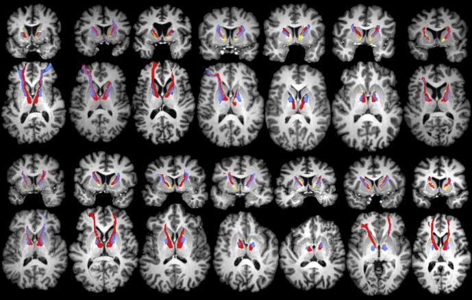
Methods: We performed diffusion magnetic resonance tractography of the superolateral branch of the medial forebrain bundle and the anterior thalamic radiation in fourteen patients that participated in our randomized clinical trial. We combined the tract reconstructions with the postoperative images to identify the DBS leads and estimated the distance between tracts and leads, which we subsequently associated with treatment response.

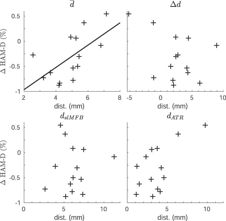
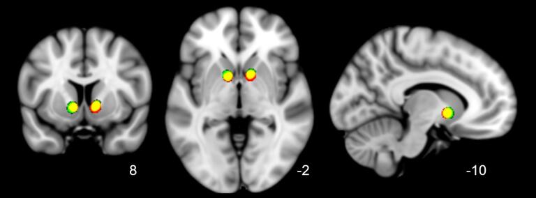
Results: Stimulation closer to both tracts was significantly correlated to a larger symptom decrease (r = 0.61, p = 0.02), suggesting that stimulation more proximal to the tracts was beneficial. Biophysical modelling indicated that 37.5% of tracts were even outside the volume of activated tissue. There was no difference in lead placement with respect to anatomical landmarks, which could mean that differences in treatment response were driven by individual differences in white matter anatomy.

Conclusions: Our results suggest that deep brain stimulation of the ventral anterior limb of the internal capsule could benefit from targeting white matter bundles. We recommend acquiring diffusion magnetic resonance data for each individual patient.

Keywords: Anterior limb of the internal capsule; Deep brain stimulation; Diffusion MRI; Tractography; Treatment-refractory depression.


Conflict of interest statement

The authors declare that they have no known competing financial interests or personal relationships that could have appeared to influence the work reported in this paper.

Figures

Fig. 1
Fig. 1
Schematic overview of analysis pipeline. (Top row) The preprocessed diffusion data were used to generate tractography results. The tractography results were affinely coregistered to the brain-extracted preoperative structural scan. The postoperative scan was rigidly coregistered to add the lead localization. (Bottom row) 3D-rendering of one patient’s structural scan, overlaid with the reconstructed anterior thalamic radiation (ATR), superolateral medial forebrain bundle (slMFB), and deep brain stimulation (DBS) leads. Views are as follows: (left) sagittal (right hemisphere), (middle) side-view (from the left), and (right) axial view (top-down). Low visualization thresholds (1–2% of the maximum in the vALIC) for the tracts were chosen here to display the full extent of the forward connectivity.
Fig. 2
Fig. 2
Overview of tractography results for all patients. Coronal and axial views of reconstructed anterior thalamic radiation (ATR), superolateral medial forebrain bundle (slMFB), and deep brain stimulation (DBS) leads, for all 14 subjects included in this study. Each coronal view corresponds to the axial view directly below. Color coding is identical to Fig. 1. It can be seen that the ATR is consistently medial to the slMFB within the anterior limb of the internal capsule (ALIC). For some subjects, the slMFB appears more dorsal in the ALIC than the ATR.
Fig. 3
Fig. 3
Distance from tracts to contacts associated with response. Scatter plots showing the relationship between distance of the anterior thalamic radiation (ATR) or superolateral medial forebrain bundle (slMFB) to the active cathodes, and percentage change on the Hamilton depression rating scale (HAM-D). The different panels include (top left) the average distance to both bundles (main effect), (top right) the difference between distances (interaction term), (bottom left) relationship to slMFB only, and (bottom right) relationship to the ATR. Only the relationship between the average distance to both bundles and treatment response (top left) was significant (r = 0.61, p = 0.02), which is indicated by the line.
Fig. 4
Fig. 4
Overlap of active stimulation sites of (non)responders in standard (MNI) space. Transformed and smoothed (4 mm full width at half max (FWHM)) stimulation sites of all subjects shown in standard MNI space (1 mm) with respective coronal, axial and sagittal views. Color coding: responders (green), nonresponders (red), overlap (yellow). Stimulation sites of responders and nonresponders were all located in the ventral anterior limb of the internal capsule, directly above the nucleus accumbens, and almost completely overlapped. This suggests that differences in treatment outcome were unrelated to stimulation with respect to anatomical landmarks. (For interpretation of the references to color in this figure legend, the reader is referred to the web version of this article.)

References

    1. Avants B.B., Epstein C.L., Grossman M., Gee J.C. Symmetric diffeomorphic image registration with cross-correlation: Evaluating automated labeling of elderly and neurodegenerative brain. Med. Image. Anal. 2008;12:26–41. doi: 10.1016/j.media.2007.06.004. - DOI - PMC - PubMed
    1. Baldermann J.C., Melzer C., Zapf A., Kohl S., Timmermann L., Tittgemeyer M. Connectivity profile predictive of effective deep brain stimulation in obsessive-compulsive disorder. Biol. Psychiatry. 2019;85:735–743. doi: 10.1016/j.biopsych.2018.12.019. - DOI - PubMed
    1. Behrens T.E.J., Berg H.J., Jbabdi S., Rushworth M.F.S., Woolrich M.W. Probabilistic diffusion tractography with multiple fibre orientations: what can we gain? Neuroimage. 2007;34:144–155. doi: 10.1016/j.neuroimage.2006.09.018. - DOI - PMC - PubMed
    1. Bergfeld I.O., Mantione M., Hoogendoorn M.L.C., Ruhé H.G., Notten P., van Laarhoven J. Deep brain stimulation of the ventral anterior limb of the internal capsule for treatment-resistant depression. JAMA Psychiatry. 2016;73:456. doi: 10.1001/jamapsychiatry.2016.0152. - DOI - PubMed
    1. Bewernick B.H., Kayser S., Gippert S.M., Switala C., Coenen V.A., Schlaepfer T.E. Deep brain stimulation to the medial forebrain bundle for depression- long-term outcomes and a novel data analysis strategy. Brain Stimul. 2017;10:664–671. doi: 10.1016/j.brs.2017.01.581. - DOI - PubMed

Publication types

MeSH terms