. 2020 Aug 3;9(8):474.
doi: 10.3390/antibiotics9080474.

Contact Effect of a Methylobacterium sp. Extract on Biofilm of a Mycobacterium chimaera Strain Isolated from a 3T Heater-Cooler System

Affiliations

Contact Effect of a Methylobacterium sp. Extract on Biofilm of a Mycobacterium chimaera Strain Isolated from a 3T Heater-Cooler System

Inés Pradal et al. Antibiotics (Basel). .

Abstract

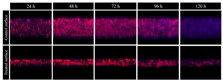
Mycobacterium chimaera is an opportunistic slowly growing non-tuberculous mycobacteriumof increasing importance due to the outbreak of cases associated with contaminated 3T heater-cooler device (HCD) extracorporeal membrane oxygenator (ECMO). The aim of this study was to evaluate the effect of pre-treating a surface with a Methylobacterium sp. CECT 7180 extract to inhibit the M. chimaera ECMO biofilm as well as of the treatment after different dehydration times. Surface adherence, biofilm formation and treatment effect were evaluated by estimating colony-forming units (CFU) per square centimeter and characterizing the amount of covered surface area, thickness, cell viability, and presence of intrinsic autofluorescence at different times using confocal laser scanning microscopy and image analysis. We found that exposing a surface to the Methylobacterium sp. CECT 7180 extract inhibited M. chimaera ECMO biofilm development. This effect could be result of the effect of Methylobacterium proteins, such as DNaK, trigger factor, and xanthine oxidase. In conclusion, exposing a surface to the Methylobacteriumsp. extract inhibits M. chimaera ECMO biofilm development. Furthermore, this extract could be used as a pre-treatment prior to disinfection protocols for equipment contaminated with mycobacteria after dehydration for at least 96 h.

Keywords: 3T HCD; Methylobacterium; Mycobacterium chimaera; antibiofilm effect; biofilm.


Conflict of interest statement

JE received travel grants from Pfizer and conference fees from Biomérieux and Heraeus.

Figures

Figure 1
Figure 1
M. chimaera ECMO adherence to a control surface (gray) and a surface treated with Methylobacterium sp. CECT 7180 extract (red). The bars represent the interquartile range.
Figure 2
Figure 2
M. chimaera ECMO biofilm formation on a control surface (gray) or on a surface treated with Methylobacterium sp. CECT 7180 extract (red). The bars represent the interquartile range. *** p-value < 0.001 for Wilcoxon test.
Figure 3
Figure 3
M. chimaera ECMO biofilm development over time on a control surface (black) and on a surface treated with Methylobacterium extract (red). The four parameters evaluated were (A) mycobacterial viability (%); (B) biofilm thickness (µm); (C) biofilm covered surface (%); and (D) relative autofluorescence (n-fold). The boxes represent the median and interquartile range and the bars indicate tenth and ninetieth percentiles. #: p-value < 0.001 for Wilcoxon test between on a control surface and on a surface treated with Methylobacterium sp. CECT 7180 extract.
Figure 4
Figure 4
3D structure of M. chimaera ECMO biofilms of different ages (24, 48, 72, 96, and 120 h), grown on a control surface (control) or on a treated surface with Methylobacterium sp. CECT 7180 (Treated). In red, the red Nile stain which corresponds to the wall of mycobacteria forming the biofilm, and in blue, the relative autofluorescence, which would correspond to the biofilm matrix.
Figure 5
Figure 5
Amount of M. chimaera ECMO per area unit after different times of dehydration and treated (red) or not (gray) with Methylobacterium extract. The bars represent the interquartile range. #: p-value < 0.001 for Wilcoxon test between a control treatment or a treatment with Methylobacterium sp. CECT 7180 extract.
Figure 6
Figure 6
Silver stained protein band pattern of pure Methylobacterium sp. extract (ME) and detached proteins from Methylobacterium sp. extract adhesion washed once (×1) or twice (×2) separated by SDS Polyacrylamide Gel Electrophoresis (PAGE) (A); Bands selected for LC-MS/MS analysis (B).

References

    1. Diggle S.P. Microbial communication and virulence: Lessons from evolutionary theory. Micobriology. 2010;156:3503–3512. doi: 10.1099/mic.0.045179-0. - DOI - PubMed
    1. Alcaide Fernández de Vega F., Moreno J.E., Martín J.G., Gutiérrez J.J.P. [(accessed on 31 December 2005)];Procedimientos en Microbiología Clínica. 2005 ISBN 8460970329. Available online: https://www.seimc.org/contenidos/documentoscientificos/procedimientosmic....
    1. Mason R.J., Slutsky A.S., Murray E.J., Nadel J.A., Gotway M.B. Murray and Nadel’s Textbook of Respiratory Medicine. Elsevier; Amsterdam, The Netherlands: 2016. Nontubercolous mycobacterial infections.
    1. Faria S., Joao I., Jordao L. General overview on nontuberculous mycobacteria, biofilms, and human infection. J. Pathog. 2015;2015:2–10. doi: 10.1155/2015/809014. - DOI - PMC - PubMed
    1. Johnson M.M., Odell J.A. Nontuberculous mycobacterial pulmonary infections. J. Thorac. Dis. 2014;6:210–220. doi: 10.3978/j.issn.2072-1439.2013.12.24. - DOI - PMC - PubMed

LinkOut - more resources