Review
. 2020 Aug 3;10(8):88.
doi: 10.3390/bios10080088.

The Current Trends of Biosensors in Tissue Engineering

Affiliations
Review

The Current Trends of Biosensors in Tissue Engineering

Yi-Chen Ethan Li et al. Biosensors (Basel). .

Abstract

Biosensors constitute selective, sensitive, and rapid tools for disease diagnosis in tissue engineering applications. Compared to standard enzyme-linked immunosorbent assay (ELISA) analytical technology, biosensors provide a strategy to real-time and on-site monitor micro biophysiological signals via a combination of biological, chemical, and physical technologies. This review summarizes the recent and significant advances made in various biosensor technologies for different applications of biological and biomedical interest, especially on tissue engineering applications. Different fabrication techniques utilized for tissue engineering purposes, such as computer numeric control (CNC), photolithographic, casting, and 3D printing technologies are also discussed. Key developments in the cell/tissue-based biosensors, biomolecular sensing strategies, and the expansion of several biochip approaches such as organs-on-chips, paper based-biochips, and flexible biosensors are available. Cell polarity and cell behaviors such as proliferation, differentiation, stimulation response, and metabolism detection are included. Biosensors for diagnosing tissue disease modes such as brain, heart, lung, and liver systems and for bioimaging are discussed. Finally, we discuss the challenges faced by current biosensing techniques and highlight future prospects of biosensors for tissue engineering applications.

Keywords: biosensors; cell-based biosensors; label-free biosensors; organs-on-chips; tissue engineering; tissue/organ-based biosensors.


Conflict of interest statement

The authors declare no conflict of interest.

Figures
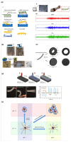
Figure 1
Figure 1
(a) Fabrication process of a curved microneedle by using an acupuncture needle as a mater structure to make a negative polydimethylsiloxane (PDMS) model. Then, filling melt poly(lactic acid( (PLA) into the negative PDMS model, a curved PLA microneedle can be obtained through the casting process. (Reprinted with permission from [30] Copyright (2015) MDPI publishing.); (b) The light-emitting diode (LED) light connected with a carbon nanotube (CNT) ink-printed circuit on a paper before and after folding. Moreover, the CNT ink-printed fibers could be encapsulated in a hydrogel and used as a 3D biosensor for the detection of cardiac electroactivity. (Reprinted with permission from [31] Copyright (2016) John Wiley & Sons publishing.); (c) The morphology and dimensions of a 3D printed ring shape graphene-based biosensor. (Reprinted with permission from [32] Copyright (2018) ACS publications.); (d) A 3D printed technique is used to fabricate a somatosensitive biosensor containing a dorsal sensor (layer 1), an actuator features and an inflation sensor (layer 2), and a contact sensor (layer 3). The resistance change could be recorded and be significant distinguished three type of resistance changed behaviors during the upward, downward, flicked movements of somatosensitive biosensor. (Reprinted with permission from [33] Copyright (2018) ACS publications.); (e) The sensing mechanism of silica/poly(methacrylic acid) (PMAA)- poly(N-isopropylacrylamide) (PNIPPAM) core-shell nanoparticles indicate that temperature and pH effects contribute to the nanoparticles with the enhanced release and moderate release behaviors, respectively. (Reprinted with permission from [34] Copyright (2017) ACS publications.).
Figure 2
Figure 2
(a)-A: A beating cardiac cluster placed on a microelectrode arrays (MEA) sensing area. (a)-B: Schematic representation of the implemented setup (Reprinted with permission from [62] Copyright (2019) ELSEVIER publishing). (b) Schematic Illustration of Detecting Hydrogen Peroxide in the Single-Cell Encapsulated Droplets in Combination with horseradish peroxidase (HRP)-AuNCs (Reprinted with permission from [64] Copyright (2018) ACS publishing). Real case and schematic of the proposed electromechanical cell-based system. (c) Schematic of surface plasmon resonance (SPR) biosensor for vascular endothelial (VE)-cadherin expression for evaluating of human mesenchymal stem cells trans-differentiation to endothelial lineage (Reprinted with permission from [70] Copyright (2017) ELSEVIER publishing). (d) Schematic of the electric cell-substrate impedance sensing (ECIS) system and representative equivalent circuit for an adherent growing cells layer. Left: Cross section of an ECIS culture well. Right: ECIS measures the sum of all individual contributions to the impedance (Reprinted with permission from [74] Copyright (2014) MyJove Corporation publishing.
Figure 3
Figure 3
(a) Schematic illustrating the design and fabrication of a biochip by using the MEA technique. The patterned channels with Indium ion oxide (ITO)-electrodes provide a connected network enabling to guide the neurite outgrowth and measure the neurological signals through impedance signals. (Reprinted with permission from [85] Copyright (2018) ACS publications). (b) Illustrate the preparation of Aβ42-immobilized graphene-based biosensors. The magnetic graphene nanomaterials assembled on an Au electrode provide a re-usable biosensor for rapidly detect the Alzheimer’s disease-related biomarker. (Reprinted with permission from [92] Copyright (2016) Springer Nature Limited.) (c) Schematic illustrating the detection of a Parkinson’s disease (PD) biomarker (α-synuclein) through a liquid crystal biosensor. After the deoxyribonucleic acid (DNA) aptamer on the biosensor surface capturing the α-synuclein, the bright view of liquid crystal could be observed in the polarized microscope. (Reprinted with permission from [93] Copyright (2020) Royal Society of Chemistry) (d) Schematic images of a soft flexible cardiac biosensor. The real-time heart rate and electrocardiogram (ECG) waveforms from this biosensor could be transmitted to smart phone to display a visible signal on an app. Additionally, the flexible polyurethane substrate endows biosensor with an ability to deform under the mechanical twist and bend force. (Reprinted with permission from [94] Copyright (2018) Springer Nature Limited.) (e) A heart-on-a-chip was assembled by an electrode and PDMS channels for culturing cardiac cells. The nanopillars on Au electrodes could be as a transducer to puncture into cellular membrane for the activation ATP-dependent K+ channels and promotion of membrane repolarization. (Reprinted with permission from [95] Copyright (2020) ACS publications) (f) Illustration of a prostate cancer biosensor integrated a microfluidic chip and vascular endothelial growth factor (VEGF)- and prostate-specific antigen (PSA)-immobilized electrodes. The biochip offers a high sensitivity for the detection of prostate cancers and their circulating tumor cells and an easy quick method to analyze by using a UV-Vis absorbance within one hour. (Reprinted with permission from [96] Copyright (2017) Ivy spring International Publisher) (g) Schematic illustrating the principle and operation of the biochip by linking neutravidin with a tether lipid-polymer hybrid nanoparticle (LPHN) and loading extracellular vesicle (EV) on an Au layer. This technique enables to distinguish patients with early- or late-stage pancreatic cancers. (Reprinted with permission from [97] Copyright (2017) Springer Nature Limited.).
Figure 4
Figure 4
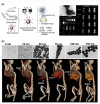
(a) Schematic illustrating ultra small iron oxide nanoparticles as T1 magnetic resonance imaging (MRI) contrast agents and possible integration with positron emission tomography (PET) and computed tomography (CT). The ultra small iron oxide nanoparticles further modified with folic acid enable to significant enhance the labels in the tumor and kidney regions in dual CT scan/MRI images, confirming that the nanoparticles could be used as contrast agents for both CT scan/MRI imaging. (Reprinted with permission from [108] Copyright (2018) Royal Society of Chemistry.) (b) The TEM images of gold nanoparticles with different particle sizes. Representative 3D CT images displayed at a window level of 1090 HU and window width of 930 HU, indicating that controlling the size of nanoparticles can enhance the image contrast at various organs. (Reprinted with permission from [109] Copyright (2019) Springer Nature Limited.).

References

    1. Bhalla N., Jolly P., Formisano N., Estrela P. Introduction to biosensors. Essays Biochem. 2016;60:1–8. - PMC - PubMed
    1. Clark L.C., Jr., Lyons C. Electrode Systems for Continuous Monitoring in Cardiovascular Surgery. Ann. N. Y. Acad. Sci. 1962;102:29–45. doi: 10.1111/j.1749-6632.1962.tb13623.x. - DOI - PubMed
    1. Turner A.P.F. Biosensors: Sense and sensibility. Chem. Soc. Rev. 2013;42:3184–3196. doi: 10.1039/c3cs35528d. - DOI - PubMed
    1. Vigneshvar S., Sudhakumari C.C., Senthilkumaran B., Prakash H. Recent Advances in Biosensor Technology for Potential Applications—An Overview. Front Bioeng. Biotech. 2016;4 doi: 10.3389/fbioe.2016.00011. - DOI - PMC - PubMed
    1. Zhang W., Li X., Zou R., Wu H., Shi H., Yu S., Liu Y. Multifunctional glucose biosensors from Fe(3)O(4) nanoparticles modified chitosan/graphene nanocomposites. Sci. Rep. 2015;5:11129. doi: 10.1038/srep11129. - DOI - PMC - PubMed

LinkOut - more resources