. 2020 Aug 3;21(15):5553.
doi: 10.3390/ijms21155553.

Inhibition of Colony-Stimulating Factor 1 Receptor by PLX3397 Prevents Amyloid Beta Pathology and Rescues Dopaminergic Signaling in Aging 5xFAD Mice

Affiliations

Inhibition of Colony-Stimulating Factor 1 Receptor by PLX3397 Prevents Amyloid Beta Pathology and Rescues Dopaminergic Signaling in Aging 5xFAD Mice

Yeonghoon Son et al. Int J Mol Sci. .

Abstract

Alzheimer's disease (AD) is a progressive neurodegenerative disease. In this study, to investigate the effect of microglial elimination on AD progression, we administered PLX3397, a selective colony-stimulating factor 1 receptor inhibitor, to the mouse model of AD (5xFAD mice). Amyloid-beta (Aβ) deposition and amyloid precursor protein (APP), carboxyl-terminal fragment β, ionized calcium-binding adaptor molecule 1, synaptophysin, and postsynaptic density (PSD)-95 levels were evaluated in the cortex and hippocampus. In addition, the receptor density changes in dopamine D2 receptor (D2R) and metabotropic glutamate receptor 5 were evaluated using positron emission tomography (PET). D2R, tyrosine hydroxylase (TH), and dopamine transporter (DAT) levels were analyzed in the brains of Tg (5xFAD) mice using immunohistochemistry. PLX3397 administration significantly decreased Aβ deposition following microglial depletion in the cortex and hippocampus of Tg mice. In the neuro-PET studies, the binding values for D2R in the Tg mice were lower than those in the wild type mice; however, after PLX3397 treatment, the binding dramatically increased. PLX3397 administration also reversed the changes in synaptophysin and PSD-95 expression in the brain. Furthermore, the D2R and TH expression in the brains of Tg mice was significantly lower than that in the wild type; however, after PLX3397 administration, the D2R and TH levels were significantly higher than those in untreated Tg mice. Thus, our findings show that administering PLX3397 to aged 5xFAD mice could prevent amyloid pathology, concomitant with the rescue of dopaminergic signaling, suggesting that targeting microglia may serve as a useful therapeutic option for neurodegenerative diseases, including AD.

Keywords: Alzheimer’s disease; Alzheimer’s disease mice; Aβ pathology; PLX3397; colony-stimulating factor 1 receptor inhibitor; dopamine D2 receptor; synaptic change.


Conflict of interest statement

The authors declare no conflict of interests.

Figures

Figure 1
Figure 1
Schematic diagram illustrating the experimental groups and protocol. When the 5xFAD mice were 9 months old, they were administered PLX3397 (50 mg·kg−1, p.o.) or vehicle for 30 days. Then, each group underwent positron emission tomography (PET) imaging targeting the dopamine D2 receptor (D2R) and metabotropic glutamate receptor 5 (mGluR5), and histopathological and molecular analyses were also performed.
Figure 2
Figure 2
The colony-stimulating factor 1 receptor (CSF1R) inhibitor PLX3397 decreased Aβ levels and amyloid precursor protein (APP) processing in the cortex of 5xFAD mice. (A) Representative immunofluorescent images of Amyloid β deposits (6E10 in red) and microglia (Iba-1 in green) in the cortex of 5xFAD mice treated with PLX3397. Arrowhead indicates microglia. Scale bar = 100 μm, inserted image scale bar = 25 μm. (B) Representative images of western blots and quantification of expression levels of full-length amyloid-precursor protein (APP-FL), CTFβ, and Aβ in the cortex of WT, Tg, and Tg + PLX3397 mice. (C) Representative images of western blots and quantification of Iba-1 expression in the cortex lysate for the indicated groups. Statistical significance has been defined as *** p < 0.001 WT vs. Tg; # p < 0.05, ## p < 0.01, ### p < 0.001 Tg vs. Tg + PLX3397 group. Error bars indicate SD (n = 4/group).
Figure 3
Figure 3
PLX3397 decreased Aβ levels and protected the neuronal cell layer in the hippocampus of 5xFAD mice. (A) Representative immunofluorescence image showing staining for Aβ (6E10 in red) and microglia (Iba-1 in green) in the hippocampus. Scale bar = 200 μm. (B) Immunoblotting for Aβ and Iba-1 using the hippocampus lysate from each group and Aβ and Iba-1 quantification (n = 4/group). (C) Representative image of the hippocampal region of 5xFAD mice treated with PLX3397 and quantification of thickness in the CA1 region (n = 4 for TG group, n = 5 for WT and Tg + PLX3397 group). Scale bar = 200 μm. Statistical significance has been defined as *** p < 0.001 WT vs. Tg; # p < 0.05, ## p < 0.01, ### p < 0.001 Tg vs. Tg + PLX3397 group. Error bars indicate SD.
Figure 4
Figure 4
Protective effect of PLX3397 treatment on the levels of synapse plasticity-related proteins in 5xFAD mice. Lysate samples of the vehicle or PLX3397-treated cortex or hippocampus were immunoblotted for synaptophysin, or postsynaptic density (PSD)-95 using specific antibodies. β-actin was used as the loading control. Statistical significance has been defined as * p < 0.05, ** p < 0.01, *** p < 0.001 WT vs. Tg; # p < 0.05, ## p < 0.01 Tg vs. Tg + PLX3397 group. Error bars indicate SD (n = 4/group).
Figure 5
Figure 5
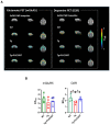
Neuro-PET imaging for evaluating dopamine D2 receptor (D2R) and metabotropic glutamate receptor 5 (mGluR5) expression following PLX3397 treatment. (A) Representative nondisplaceable binding potential (BPND) parametric images for WT and Tg mice. Regional brain BPND levels in Tg mice after PLX3397 administration are shown. (B) Quantification of BPND for mGluR5 and D2R in the WT, Tg, and Tg + PLX3397 groups. * p < 0.05 WT vs. Tg; # p < 0.05 Tg vs. Tg + PLX3397 group. Error bars indicate SD (n = 4/group).
Figure 6
Figure 6
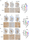
Effects of PLX3397 on the dopaminergic pathway. Representative immunohistochemical images for WT, Tg, and Tg + PLX3397 mice showing staining for dopamine D2 receptor (D2R) (A), tyrosine hydroxylase (TH) (B), and dopamine transporter (DAT) (C) and the quantification of D2R, TH, and DAT levels in the striatum region of the mouse brain (n = 4 for TG group, n = 5 for WT and Tg + PLX3397 group). * p < 0.05 WT vs. Tg; # p < 0.05, ## p < 0.01 Tg vs. Tg + PLX3397 group. Error bars indicate SD.

References

    1. A DeTure M., Dickson D.W. The Neuropathological Diagnosis of Alzheimer’s Disease. Mol. Neurodegener. 2019;14:32. doi: 10.1186/s13024-019-0333-5. - DOI - PMC - PubMed
    1. Hsia A.Y., Masliah E., McConlogue L., Yu G.Q., Tatsuno G., Hu K., Kholodenko D., Malenka R.C., Nicoll R.A., Mucke L. Plaque-independent disruption of neural circuits in Alzheimer’s disease mouse models. Proc. Natl. Acad. Sci. USA. 1999;96:3228–3233. doi: 10.1073/pnas.96.6.3228. - DOI - PMC - PubMed
    1. Folch J., Petrov D., Ettcheto M., Abad S., Sanchez-Lopez E., Garcia M.L., Olloquequi J., Beas-Zarate C., Auladell C., Camins A. Current Research Therapeutic Strategies for Alzheimer’s Disease Treatment. Neural Plast. 2016;2016:8501693. doi: 10.1155/2016/8501693. - DOI - PMC - PubMed
    1. Khan A., Corbett A., Ballard C. Emerging treatments for Alzheimer’s disease for non-amyloid and non-tau targets. Expert Rev. Neurother. 2017;17:683–695. doi: 10.1080/14737175.2017.1326818. - DOI - PubMed
    1. Hansen D.V., Hanson J.E., Sheng M. Microglia in Alzheimer’s disease. J. Cell Biol. 2018;217:459–472. doi: 10.1083/jcb.201709069. - DOI - PMC - PubMed

MeSH terms