Review
. 2020 Aug 3;21(15):5563.
doi: 10.3390/ijms21155563.

A Re-Appraisal of Pathogenic Mechanisms Bridging Wet and Dry Age-Related Macular Degeneration Leads to Reconsider a Role for Phytochemicals

Affiliations
Review

A Re-Appraisal of Pathogenic Mechanisms Bridging Wet and Dry Age-Related Macular Degeneration Leads to Reconsider a Role for Phytochemicals

Roberto Pinelli et al. Int J Mol Sci. .

Abstract

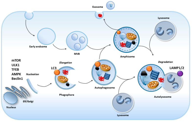
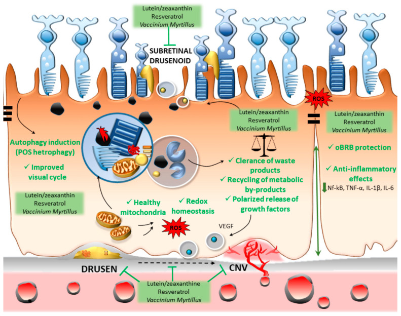
Which pathogenic mechanisms underlie age-related macular degeneration (AMD)? Are they different for dry and wet variants, or do they stem from common metabolic alterations? Where shall we look for altered metabolism? Is it the inner choroid, or is it rather the choroid-retinal border? Again, since cell-clearing pathways are crucial to degrade altered proteins, which metabolic system is likely to be the most implicated, and in which cell type? Here we describe the unique clearing activity of the retinal pigment epithelium (RPE) and the relevant role of its autophagy machinery in removing altered debris, thus centering the RPE in the pathogenesis of AMD. The cell-clearing systems within the RPE may act as a kernel to regulate the redox homeostasis and the traffic of multiple proteins and organelles toward either the choroid border or the outer segments of photoreceptors. This is expected to cope with the polarity of various domains within RPE cells, with each one owning a specific metabolic activity. A defective clearance machinery may trigger unconventional solutions to avoid intracellular substrates' accumulation through unconventional secretions. These components may be deposited between the RPE and Bruch's membrane, thus generating the drusen, which remains the classic hallmark of AMD. These deposits may rather represent a witness of an abnormal RPE metabolism than a real pathogenic component. The empowerment of cell clearance, antioxidant, anti-inflammatory, and anti-angiogenic activity of the RPE by specific phytochemicals is here discussed.

Keywords: autophagy; immunoproteasome; inflammation; lutein; oxidative stress; proteasome; resveratrol; retinal pigment epithelium; retinopathy.


Conflict of interest statement

The authors declare no conflict of interest.

Figures

Figure 1
Figure 1
Schematic overview of the autophagy machinery progression. The recruitment of Atg proteins to the phagophore assembly site (nucleation) is followed by the formation of the phagophore, which engulfs various intracellular substrates. The phagosome gives birth to the autophagosome, which may either fuse with late endosomes (multivesicular bodies, MVB), leading to the formation of the amphisome, or fuse directly with lysosomes. The autolysosome, where cargo degradation eventually occurs, derives from the fusion of the amphisome or autophagosome with the lysosome. When amphisomes do not fuse with lysosomes, partially indigested cargos can be spread extracellularly via exosomes.
Figure 2
Figure 2
Autophagy grants retinal pigment epithelium (RPE) functions and homeostasis. Within the RPE, Beclin 1/Rubicon, AMPK, and Nrf2 trigger heterophagy, which is the concerted action of LC3-associated autophagy and phagocytosis, which is crucial for the degradation and renewal of the outer segments of photoreceptors. A functional autophagy flux within RPE cells also grants redox and energy homeostasis by degrading altered mitochondria and oxidized material, including melanin granules, lipids, and lipofuscin. At the same time, some metabolic by-products, including retinoids and growth factors, are delivered to the photoreceptors or the inner choroid. A functional autophagy status goes along with the maintenance of the outer retinal–blood barrier integrity and a balanced polarity of RPE cells.
Figure 3
Figure 3
Autophagy and proteasome failure within RPE cells may foster AMD onset and progression. In aged RPE cells, impaired heterophagy due to the downregulation of Beclin 1, AMPK, Nrf2/PGC1a, melanoregulain, and crystallin co-chaperones occludes the digestion of the outer segments of photoreceptors, thus promoting the accumulation of damaged mitochondria and oxidized substrates such as lipofuscin and melanin. This further sensitizes RPE cells to light-induced oxidative stress, proteasome impairment, and protein misfolding. This goes along with an altered polarity of RPE cells due to abnormal, exosomal secretion of waste products (melano-lipofuscin, AGEs, oxidized lipids, and beta-amyloid), growth factors (VEGF and crystallins), and cathepsin D at either RPE sides. These events eventually contribute to impairing the visual cycle and photoreceptor’s metabolism, while promoting the formation of drusen and/or subretinal drusenoid deposit, the leakage of the outer blood–retinal barrier (oRBR), the recruitment of inflammatory mediators, and, eventually, CNV and angiogenesis fostering transition from dry to wet AMD.
Figure 4
Figure 4
The effects of phytochemicals within the RPE. Lutein/zeaxanthin, resveratrol, and Vaccinium myrtillus may act through a synergistic approach involving autophagy-inducing, antioxidant, and anti-inflammatory effects. This is expected to produce an empowering of the RPE cells to metabolize properly the excess of waste substrates, while restoring the paracrine homeostasis bridging the REP with photoreceptors, the Bruch’s membrane, and the choriocapillaris. By targeting the autophagy-related dysfunction in the clearance of RPE intracellular material (including oxidized/glycated proteins/lipids, altered mitochondria, and inflammatory molecules) and by restoring the physiological RPE polarity, these phytochemicals may prevent the accumulation of extracellular polymorphic debris (drusen and subretinal drusenoid), as well as the development of choroidal neovascularization (CNV).

References

    1. Pascolini D., Mariotti S.P., Pokharel G.P., Pararajasegaram R., Etya’ale D., Négrel A.D., Resnikoff S. 2002 Global update of available data on visual impairment: A compilation of population-based prevalence studies. Ophthalmic Epidemiol. 2004;11:67–115. doi: 10.1076/opep.11.2.67.28158. - DOI - PubMed
    1. Congdon N., O’Colmain B., Klaver C.C., Klein R., Muñoz B., Friedman D.S., Kempen J., Taylor H.R., Mitchell P. Causes and prevalence of visual impairment among adults in the United States. Arch. Ophthalmol. 2004;122:477–485. - PubMed
    1. Jager R.D., Mieler W.F., Miller J.W. Age-related macular degeneration. N. Engl. J. Med. 2008;358:2606–2617. doi: 10.1056/NEJMra0801537. - DOI - PubMed
    1. De Jong P.T. Age-related macular degeneration. N. Engl. J. Med. 2006;355:1474–1485. doi: 10.1056/NEJMra062326. - DOI - PubMed
    1. Wong W.L., Su X., Li X., Cheung C.M., Klein R., Cheng C.Y., Wong T.Y. Global prevalence of age-related macular degeneration and disease burden projection for 2020 and 2040: A systematic review and meta-analysis. Lancet Glob. Health. 2014;2:CE106–CE116. doi: 10.1016/S2214-109X(13)70145-1. - DOI - PubMed

MeSH terms