Review
. 2020 Aug 5;11(1):89.
doi: 10.1186/s13244-020-00896-1.

Multimodality approach to the nipple-areolar complex: a pictorial review and diagnostic algorithm

Affiliations
Review

Multimodality approach to the nipple-areolar complex: a pictorial review and diagnostic algorithm

Javier Del Riego et al. Insights Imaging. .

Abstract

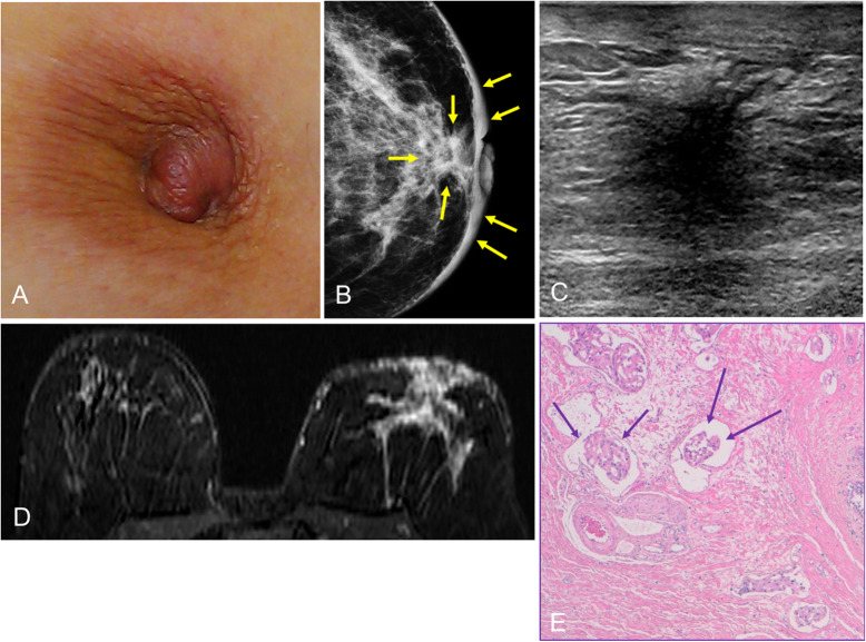
The anatomic and histologic characteristics of the nipple-areolar complex make this breast region special. The nipple-areolar complex can be affected by abnormal development and a wide spectrum of pathological conditions, many of which have unspecific clinical and radiological presentations that can present a challenge for radiologists. The nipple-areolar complex requires a specific imaging workup in which a multimodal approach is essential. Radiologists need to know the different imaging modalities used to study the nipple-areolar complex, as well as their advantages and limitations. It is essential to get acquainted with the acquisition technique for each modality and the spectrum of findings for the different conditions. This review describes and illustrates a combined clinical and radiological approach to evaluate the nipple-areolar complex, emphasizing the findings for the normal morphology, developmental abnormalities, and the most common benign and malignant diseases that can affect this region. We also present a diagnostic algorithm that enables a rapid, practical approach to diagnosing condition involving the nipple-areolar complex.

Keywords: Breast disease; Contrast-enhanced magnetic resonance imaging; Mammography; Nipple-areolar complex; Sonography.


Conflict of interest statement

The authors declare that they have no competing interests.

Figures

Fig. 1
Fig. 1
Anatomy of the nipple-areolar complex
Fig. 2
Fig. 2
Morgagni tubercles. Photograph of a nipple-areolar complex. Note the small bumps in the skin (arrow)
Fig. 3
Fig. 3
Milk lines. Placodes form along the milk lines, which extend from the axillae to the inguinal region (arrow)
Fig. 4
Fig. 4
Developmental abnormalities. a Photograph of polythelia in the right breast of a 45-year-old woman. b Craniocaudal and mediolateral oblique 2D mammograms show a nodule with well-defined margins in the posterior region of the junction between the lower quadrants. c Repeat craniocaudal mammogram with a cutaneous marker confirms that the nodule corresponds to the accessory nipple
Fig. 5
Fig. 5
Mammography positioning. Craniocaudal and mediolateral oblique 2D mammograms show the nipples are perfectly tangential
Fig. 6
Fig. 6
Projection centered in the anterior region. A 56-year-old woman with voluminous breasts. a Craniocaudal 2D mammogram: the nipple is not tangential and is hidden in the lower part of the breast, producing a false image of a nodule (arrow). b Repeat craniocaudal view with the nipple tangential
Fig. 7
Fig. 7
Inverted nipples. A 60-year-old woman with inverted nipples. a Craniocaudal 2D mammograms show bilateral inverted nipples that are perfectly tangential and symmetrical. b Photograph of the same patient
Fig. 8
Fig. 8
Pitfall. a Synthesized craniocaudal 2D mammogram shows multiple dense punctiform images at the level of the nipple mimicking calcifications; these artifacts were caused by remnants of body cream. b Repeat image after cleansing the nipple shows the artifacts have disappeared
Fig. 9
Fig. 9
Nipple calcifications. Screening mammogram in a 54-year-old woman. a Craniocaudal view of the left breast shows a group of calcifications in the retroareolar region (note that the nipple is not tangential). b Magnified view with the nipple perfectly tangential, confirming that the calcifications have benign characteristics and are located in the nipple
Fig. 10
Fig. 10
Periareolar calcifications. A 47-year-old woman with a history of breast reduction surgery. a Synthesized craniocaudal 2D mammogram shows bilateral periareolar calcifications. b Photograph shows the periareolar scar
Fig. 11
Fig. 11
US techniques. Stavros ultrasound techniques to best demonstrate the subareolar and intranipple ducts
Fig. 12
Fig. 12
Physiological enhancement in the nipple-areolar complex. Axial contrast-enhanced T1-weighted spoiled gradient-echo (subtracted) images show various degrees of enhancement in a normal nipple, including none (a), mild symmetric enhancement (b), intense symmetric enhancement (c), a thin symmetric ring of enhancement (d), and asymmetric early enhancement with symmetric late enhancement (e)
Fig. 13
Fig. 13
Pathological enhancement in the nipple-areolar complex. A 71-year-old woman. Axial contrast-enhanced T1-weighted spoiled gradient-echo (subtracted) images show asymmetric irregular nodular early enhancement (a) that is maintained in late phases (b) secondary to involvement by invasive ductal carcinoma. Irregular-shaped masslike enhancement in the middle third of the junction of the outer quadrants in the right breast with linear uptake and segmental distribution to the nipple-areolar complex, compatible with an intraductal component (arrows)
Fig. 14
Fig. 14
Galactography technique. 30-gauge cannula (a); the nipple must be firmly stabilized between the thumb and forefinger (b); canalization of the discharging orifice and contrast injection (c). A magnified craniocaudal view is obtained with the cannula taped in place and the breast compressed
Fig. 15:
Fig. 15:
Ductograms. a Normal ductogram, craniocaudal view. Note the normal “lobular blush” in (b) (arrows), caused by the contrast material filling the lobular portion of the terminal ductal lobular unit. Ninety-degree mediolateral ductogram (c) shows delayed extravasation from excess injection pressure (arrow)
Fig. 16
Fig. 16
Ductal cutoff on galactography. A 65-year-old woman with spontaneous bloody discharge from a single orifice in the right breast and negative findings at mammography and ultrasound (images not shown). a Craniocaudal ductogram shows a concave filling defect situated 2 cm behind the nipple. b Ultrasound obtained after galactography shows ductal ectasia with an intraductal lesion (arrow). c Sagittal T2-weighted MRI shows hyperintense ductal ectasia with an intraductal mass, which on (d) sagittal contrast-enhanced T1-weighted MRI (subtracted image obtained 120 s after contrast injection) corresponds to a mass with differential contrast enhancement (arrow). Histologic study: solitary intraductal papilloma
Fig. 17
Fig. 17
Galactography-guided percutaneous excision biopsy. A 59-year-old woman with a 1-week history of serous discharge from a single orifice in her right nipple. a Ultrasound shows retroareolar ductal ectasia without apparent intraductal lesions (arrows). b Craniocaudal ductogram shows a concave cutoff situated 2.5 cm behind the nipple. c Image of the lesion at the level of the cutoff obtained with the patient positioned prone on the stereotactic table. d The lesion was excised with the Intact-BLES™ biopsy system (Medtronic Inc., Dublin, Ireland). Histology diagnosed intraductal papilloma
Fig. 18
Fig. 18
Ductal ectasia. Different examples of ductal ectasia without interior contents seen on mammography (a) and ultrasound (b, c)
Fig. 19
Fig. 19
Ductal ectasia with intraductal contents. Solitary intraductal papilloma. A 47-year-old woman with serous secretion from a single orifice in the right nipple. a Craniocaudal tomosynthesis slice shows a nodular image with well-defined borders in the retroareolar region of the outer quadrants with a segmental distribution (arrows). b Ultrasound shows ductal ectasia with a well-defined solid nodular lesion (arrows) adjacent to the nipple (N). c Doppler signal due to flow inside the intraductal lesion
Fig. 20
Fig. 20
Ectasia and MRI. Screening MRI in an asymptomatic high-risk 38-year woman 6 months after lactation. a Unenhanced axial and sagittal T1-weighted images. b Contrast-enhanced axial and sagittal T1-weighted MRI (subtracted image obtained 120 s after contrast injection). Note the tubular structures in the retroareolar region of the left breast with a segmental distribution; proteinaceous material causes increased signal intensity on T1-weighted sequences, but not intraductal enhancement
Fig. 21
Fig. 21
Periductal mastitis. Photograph of a 59-year-old smoker with a painful erythematous area in the upper outer quadrant of her right breast (a). Ultrasound shows skin thickening and retroareolar ductal ectasia with echogenic contents (b) and increased periductal Doppler signal (c). Fine-needle aspiration obtained purulent material (pus) (d, e)
Fig. 22
Fig. 22
Subareolar abscess. Photograph of a 42-year-old woman with a painful erythematous palpable areolar mass in her left breast with mild involvement of the adjacent skin (a). Synthesized 2D mammogram shows marked skin thickening of the nipple-areolar complex without other underlying findings (b). Ultrasound shows a heterogeneous hypoechogenic intradermal collection compatible with an abscess (c). Fine-needle aspiration was able to drain the collection completely (purulent material), and the patient was prescribed antibiotics and follow-up (d, e)
Fig. 23
Fig. 23
Zuska’s disease. a Photograph of a 47-year-old woman with a history of smoking who had a recurrent painful erythematous palpable subareolar mass with a small secreting ulcerous lesion. b Ultrasound shows skin thickening with a small hypoechogenic collection below the lesion (arrows), with increased peripheral color-Doppler signal (c)
Fig. 24
Fig. 24
Solitary intraductal papilloma. A 68-year-old woman with a palpable retroareolar nodule in her left breast. a Normal findings on craniocaudal and mediolateral oblique 2D mammograms. b Ultrasound shows a cystic lesion with a solid nodule inside it (arrow) adjacent to the nipple (N). c Hematoxylin-eosin stain (× 4) shows branching intraductal structures consisting of a central fibrovascular axis surrounded by epithelial and myoepithelial cells
Fig. 25
Fig. 25
Intraductal filling defect. A 48-year-old woman with bloody discharge from a single orifice in the right breast and negative findings at mammography and ultrasound (images not shown). a Craniocaudal ductogram shows a filling defect just behind the secreting orifice (arrow). Histologic study: solitary intraductal papilloma. b Photograph of a bluish nodule that appeared in the same breast five years later; c ultrasound shows the lesion as a solid nodule (arrow). Histologic study: solitary intraductal papilloma (inverted)
Fig. 26
Fig. 26
Nipple adenoma. An 81-year-old woman with a several-week history of pain and swelling of the right breast. a Mammogram shows an isodense rounded retroareolar mass with slightly irregular margins (arrows); b on ultrasound, it is seen as a solid nodular lesion (N: nipple). c Immunohistochemistry stain with p63 (× 4) shows glandular and ductal proliferation consisting of epithelial and myoepithelial cells, which express p63
Fig. 27
Fig. 27
Syringomatous tumor of the nipple. A 47-year-old woman with retraction and hardening of the left nipple. a Mammogram shows retraction of the left nipple and asymmetric retroareolar density. b On ultrasound, the lesion is solid and hypoechogenic with ill-defined borders and increased peripheral color-Doppler signal. c Contrast-enhanced coronal T1-weighted MRI (subtracted image obtained 120 s after contrast injection) shows mass-type uptake with pronounced early enhancement in a lesion with hazy borders that retracts the nipple-areolar complex. d Hematoxylin-eosin stain (× 4) shows a proliferation of elongated glandular structures like strings of cells in the dermis, with the formation of keratin cysts (star)
Fig. 28
Fig. 28
Epidermal inclusion cyst. A 37-year-old woman with a palpable retroareolar nodule in her right breast, without inflammation. a Synthesized 2D mammogram (craniocaudal view) shows a well-defined isodense nodule measuring 25 mm (arrows) adjacent to the nipple. b Ultrasound shows a well-defined hypoechogenic nodule below the skin of the right areola (N nipple)
Fig. 29
Fig. 29
Retroareolar cyst in an adolescent. A 15-year-old girl presented with a palpable retroareolar mass in her left breast. Ultrasound shows a large thin-walled cystic lesion (a). Follow-up ultrasound 6 months later shows a marked decrease in the size of the lesion, which is oval and elongated (b)
Fig. 30
Fig. 30
Radiologic patterns in gynecomastia. a Nodular. b Dendritic. c Diffuse
Fig. 31
Fig. 31
Cutaneous manifestations of Paget’s disease of the nipple. a Crusted ulcerated papule in the center of the nipple. b Scaly erythematous plaque with erosions that destroys the nipple. c Scaly erythematous plaque that covers the entire nipple
Fig. 32
Fig. 32
Paget’s disease (I). A 56-year-old woman with right nipple retraction. a 2D mammogram shows a spiculated retroareolar mass in the right breast with nipple retraction and skin thickening; b on ultrasound, it is seen as a solid lesion with ill-defined borders. c MRI shows the retroareolar lesion extending to the nipple-areolar complex. Histologic study revealed infiltrating ductal carcinoma extending to the epidermis
Fig. 33
Fig. 33
Paget’s disease (II). An 81-year-old woman with an ulcerated lesion in her left nipple. a Synthesized 2D mammogram shows slight skin thickening in the left nipple-areolar complex (arrows). b MRI shows differential pathologic enhancement of the left nipple-areolar complex. c, d Hematoxylin-eosin-stained punch-biopsy specimen (× 0.3 and × 4, respectively) shows ductal carcinoma with extensive infiltration of the nipple and ulceration of the epidermis (Paget’s disease)
Fig. 34
Fig. 34
Ductal carcinoma in situ. a Photograph of an 85-year-old woman with a large palpable retroareolar mass with marked involvement of the right nipple-areolar complex. b Synthesized 2D mammogram shows a circumscribed retroareolar mass with associated pleomorphic calcifications. c Ultrasound shows a cystic mass with debris inside and a solid mural nodule with a penetrating vessel in the power-Doppler study. d Hematoxylin-eosin stain (× 10). Fragment of the cyst wall with slight chronic inflammatory involvement and vascular congestion and epithelial lining made up of few layers of markedly pleomorphic elements and dense eosinophilic cytoplasm. Note the proliferation of the papillary pattern, with fronds with wide fibroconnective stems lined with similar pleomorphic elements (intraductal carcinoma within the cyst). e Close-up (Hematoxylin-eosin stain × 20)
Fig. 35
Fig. 35
Invasive ductal carcinoma (I). An 88-year-old woman with a palpable nodule in the lower inner quadrant of her left breast. a Synthesized 2D mammogram shows a spiculated nodule (circle), classified as infiltrating lobular carcinoma at histology. Note the thickening of the skin and of the nipple-areolar complex (arrows). b MRI shows pathologic asymmetric enhancement of the left nipple-areolar complex. c Hematoxylin-eosin stain of punch-biopsy specimen (× 10) shows extensive dermal infiltration by infiltrating ductal carcinoma. d Immunohistochemistry (× 4) shows diffuse nuclear expression of estrogen receptors. These are two synchronous tumors
Fig. 36
Fig. 36
Invasive ductal carcinoma (II). a Photograph of a 54-year-old woman with left nipple retraction. b Mammogram shows skin thickening and increased retroareolar density; c on ultrasound, the lesion is hypoechogenic and ill-defined. d MRI shows a large lesion in the left breast involving the nipple-areolar complex. e Hematoxylin-eosin stain (× 4) shows lymphatic invasion of the dermis of the nipple by infiltrating ductal carcinoma (arrows)
Fig. 37
Fig. 37
Radiation-associated angiosarcoma. A 76-year-old woman with a history of breast cancer treated with conservative surgery and whole-breast radiation therapy 6 years prior. a Photograph shows an ill-defined erythematous-violaceous infiltrated plaque with an eroded area occupying part of the areola. b Synthesized 2D mammogram shows skin thickening and interstitial edema in the retroareolar region of the left breast. c Ultrasound shows an ill-defined hypoechoic skin lesion with an internal Doppler signal. Punch biopsy diagnosed radiation-associated angiosarcoma, and the patient underwent mastectomy
Fig. 38
Fig. 38
Diagnostic imaging algorithm

References

    1. Stone K, Wheeler A. A Review of anatomy , physiology , and benign pathology of the nipple. Ann Surg Oncol. 2015;22:3236–3240. doi: 10.1245/s10434-015-4760-4. - DOI - PubMed
    1. Reisenbichler E, Hanley KZ. Seminars in diagnostic pathology developmental disorders and malformations of the breast. Semin Diagn Pathol. 2019;36:11–15. doi: 10.1053/j.semdp.2018.11.007. - DOI - PubMed
    1. Pasquali P, Freites-Martinez A, Fortuño A. Nipple adenoma: new images and cryosurgery treatment. Breast J. 2016;22:584–585. doi: 10.1111/tbj.12636. - DOI - PubMed
    1. Nicholson BT, Harvey JA, Cohen MA. Nipple-areolar complex : normal anatomy and benign and malignant processes. Radiographics. 2009;29:509–523. doi: 10.1148/rg.292085128. - DOI - PubMed
    1. Geffroy D, Doutriaux-Dumoulins I. Clinical abnormalities of the nipple-areola complex: the role of imaging. Diagn Interv Imaging. 2015;96:1033–1044. doi: 10.1016/j.diii.2015.07.001. - DOI - PubMed

LinkOut - more resources