. 2020 Nov:95:56-68.
doi: 10.1016/j.neurobiolaging.2020.06.018. Epub 2020 Jul 3.

Apolipoprotein E isoforms differentially regulate matrix metallopeptidase 9 function in Alzheimer's disease

Affiliations

Apolipoprotein E isoforms differentially regulate matrix metallopeptidase 9 function in Alzheimer's disease

Charis Ringland et al. Neurobiol Aging. 2020 Nov.

Abstract

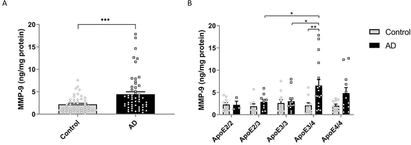
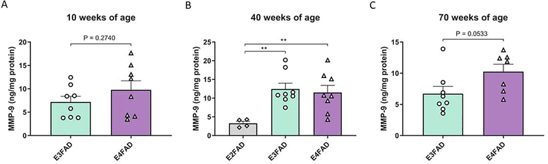
Apolipoprotein E (APOE) has been shown to influence amyloid-β (Aβ) clearance from the brain in an isoform-specific manner. Our prior work showed that Aβ transit across the blood-brain-barrier was reduced by apoE4, compared to other apoE isoforms, due to elevated lipoprotein receptor shedding in brain endothelia. Recently, we demonstrated that matrix metallopeptidase 9 (MMP-9) induces lipoprotein receptor proteolysis in an apoE isoform-dependent manner, which impacts Aβ elimination from the brain. The current studies interrogated the relationship between apoE and MMP-9 and found that apoE impacted proMMP-9 cellular secretion from brain endothelia (apoE2 < apoE3 = apoE4). In a cell-free assay, apoE dose-dependently reduced MMP-9 activity, with apoE4 showing a significantly weaker ability to inhibit MMP-9 function than apoE2 or apoE3. Finally, we observed elevated MMP-9 expression and activity in the cerebrovasculature of both human and animal AD brain specimens with an APOE4 genotype. Collectively, these findings suggest a role for apoE in regulating MMP-9 disposition and may describe the effect of apoE4 on Aβ pathology in the AD brain.

Keywords: Alzheimer’s disease; Apolipoprotein E; Binding affinity; Cerebrovasculature; Enzyme regulation; Matrix metallopeptidase 9.


Figures

Figure 1:
Figure 1:. ApoE genotype effect on total MMP-9 levels in the cerebrovasculature of human brain tissue in AD and control subjects.
Levels of total MMP-9 were found to be elevated in (A) Alzheimer’s disease (AD) subjects compared to non-demented controls and (B) apoE4 carriers with AD. MMP-9 levels in the cerebrovasculature were determined using an ELISA. Values were normalized to total protein concentration and represent mean ± SEM. (A) n = 49 (control) n = 55 (AD). ***p<0.001 as determined by an unpaired t-test. (B) Control subjects: apoE2/2: N = 10, apoE2/3: N = 9, apoE3/3: N = 10, apoE3/4: N = 10, apoE4/4: N = 10. AD subjects: apoE2/2: N = 3, apoE2/3: N = 10, apoE3/3: N = 13, apoE3/4: N = 17, apoE4/4: N = 12. *p<0.05, **p<0.01 as determined by two-way ANOVA and the BKY procedure.
Figure 2:
Figure 2:. ApoE genotype effect on active MMP-9 levels in the cerebrovasculature of human brain tissue in AD and control subjects.
Using a quantitative substrate-based MMP-9 activity assay, levels of endogenously active MMP-9 were found to be elevated in (A) Alzheimer’s disease (AD) subjects compared to non-demented controls and (B) apoE4 carriers with AD. Values were normalized to total protein concentration and represent mean ± SEM. (A) n = 44 (control) n = 48 (AD). *p<0.05 as determined by an unpaired t-test. (B ) Control subjects: apoE2/2: N = 8, apoE2/3: N = 8, apoE3/3: N = 13, apoE3/4: N = 8, apoE4/4: N = 7. AD subjects: apoE2/2: N = 3, apoE2/3: N = 9, apoE3/3: N = 11, apoE3/4: N = 15, apoE4/4: N = 10. *p<0.05 as determined by two-way ANOVA and the BKY procedure.
Figure 3.
Figure 3.. MMP-9 expression in mouse cerebrovasculature tissue from EFAD mice at 10, 40 and 70 weeks of age.
Levels of MMP-9 were measured in (A) E3FAD and E4FAD mice at 10 weeks of age, (B) E2FAD, E3FAD and E4FAD mice at 40 weeks of age and (C) E3FAD and E4FAD mice at 70 weeks of age. MMP-9 levels were found to be higher in E3FAD and E4FAD mice relative to E2FAD mice at 40 weeks of age. MMP-9 levels were determined using an ELISA. Values were normalized to total protein concentration and represent mean ± SEM (E2FAD: N=4, E3FAD & E4FAD: N=8 per age group). (B) **p<0.01 as determined by ANOVA and the BKY procedure. (A, C) No significant differences as determined by an unpaired t-test.
Figure 4.
Figure 4.. ApoE isoform differences in MMP-9 secretion in conditioned media from apoE-treated HBMECs.
Using a quantitative substrate-based MMP-9 activity assay, levels of (A, D, G) endogenously active MMP-9, (B, E, H) proMMP-9 and (C, F, I) total MMP-9 were analyzed in the presence of apoE2, 3 or 4 in the conditioned extracellular media of HBMECs. Cells were stimulated with either (A, B, C) apoE alone, (D, E, F) PMA and apoE or (G, H, I) Aβ-42 and apoE. ApoE isoform effects on secreted proMMP-9 levels and total secreted MMP-9 were apparent upon stimulation/insult. Levels of active MMP-9 were elevated in an apoE isoform dependent manner (apoE2<apoE3<apoE4) when added alone and after stimulation with PMA. Values represent mean ± SEM. (A, B, C) n = 14, (D, E, F) n = 10, (G, H, I) n = 8. $$p<0.01, $$$$p<0.0001 indicates significant differences relative to all other conditions and *p<0.05, **p<0.01, ***p<0.001, ****p<0.0001 indicates significant differences relative to indicated bars as determined by ANOVA and the BKY procedure.
Figure 5.
Figure 5.. Effect of apoE isoform on the conversion of MMP-9 to the active form.
Recombinant human MMP-9 was incubated with (A, B) recombinant (C, D) glia-lipidated (GCM) or (E, F) artificially lipidated recombinant human apoE2, 3 or 4 or no apoE (control) before being activated with APMA. (A, C, E) Gelatin zymography demonstrating pro and active MMP-9 bands. (B, D, F) Quantification of zymogram bands (ratio of active to proMMP-9). (B) n = 17, (D) n = 13, (F) n = 5–6. Values represent mean ±± SEM. N=3. *p<0.05, ***p<0.001, ****P<0.0001 as determined by ANOVA and the BKY procedure.
Figure 6:
Figure 6:. Differential modulation of MMP-9 activity by apoE isoforms.
MMP-9 activity was significantly modulated by apoE in an isoform and dose-dependent manner (apoE2>apoE3>apoE4). Differences between each of the apoE isoforms were statistically significant at concentrations ≥ 5 ng/ml. Treatment of 1 μM SB-3CT was included as a positive control and showed considerable attenuation of MMP-9 activity. Values represent mean ± SEM (N = 3) and are expressed as fluorescent units. Statistical significance was determined by two-way ANOVA followed by the BKY procedure. *p<0.05, **p<0.01, ***p<0.001, ****p<0.0001. Asterisk symbols above bars indicates significant differences compared to MMP-9 alone. Asterisk symbols above brackets indicates significant differences from other apoE isoforms at that concentration.
Figure 7:
Figure 7:. Kinetic binding studies of MMP-9 and apoE2, apoE3 or apoE4 using Bio-Layer Interferometry.
Biotinylated recombinant human MMP-9 was immobilized to streptavidin biosensors (1800 seconds). Recombinant apoE was then added in increasing concentrations (dark blue: 0.25 μg/ml, red: 0.5 μ/ml, light blue: 1 μg/ml, green: 2.5 μg/ml, orange: 5 μg/ml, purple: 10 μg/ml) to separate sensors then allowed to dissociate in PBS (A: apoE2. Association time: 1200s, Dissociation time: 1200s, B: apoE3. Association time: 1800s, Dissociation time: 3800s, C: apoE4. Association time: 1200s, Dissociation time: 1200s). Individual binding affinities were assessed by measuring the dissociation constant (KD) (G, H) determined from the steady state (D: apoE2, E: apoE3, F: apoE4). ApoE4 demonstrated the weakest binding affinity; the KD for apoE4 was greater compared to apoE3 and apoE2. Values represent mean ± SD. N=5–6.
Figure 8:
Figure 8:. MMP-9 and apoE immunoreactivities in cortices of E3FAD and E4FAD mice.
Representative confocal images depicting 6-month-old female (A) E3FAD and (B) E4FAD mice stained with antibodies against CD31/PECAM-1 (AF3628, red), apoE [E6D7], (AB1907, purple) MMP-9 (AB38898, green). Both E3FAD and E4FAD mice exhibited a prominent MMP-9 signal that overlapped with apoE in endothelial cells surrounding blood vessels. (C) Quantification of MMP-9 immunofluorescent intensity within endothelial cells (CD31/PECAM-1). (D) Quantification of MMP-9 area within endothelial cells (CD31/PECAM-1). (E) Percentage overlap of MMP-9 and apoE areas normalized to total MMP-9 area. The scale bars represent 50μm.

References

    1. Aleshkov S, Abraham CR, Zannis VI, 1997. Interaction of nascent ApoE2, ApoE3, and ApoE4 isoforms expressed in mammalian cells with amyloid peptide beta (1–40). Relevance to Alzheimer’s disease. Biochemistry 36, 10571–10580. 10.1021/bi9626362 - DOI - PubMed
    1. Bachmeier C, Paris D, Beaulieu-Abdelahad D, Mouzon B, Mullan M, Crawford F, 2013. A multifaceted role for apoE in the clearance of beta-amyloid across the blood-brain barrier. Neurodegener. Dis. 11, 13–21. 10.1159/000337231 - DOI - PubMed
    1. Bachmeier C, Shackleton B, Ojo J, Paris D, Mullan M, Crawford F, 2014. Apolipoprotein E isoform-specific effects on lipoprotein receptor processing. Neuromolecular Med. 16, 686–696. 10.1007/s12017-014-8318-6 - DOI - PMC - PubMed
    1. Backstrom J, Miller C, Tökés Z, 1992. Characterization of neutral proteinases from Alzheimer-affected and control brain specimens: identification of calcium-dependent metalloproteinases from the hippocampus. J. Neurochem. 58, 983–992. 10.1111/j.1471-4159.1992.tb09352.x - DOI - PubMed
    1. Backstrom JR, Lim GP, Cullen MJ, Tökés ZA, 1996. Matrix metalloproteinase-9 (MMP-9) is synthesized in neurons of the human hippocampus and is capable of degrading the amyloid-beta peptide (1–40). J. Neurosci. Off. J. Soc. Neurosci. 16, 7910–7919. - PMC - PubMed

Publication types

MeSH terms